

Therapeutic Micro-Nano Technology BioMEMs - Tejlal Desai & Sangeeta Bhatia
.pdf254 |
SARAH L. TAO AND TEJAL A. DESAI |
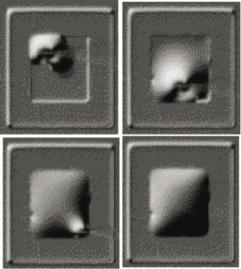
FIGURE 14.15. AFM scanning images of surfaces before and after surface modification. A) PMMA. B) Amine functionalized PMMA. C) Porous silicon. B) Lectin-modified porous silicon.
14.6.1. Welled Silicon Dioxide and PMMA Microdevices
These reservoirs were filled with picoto nanoliters of a polymeric solution using microinjectors (Figure 14.16). Water quickly evaporates from these reservoirs leaving behind the drug contained in polymer which acts as a timed-release plug. Using a specific type of polymer predetermines the time and rate of release of drug from the reservoir; for example, a hydrogel that swells in response to a specific pH, solvent or temperature or a polymer with a known dissolution rate. Different polymers with various dissolution rates can then be used in separate reservoirs to obtain controlled release of several compounds compounds.
14.6.2. Porous Silicon Microdevices
Capillary action was exploited in order to load the porous silicon microdevices. Homogeneous liquid formulations were prepared and slowly loaded onto the porous silicon particles using a micropipettor. The solution was then taken into the porous particles by
MICRODEVICES FOR ORAL DRUG DELIVERY |
255 |
FIGURE 14.16. Time sequence of a microdevice reservoir filled by microinjection.
capillary action. The microdevices were then dried by applying vacuum pressure until any air trapped within the pores was released.
14.6.3. Caco-2 In Vitro Studies
The Caco-2 cell line, derived from human colorectal carcinoma cells, is one of the most useful in vitro models for the study of intestinal epithelial permeability and function as well as a model for drug delivery. The Caco-2 cell line is known to differentiate spontaneously into enterocytes to form polarized monolayers in culture [50]. When grown to confluency these cells highly resemble the small intestinal epithelium both, structurally and functionally. The microvilliated cells in the monolayer maintain tight intercellular junctions and expose only their apical, brush border membranes [51]. The cells also maintain transport systems, enzymes, and ion channels similar to those of the intestinal epithelium. The glycosylation pattern of the Caco-2 cells is characterized by high amounts of N-acetylglucosamine containing oligosaccharides followed by fucosyland mannosyland minor amounts of galactosamineand N-acetyl-galactosamine-residues [52].
14.6.4. Cell Culture Conditions
Caco-2 cells were grown in culture medium consisting of Eagle’s minimum essential media supplemented with Earle’s Balanced Salt Solution, 2.0 mM L-glutamine, 1.0 mM sodium pyruvate, 0.1 mM nonessential amino acids, 1.5 g/L sodium bicarbonate, 20% FBS and 20 μg/ml gentamycin sulfate antibiotic. Cells were maintained in a humidified 5% CO2/95% air atmosphere at 37◦C while renewing media every second day. Cells were subcultured by trypsinization with 0.25% trypsin-EDTA. For experiments, cells were seeded into either six-well plates or Costar Transwell inserts (0.4 μm pore size, and 4.7 cm2 surface
256 |
SARAH L. TAO AND TEJAL A. DESAI |
area). Before seeding, culture surfaces were coated with rat collagen in order to promote cell adhesion. Caco-2 monolayers grown between 14 and 30 days were used in all experiments.
14.6.5. Assessing Confluency and Tight Junction Formation
Cell confluency and tight junction formation, as a function of time, can be assessed using a number of techniques. All techniques rely on the measurement of selected substance from one tissue compartment to the opposing compartment. The most frequently used method is by measuring transepithelial electrical resistance. A square current pulse ( I) is passed across the epithelium and the resulting voltage response ( V) is measured. Using Ohm’s law, the conductance can be calculated ( I/ V). Here, cell monolayers exhibiting TEER values of at least 220 cm2 were used in transport studies. Although this method is rapid and simple to implement, it can often be inaccurate due to nonuniform current fields caused by the point source nature of the current passing electrodes and can increase infection of the culture system [53]. A simple, inexpensive spectroscopic method can instead be used to assess cell confluency, tight junction formation, and fluid flow [54]. Using this method, the flux of phenol red across a cell monolayer when grown on a transwell permeable support is observed. Phenol red is a pH indicator used in commercially available media. It is nontoxic to epithelial cells and is not metabolized or synthesized by the cells. Because phenol red is a pH indicator, its absorbance properties are a function of pH; however at a wavelength of 479 nm, its isosbestic point, it is not pH sensitive. The concentration of phenol red in a solution can be calculated by applying the Beer-Lambert Law using the extinction coefficient for phenol red (8450 M−1 cm−1). The flux of phenol red and the permeability of the monolayer can be calculated using the following equations:
J = (A479b × Volb)/(t × A × EC × Pl) |
(14.4) |
P = J/[PR] |
(14.5) |
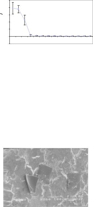
Where J is the phenol red flux, A479b is the absorbance of the basolateral solution at 479 nm, Volb is the volume of the basolateral solution at the time of measurement, t is time, A is the surface area of the epithelium, EC is the extinction coefficient, and Pl is the light path. P denotes the permeability to phenol red and [PR] is the phenol red concentration in the apical compartment. Days 1–4 represent cell confluence, days 4–8 represents complete formation of tight junction and inhibition of cell proliferation, and days 8–21 represent a small but marginally significant further decrease in permeability (Figure 14.17).
14.6.6. Adhesion of Lectin-Modified Microdevices
In vitro studies were performed using the Caco-2 cell line to measure the bioadhesive properties of lectin-conjugated microdevices (Figure 14.18). The binding characteristics of microdevices modified with two types of lectin (tomato, known to agglutinate Caco-2 cells, and peanut, an unrelated lectin) were observed as a function of time. Although both lectin conjugates produce a higher degree of binding than the pristine microdevices, a marked difference still remains between the peanut and tomato lectin conjugates. The amount of binding associated with tomato lectin-modified microdevices is thought to be a direct result of the high amounts of N-acetylglucosamine-containing oligosaccharides which are
MICRODEVICES FOR ORAL DRUG DELIVERY |
257 |
|
250 |
|
|
|
|
|
|
|
|
|
|
|
|
|
|
200 |
|
|
|
|
|
|
|
|
|
|
|
|
|
(cm/s) |
150 |
|
|
|
|
|
|
|
|
|
|
|
|
|
|
|
|
|
|
|
|
|
|
|
|
|
|
|
|
-7 |
100 |
|
|
|
|
|
|
|
|
|
|
|
|
|
x 10 |
|
|
|
|
|
|
|
|
|
|
|
|
|
|
|
|
|
|
|
|
|
|
|
|
|
|
|
|
|
app |
50 |
|
|
|
|
|
|
|
|
|
|
|
|
|
|
|
|
|
|
|
|
|
|
|
|
|
|
|
|
P |
|
|
|
|
|
|
|
|
|
|
|
|
|
|
|
0 |
|
|
|
|
|
|
|
|
|
|
|
|
|
|
1 |
2 |
3 |
4 |
5 |
6 |
7 |
8 |
9 |
12 |
14 |
16 |
19 |
21 |
|
- 50 |
|
|
|
|
|
|
|
|
|
|
|
|
|
|
|
|
|
|
|
|
|
Day |
|
|
|
|
|
|
FIGURE 14.17. Change in apparent permeability as the Caco-2 cells become confluent and form tight junctions.
associated with Caco-2 cells. These oligosaccharides provide specific sites for the tomato lectins to bind. In contrast, only minor amounts of galactosamine residues are present on Caco-2 cells, limiting the number of peanut lectin specific binding sites. Figure 14.19 shows the binding trend of unmodified, peanut lectin-modfied, and tomato lectin-modified PMMA microdevices.
14.6.7. Bioavailibility Studies
The Caco-2 model was used to test drug transport through paracellular tight junctions, similar to those found in the intestinal lining, as a model for systemic bioavailability. Upon microdevice application, the rate of permeation of a model drug, FITC-labeled insulin, through the monolayer was measured using fluorescence spectroscopy. Concurrent measurement of the total resistance across the membrane was also measured to determine the magnitude of the physiologic response of tight junction opening. The effect of transport by microdevice delivery was compared to that of a liquid formulation. It was found that drug transport efficiency was augmented when drug formulations were delivered from
FIGURE 14.18. Scanning electron micrograph of adherent PMMA microdevices on a Caco-2 monolayer.

258 |
SARAH L. TAO AND TEJAL A. DESAI |
|
100 |
|
|
|
|
|
90 |
|
|
|
|
|
80 |
|
|
|
|
(%) |
70 |
|
|
|
|
|
|
|
|
|
|
Bound |
60 |
|
|
|
|
50 |
|
|
|
|
|
|
|
|
|
|
|
Percent |
40 |
|
|
|
|
30 |
|
|
|
|
|
20 |
|
|
|
|
|
|
|
|
|
|
|
|
10 |
|
|
|
|
|
0 |
|
|
|
|
|
0 |
15 |
30 |
60 |
120 |
Time (Minutes)
 Tomato Lectin Microdevice
 Peanut Lectin Microdevice

 Control Microdevice
FIGURE 14.19. Binding of PMMA microdevices to Caco-2 cell monolayers as a function of incubation time.
microdevices as compared to liquid formulations. This efficiency increased further with the additional use of a permeation enhancer (Figure 14.20). This dramatically augmented flux when utilizing microdevice delivery is a result of a very high concentration of both permeation enhancer (sodium laurate) and FITC-insulin directly at tight junctions.
% FITC-insulin transported / h
12
10
8
6
4
2
0
-2 0
-4
- Constant number of particles (monolayer)
- Constant dose per particle (insulin)
 % transported / h (uncapped)
 % transported / h (liquid)
|
100 |
200 |
300 |
|
sodium laurate (ug)
FIGURE 14.20. Transport rate of FITC-insulin across Caco-2 monolayers when delivered from porous silicon microdevices and in liquid formulation in the presence of encorporated sodium laurate permeation enhancer.
ACKNOWLEDGEMENTS
Funding is gratefully acknowledged from iMEDD, Inc., The Whitaker Foundation, The National Science Foundation, The Center for Integrated Medicine and Technology, and The National Aeronautic and Space Administration. Also, special thanks to those who
MICRODEVICES FOR ORAL DRUG DELIVERY |
259 |
have contributed to this work: Christopher Bonner, Aamer Ahmed, and Michael Lubeley or the University of Illinois-Chicago; Ketul Popat and Simon Su of Boston University; colleagues from iMEDD, Inc.
REFERENCES
[1]S. Henry, D.V. McAllister, M.G. Allen, and M.R. Prausnitz. Microfabricated microneedles: a novel approach to transdermal drug delivery. J. Pharm. Sci., 87:922–925, 1998.
[2]D.V. McAllister, M.G. Allen, and M.R. Prausnitz. Microfabricated microneedles for gene and drug delivery. Annu. Rev. Biomed., 2:289–313, 2000.
[3]L. Leoni and T.A. Desai. Nanoporous biocapsules for the encapsulation of insulinoma cells: biotransport and biocompatibility considers. IEEE Trans. Biomed. Eng., 48:1335–1341, 2001.
[4]L. Leoni, T. Boriarski, and T.A. Desai. Characterization of nonporous membranes for immunoisolation: diffusionproperties and tissue effects. J. Biomed. Microdev., 4:131–139, 2001.
[5]J.T. Santini, M.J. Cima, and R. Langer. A controlled-release microchip. Nature, 397:335–338, 2000.
[6]J.T. Santini, A.C. Richards, R. Scheidt, M.J. Cima, and R. Langer. Microchips as controlled drug-delivery devices. Agnew. Chem. Int. Ed. Engl., 39:2396–2407, 2000.
[7]G. Orive, R.M. Hernandez, A.R. Gascon, A. Dominguez-Gil, and J.L. Pedraz. Drug delivery in biotechnology: present and future. Curr. Opin. Biotechnol., 14:659–664, 2003.
[8]S.S. Davis and L. Illum. Drug delivery systems for challenging molecules. Int. J. Pharm., 176:1–8, 1998.
[9]M. Saffran, G.S. Kumar, D.C. Neckers, J. Pena, R.H. Jones, and J.B. Field. Biodegradable azopolymer coating for oral delivery of peptide drugs. Biochem. Soc. Trans., 18:752–754, 1990.
[10]K. Iwanaga, S. Ono, K. Narioka, M. Kakemi, K. Morimoto, S. Yamashita, Y. Namba, and N. Oku. Application of surface-coated liposomes for oral delivery of peptide: effect of coating the liposome’s surface on the GI transit of insulin. J. Pharm. Sci., 88:248–252, 1999.
[11]G. Ponchel and J. Irache. Specific and non-specific bioadhesive particulate systems for oral delivery to the gastrointestinal tract. Adv. Drug Deliv. Rev., 34:191–219, 1998.
[12]M.A. Arrangoa, G. Ponchel, A.M. Orecchioni, M.J. Renedo, D. Duchene, and J.M. Irache. Bioadhesive potential of gliadin nanoparticulate systems. Eur. J. Pharm. Sci., 11:333–341, 2000.
[13]C.M. Lehr. Lectin-mediated drug delivery: the second generation of bioadhesives. J. Control. Rel., 65:19–29, 2000.
[14]A. Fasano and S. Uzzau. Modulation of intestinal tight junctions by Zonula occludens toxin permits enteral administration of insulin and other macromolecules in an animal model. J. Clin. Invest., 99:1158–1164, 1997.
[15]U.I. Schwarz, T. Gramatte, J. Krappweis, R. Oertel, and W. Kirch. P-glycoprotein inhibitor erythromycin increasesoral bioavailability of talinolol in humans. Int. J. Clin. Pharmacol. Ther., 38:161–167, 2000.
[16]J. Woodley. Bioadhesion: new possibilities for drug administration? Clin. Pharmacokinet., 40:77–84, 2001.
[17]E.C. Lavelle. Targeted delivery of drugs to the gastrointestinal tract. Crit. Rev. Ther. Drug Carrier Syst., 18:341–386, 2001.
[18]M. Montisci, A. Dembri, G. Giovannuci, H. Chacun, D. Duchene, and G. Ponchel. Gastrointestinal transit and mucoadhesion of colloidal suspensions of Lycopersicon esculentum L. and Lotus tetragonolobus lectin-PLA microsphere conjugates in rats. Pharm. Res., 18:829–837, 2001.
[19]M.A. Clark, B.H. Hirst, and M.A. Jepson. Lectin-mediated mucosal delivery of drugs and microparticles. Adv. Drug Deliv. Rev., 43:207–223, 2000.
[20]R. Mody, S. Joshi, and W. Chaney. Use of lectins as diagnostic and therapeutic tools for cancer. J. J. Pharm. Toxic. Methods., 33:1–10, 1995.
[21]A. Putzai. Lectin-targeting of microparticles to different parts of the gastrointestinal tract. Proc. Intern. Symp. Control. Rel Bioact. Mater., 22:161–162, 1995.
[22]F.J. Martin and C. Grove. Microfabricated drug delivery systems: concepts to improve clinical benefit.
Biomed. Microdev., 3:97–108, 2001.
[23]A. Ahmed, C. Bonner, and T.A. Desai. Bioadhesive microdevices with multiple reservoirs: a new platform fororal drug delivery. J Control Rel., 3:291–306, 2002.
260 |
SARAH L. TAO AND TEJAL A. DESAI |
[24]S.L. Tao, M.W. Lubeley, and T.A. Desai. Bioadhesive poly(methyl methacrylate) microdevices for controlled drug delivery. J. Control Rel., 2215–2228, 2003.
[25]A.B. Foraker, R.J. Walczak, M.H. Cohen, T.A. Boiarski, C.F. Grove, and P. Swann. Microfabricated porous silicon particles enhance paracellular delivery of insulin across intestinal Caco-2 cell monolayers. Pharmaceut. Res., 20:110–116, 2003.
[26]D.L. Polla, A.G. Erdman, W.P. Robbins, D.T. Markus, J. Diaz-Diaz, R. Rizq, Y. Nam, and H.T. Brickner. Microdevices in medicine. Annu. Rev. Biomed. Eng., 2:551–576, 2000.
[27]M. Madou. Fundamentals of Microfabrication. Boca Raton, CRC 2003.
[28]N. Maluf. An Introduction to Microelectromechanical Systems Engineering. Boston, Artech House, 2000.
[29]A. Uhlir. Electrolytic shaping of germanium and silicon. Bell. Syst. Tech. J., 35:333–347, 1956.
[30]A. Angelescu, I. Kleps, M. Mihaela, M. Simion, T. Neghina, S. Petrescu, N. Moldovan, C. Paduraru, and A. Raducanu. Porous silicon matrix for applications in biology. Rev. Adv. Mater. Sci., 5:240–449, 2003.
[31]L.T. Canham. Properties of porous silicon. EMIS Datare View Series. London, INSPEC, 1997.
[32]F. Cunin, T.A. Schmedake, J.R. Link, Y.Y. Li, J. Koh, S.N. Bhatia, and M.J. Sailor. Biomolecular screening with encoded porous-silicon photonic crystals. Nat. Mater., 1:39–41, 2002.
[33]A. Henry, T. Tutt, M. Gallowaty, Y. Davidson, S. McWhorter, S. Soper, and R. McCarley. Surface modification of poly(methyl methacrylate) used in the fabrication of microanalytical devices. Anal. Chem., 72:5331–5337, 2000.
[34]M. Jager and A. Wilke. Comprehensive biocompatibility testing of a new PMMA-hA bone cement versus conventional PMMA cement in vitro. J. Biomater. Sci. Polym. Ed., 11:1283–1298, 2003.
[35]K.T. Chu, Y. Oshida, E.B. Hancock, M.J. Kowolik, T. Barco, and S.L. Zunt. Hydroxyapatite/PMMA composites as bone cements. Biomed. Mater. Eng., 1:87–105, 2004.
[36]M.G. Harris, N.H. Wong, and A.W. Low. Patient response to PMMA contact lenses. J. Am. Optom. Assoc., 46:1184–1187, 1975.
[37]G.N. Papaliodis, Q.D. Nguyen, C.M. Samson, and C.S. Foster. Intraocular lens tolerance in surgery for cataracta complicata: assessment of four implant materials. Semin. Ophthalmol.,17(3–4):120–123, Sep-Dec, 2002.
[38]V.J. Tomazic-Jezic, K. Merritt, and T.H. Umbreit. Significance of the type and the size of biomaterial particles on phagocytosis and tissue distribution. J. Biomed. Mater. Res., 55:523–529, 2001.
[39]Y. Tabata, Y. Inoue, and Y. Ikada. Size effect on systemic and mucosal immune responses induced by oral administration of biodegradable microspheres. Vaccine, 14:1677–1685, 1996.
[40]Y. Tabata and Y. Ikada. Phagocytosis of polymer microspheres by macrophages. Adv. Poly. Sci., 94:107–141, 1990.
[41]I.H. Al-Abdulla, G.W. Mellor, M.S. Childerstons, A.M. Sidki, and D.S. Smith. Comparison of three different activation methods for coupling antibodies to magnetisable cellulose particles. J. Immunol. Meth., 122:253– 258, 1989.
[42]A. Rembaum, S.P. Yen, and E. Cheong. Functional polymeric microspheres based on 2-hydroxyethyl methacrylate for immunocheical studies. Macromolecules, 9:328–336, 1976.
[43]J.H. Bowes and C.W. Cater. The reaction of gluteraldehyde with proteins and other biological materials. J. Roy. Microsc. Soc., 85:193–200, 1966.
[44]H. Otto, H.Takamiya and A.Vogt. A two stage method for crosslinking antibody globulin to ferritin by glutaraldehyde. Comparison between the one-stage and the two-stage method. J. Immuno. Meth., 2:127– 146, 1973.
[45]K.J. Sultzbaugh and T.J. Speaker. A method to attach lectins to the surface of spermine alginate microcapsules based on the avidin biotin interaction. J. Microencapsul., 13:363–375, 1996.
[46]S.L. Tao, M.W. Lubeley, and T.A. Desai. Synthesis of cytoadhesive poly(methylmethacrylate) for applications in targeted drug delivery. J. Biomed. Mater. Res., 67:369–75, 2003.
[47]B. Karandikar, J. Puschett, and K. Matyjaszewski. Homogeneous and heterogeneous modificatin of poly(methyl methacrylate) with ethylene diamine. Polym. Prepr. (Am. Chem. Soc., Div. Polym. Chem.), 30:250–251, 1989.
[48]A.C. Henry, T.J. Tutt, M. Galloway, Y. Davidson, C.S. McWhorter, S. Soper, and R. McCarley. Surface odificatin of poly(methyl methacrylate) used in the fabrication of microanalytical devices. Anal. Chem., 72:5331–5337, 2000.
[49]R.K. Gaur and K.C. Gupta. A spectrophotometric method for the estimation of amino groups on polymer supports. Anal. Biochem., 180:253–258, 1989.
MICRODEVICES FOR ORAL DRUG DELIVERY |
261 |
[50]C.M. Lehr, J. Bouwstra, W. Kok, A. Noach, A. Boer, and H. Junginger. Bioadhesion by means of specific binding to tomato lectin. Pharm. Res., 9:547–553, 1992.
[51]G.J. Russell-Jones, H. Veitch, and L. Arthur. Lectin-mediated transport of nanoparticles across Caco-2 and OK cells. Int. J. Pharm., 190:165–174, 1999.
[52]F. Gabor, M. Stangl, and M. Wirth. Lectin-mediated bioadhesion: binding characteristics of plant lectins on the enterocyte-like cell lines Caco-2, HT-29 and HCT-8. J. Control. Rel., 55:131–142, 1998.
[53]S.A. Lewis. Assessing epithelial cell confluence by spectroscopy. Methods Mol. Biol., 188:329–336, 2002.
[54]B. Jovov, N.K. Wills, and S.A. Lewis. A spectroscopic method for assessing confluence of epithelial cell cultures. Am. J. Physiol., 261:C1196–C1203, 1991.
15
Nanoporous Implants for
Controlled Drug Delivery
Tejal A. Desai1, Sadhana Sharma2, Robbie J. Walczak3, Anthony Boiarski3, Michael Cohen3, John Shapiro2, Teri West3, Kristie Melnik3, Carlo Cosentino4, Piyush M. Sinha, and Mauro Ferrari5
1Department of Bioengineering and Physiology, University of California, San Francisco, CA 2Department of Physiology and Biophysics, University of Illinois at Chicago, Chicago, IL
3IMEDD Inc., Foster City, CA
4Department of Experimental and Clinical Medicine, University of Catanzaro “Magna Græcia”, Catanzaro, Italy
5Professor, Brown Institute of Molecular Medicine Chairman, Department of Biomedical Engineering, University of Texas Health Science Center, Houston, TX; Professor of Experimental Therapeutics, University of Texas M.D. Anderson Cancer Center, Houston, TX; Professor of Bioengineering, Rice University, Houston, TX; Professor of Biochemistry and Molecular Biology, University of Texas Medical Branch, Galveston, TX; President, the Texas Alliance for NanoHealth, Houston, TX
15.1. INTRODUCTION
15.1.1. Concept of Controlled Drug Delivery
Over the last three decades considerable advances have marked the field of drug delivery technology, resulting in many breakthroughs in clinical medicine. However, major unmet needs remain. Among these are broad categories of: 1) Continuous release of therapeutic agents over extended time periods and in accordance to a pre-determined temporal profile [38, 60]; 2) Local delivery at a constant rate to the tumor microenvironment, to overcome much of the systemic toxicity and improve anti-tumor efficacy; 3) Improved ease of administration, increasing patient compliance, while minimizing the needed intervention of healthcare personnel and decreasing the length of hospital stays [6, 46]. Success in addressing some or all of these challenges would potentially lead to improvements in efficacy and patient compliance, as well as minimization of side effects [66].
264
Plasma
in
Concentration
Drug
1
Oral/ IV
Bolus
TEJAL A. DESAI ET AL.
Overdose
Toxicity
Drug Waste
Membrane |
Therapeutic |
Implant |
Window |
|
Under-dose
Sub therapeutic
2 |
3 |
4 |
5 |
6 |
7 |
Days
Oral/ IV
Bolus
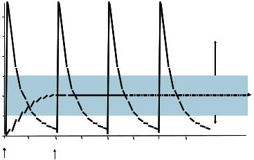
FIGURE 15.1. Concept of Controlled Drug Delivery.
The importance of controlled-release drug delivery systems may be argued with reference to the goal of achieving a continuous drug release profile consistent with zero-order kinetics, wherein blood levels of drugs would remain constant throughout the delivery period. By contrast, injected drugs follow first-order kinetics, with initial high blood levels of the drug after initial administration, followed by an exponential fall in blood concentration [65]. Toxicity often occurs when blood levels peak, while efficacy of the drug diminishes as the drug levels fall below the therapeutic range (Figure 15.1). The potential therapeutic advantages of continuous-release drug delivery systems are thus significant, and encompass: In vivo predictability of release rates on the basis of in vitro data, minimized peak plasma levels and thereby reduced risk of adverse reactions, predictable and extended duration of action, reduced inconvenience of frequent dosing and thereby improved patient compliance [7, 39].
15.1.2. Nanopore Technology
Nanopore technology provides excellent opportunities for controlled drug delivery through the fabrication of microfabricated nanoporous membranes. Unlike conventional polymeric membranes, silicon membranes are biologically, thermally, chemically, and mechanically stable in vivo-like environments. Furthermore, compared to alternate nanopore fabrication technology such as ion track-etched polycarbonate (e.g. Osmonics, Minnetonka, MN) and porous alumina (e.g. Whatmann Ann Arbor, Michigan), microfabricated nanoporous membranes have uniform pore size and very low thickness. These membranes are ideally suited for drug delivery applications, including controlled diffusion and sustained release. It is possible to design nanopore membranes, which achieve almost constant rate of drug delivery, avoiding the “burst effect”. By precisely controlling pore size, pore length and pore density, the nanopore membrane fitted with a drug reservoir suitable