

Therapeutic Micro-Nano Technology BioMEMs - Tejlal Desai & Sangeeta Bhatia
.pdf214 |
´ ´ |
FREDERIQUE CUNIN, YANG YANG LI, AND MICHAEL J. SAILOR |
12.1. SYSTEM DESIGN CONSIDERATIONS
The most common forms of drug delivery are oral injested tablets, injection, transdermal patches and implantable fixtures. Each of these modes of delivery imposes its own set of requirements. For example, the rate of release of a drug depends on numerous bioenvironmental conditions such as pH, circulation of fluid, viscosity, temperature, ionic strength, adsorption of specific or non-specific biomolecules, and local redox potential of the surrounding medium. Some general requirements include:
Either before or after release, the drug must display some preferential targeting or partitioning to the intended tissue or organ.
The system must protect the rest of the organism from intoxication, which generally means that it should avoid any release during transport to the target.
It must load the appropriate amount of active molecule for optimal availability and therapeutic efficacy upon arrival at the target.
It must protect the incorporated active molecule from degradation before reaching the target. This implies that the drug delivery system is chemically inert to the entrapped drug.
It must control the rate of delivery, which is specific to each therapeutic case. For example, one might want to obtain a sustained profile of release over an extended period of time for prolonged therapeutic effect. A pulsative release that mimics physiological secretion behaviour is preferred in the case of delivery of insulin for the treatment of diabetes, for example.
In some cases it is desirable that the system initiates release in response to an external stimulus (magnetic, electrical, ultrasonic, photonic, thermal, etc.).
In the case of a self-regulated system, release in response to changes in the local bio-environment such as pH, temperature, enzyme activity, antibody concentration, etc. This implies a biosensing feature in addition to release.
The fixture must disintegrate in the body for biocompatibility and biodegradability purposes.
Finally, the drug delivery system must satisfy long term toxicological requirements while respecting the patient’s compliance with the regimen.
Traditional controlled-release drug delivery systems have used polymer materials as key components because of the particular range of physical and chemical properties they offer such as diffusivity, permeability, biocompatibility, solubility, and their response to pH or temperature changes. The diffusion, dissolution, permeation and swelling characteristics of these materials have been utilized to obtain constant release of entrapped molecules [2, 3]. In most cases, polylactide (PLA), and polyglycolide (PGA) have been used in either homopolymer or copolymer form for in vivo clinical applications because of their low toxicity.
12.2. POROUS MATERIAL-BASED SYSTEMS
Porous nanostructured materials (with pore sizes ranging from a few nanometers to several microns) have emerged as a new class of efficient vehicles for drug delivery. Made of various components including polymers [4], lipids (for example liposomes, micelles,

NANODESIGNED PORE-CONTAINING SYSTEMS FOR BIOSENSING |
215 |
TABLE 12.1. Common “Soft” materials used in controlled drug release.
Material |
Comments |
|
|
Polymer nanocapsules [7] |
—provides efficient drug protection |
SIZE: Smaller than 1000 nm |
—provides efficient controlled release of entrapped drug |
DESCRIPTION: Single polymeric |
—biodegradable |
membrane enclosing an aqueous |
—tunable surface chemistry for targeting |
or oily cavity |
—suitable for cancer therapy (accommodates multiple drug resistance). |
Liposomes [8] |
—well tolerated excipient, safe for medical applications |
SIZE: 25–2500 nm |
—provides efficient drug protection |
DESCRIPTION: vesicles made from |
—Biomimetic artificial system (model for cell membranes when made |
single or multi bilayered |
of natural phospholipids) |
phospholipid membranes |
—can be made sensitive to pH changes, light, magnetic fields. |
enclosing an aqueous cavity. |
—can entrap substances in either the membrane or in the cavity |
|
—flexibility in formulation of chemotherapeutic agents |
|
—amenable to mass production |
|
—poor storage stability |
|
—faster degradation rate in vivo than polymers |
Solid lipid nanoparticles [6] SIZE: 80–1000 nm DESCRIPTION: particles made
from lipids that are solid at room temperature and body temperature (in contrast to emulsions, where the lipid phase is liquid).
—more stable in biological fluid and during storage than liposomes —good protection against drug degradation
“Liposil”: silicic |
—very stable formulation |
liposome-templated nanocapsules |
—silica shell resistant to low pH (gastric conditions) |
[9, 10] |
—promising for oral administration drug delivery systems |
SIZE: 1 to several microns |
—possibility of long-term storage and protection of molecules |
DESCRIPTION: unilamellar |
—slow release rates-amenable to longer term regimen |
phospholipid membrane vesicle |
—dry formulation |
encapsulated in a non porous |
|
silica shell |
|
|
|
microemulsions, nanoemulsions, solid lipid nanoparticles) [5, 6], and inorganic materials, reservoir-containing structures in general have the advantage of providing flexibility in preparation. One can control morphological and chemical parameters such as the size and shape of the material, the number of “reservoirs” and their volume, the wall thickness as well as the surface chemistry, permeability and resorption rate. Porous materials allow the design of more sophisticated drug carrier systems for better control of the vectorisation and the release kinetics of the drug. A variety of soft materials are under study, outlined in Table 12.1.
12.3. SILICON-BASED POROUS MATERIALS
A recent approach in the development of controllable reservoir-based drug delivery systems involves the use of more rigid inorganic porous solids as substrate materials for the preparation of functionalized organic/inorganic nanostructured drug carriers [11]. In particular, nanostructured materials based on Si are very promising platforms for pharmaceutical
216 |
´ ´ |
FREDERIQUE CUNIN, YANG YANG LI, AND MICHAEL J. SAILOR |
applications. Porous silica [12, 13] and porous Si [14] are good examples of this class. Their more complex architectural and chemical structures provide these hybrid systems with specific functionalities that allow them to respond to a designated stimulus. Widely studied and characterized, mesoporous silica from the M41S family are templated materials made by a chemical route. The synthesis typically involves self assembly of silica using surfactant micelles as the structuring agent [15–21]. These non-toxic materials exhibit well-defined, ordered porosity with a large specific surface area (up to 1000 m2 g−1), a large mesoporous volume, and thermal stability. The pore sizes can be controlled during the synthesis, and typically range from 15 Å to 100 Å. The silanol-terminated pore walls can be functionalized using convenient chemistry to provide specificity for drug absorption and release schemes [13, 22].
12.4. “OBEDIENT” MATERIALS
Like electronically engineered microchips, some hybrid drug delivery systems can be designed to respond to external commands. For example “gates” that will open and close the pores can be installed, releasing the right amount of drug when desired. Tanaka and coworkers have developed a system where coumarin ligands are attached at the entrance of the pores of the mesoporous silica MCM41 [23, 24]. Irradiation with light causes dimerization of the coumarin and closes the pores of the matrix. The process is reversible, allowing the pores to open and release the stored active compound. In a similar manner Lin and coworkers have shown that nanocrystals made of CdS can be functionalized and attached to the surface of the pores of porous silica MCM41 and play the role of caps to trap drugs in the porous matrix. Release of the drug is then triggered by a specific chemical or enzymatic reaction that removes the CdS caps [12].
12.5. POROUS SILICON
The attraction of placing active electronic circuit components into in-vivo drug delivery materials led to the exploration of elemental silicon as a biomaterial. In particular a porous form of Si produced by an electrochemical corrosion reaction has been of interest. Since the pioneering work of Canham and others in the late 90’s demonstrating the biocompatibility and biodegradability of porous Si in vitro and in vivo [25–35], this material has been under intensive investigation for controlled drug delivery applications. Like mesoporous silica of the MCM41 class, porous Si offers tuneable structural properties: a large specific surface area, large free volume, and pore sizes that can be controlled from a few nanometers to several hundreds of nanometers depending on the preparation conditions. The surface of freshly prepared porous Si is easily modified via convenient chemistry with a large range of organic or biological molecules (ex: antibody, proteins, etc.) [36]. Recently Swaan and coworkers have performed in vitro experiments showing that porous Si particles can be used as efficient delivery vehicles of insulin across intestinal epithelial cells [37]. The drug permeation rate through the membrane is dramatically enhanced when delivered via porous Si particles compared with conventional liquid formulations.
Like other Si-based materials, porous Si offers attractive morphological and chemical properties for biomedical applications but it has one supplementary dimension: its optical
NANODESIGNED PORE-CONTAINING SYSTEMS FOR BIOSENSING |
217 |
properties. Porous Si displays fluorescence deriving from Si quantum dot structures that are produced during the etch [38], and it can also display unique optical reflectivity spectra [39, 40]. Both of these features allow porous Si to exhibit a signal that is affected in a predictable way when exposed to environmental changes [32, 41–46]. This presents new possibilities for the development of more advanced functional systems, referred to as “intelligent systems,” that incorporate a sensor for either diagnostic or therapeutic functions. The ease with which porous Si can be integrated into well-established Si microelectronics fabrication techniques should lead to more sophisticated, active devices for medical applications [35, 47–48].
12.6. TEMPLATED NANOMATERIALS
Any porous solid can act as a structural template, and the fabrication of ordered nanostructures using templates has been investigated extensively [49]. Porous alumina membranes [50–51], zeolites [20], and crystalline colloidal arrays [52–54] are commonly used as templates to construct elaborate electronic, mechanical, or optical structures. Recent work has employed other template materials such as porous Si [55–56], and the use of templated materials for biosensing [57] and drug delivery [56] has been demonstrated. For in-vivo applications, templated structures allow one to impart a unique microor nanostructure into a recognized biocompatible and therapeutically useful material. One obvious advantage of such an approach is that it can provide an additional degree of control over the dissolution rate of the fixture and the drug release profile. Most recently, the possibility of incorporating a unique optical signature such as a photonic crystal has emerged as an additional feature of the template approach.
12.7. PHOTONIC CRYSTALS AS SELF-REPORTING BIOMATERIALS
Photonic crystals are materials whose index of refraction varies with a periodicity on the order of a few hundred nanometers over length scales of at least a few microns. Such structures can diffract visible light. Familiar examples from Nature include opals, the inside of abalone shells [58], and the carapace of many species of beetle [59]. Photonic crystals constructed from inorganic materials are an active area of research for optical switching, optical computing, and other optoelectronics applications [60], and the capabilities of these materials to act as sensors for chemical or biological compounds has led to a series of developments in the biomedical field. One of the early demonstrations of the potential for photonic crystals in medicine came from the laboratories of Sanford Asher at the University of Pittsburgh. By incorporating a photonic crystal into a biocompatible hydrogel matrix, the Asher group developed contact lenses that change color depending on the concentration of glucose in the wearer’s blood [57, 61]. Sensors for proteins, DNA, and small molecules have also been developed based on these and related photonic crystals [44, 62–64].
12.8. USING POROUS SI AS A TEMPLATE FOR OPTICAL NANOSTRUCTURES
As mentioned above, porous Si offers tuneable structural properties, which makes it an interesting candidate material for controlled drug delivery. In addition, with its easily

218 |
´ ´ |
FREDERIQUE CUNIN, YANG YANG LI, AND MICHAEL J. SAILOR |
|
Infuse polymer |
Remove |
|
|
template |
porous Si photonic |
porous Si-polymer |
polymer film |
crystal template |
composite |
photonic crystal |
FIGURE 12.1. Template approach for constructing photonic materials for controlled release and monitoring of drugs.
controlled nanostructure, porous Si has been demonstrated to be an excellent template material for construction of elaborate photonic organic and biological polymers [56]. The advantage of such an approach is that it allows one to impart the desirable optical features of the porous Si master to a polymer that possesses the required biocompatibility, resorbability, or drug solubility parameters.
A schematic of the templating method used to produce an optical structure suitable for medical applications is shown in Figure 12.1. This approach was first demonstrated using a rugate dielectric mirror as the template [56]. A rugate dielectric mirror is a structure that contains a sinusoidal refractive index variation, producing a sharp spectral feature in the optical spectrum. The multilayered porous Si template containing nanometer-scale pores is prepared by anodic electrochemical etch of a crystalline Si wafer using a pseudo-sinusoidal current-time waveform [39, 62, 65–69]. The sharp features in the optical reflectivity spectrum are controlled by adjustment of the frequency and amplitude of the sinusoidal currenttime waveform [70].
One challenge of the templating approach is to efficiently remove the master from the templated biomaterial, particularly if the biocompatibility of the master is an issue. The porous Si multilayer masters can be converted to SiO2 by thermal oxidation prior to solution-casting or injection-molding of the daughter material. Removal of the porous SiO2 template from the polymer or biopolymer imprint can then be achieved by exposure to a dilute solution of HF. Alternatively, the porous Si template dissolves in strong aqueous base. In either case, chemical dissolution of the template provides a freestanding porous polymer film [56]. The polymer replicas inherit an inverse of the optical structure of the template.
It is possible to fabricate more sophisticated optical structures from porous Si films. In 1995, Pavesi and Mazzoleni reported the first microcavity made entirely out of porous Si [40]. These were luminescent structures comprised of planar microcavities with a luminescent porous Si active medium sandwiched between two distributed Bragg reflectors. Recently the superposition of multiple spectral features in one monolithic film has been demonstrated [70–71]. By constructing more elaborate optical structures, one can design into the material characteristic spectral “bar codes” that allow the fixture to be distinguished from tissue, light scattering centers, or highly colored materials in the body [56, 66].
A self-reporting drug delivery matrix is desired for various non-invasive or minimally invasive applications. Implants that change their spectral properties as they degrade or as they release a loaded drug could be imaged using visible light if implanted in transparent media such as the vitreous body of the eye. Alternatively, if near-infrared, tissue-penetrating
NANODESIGNED PORE-CONTAINING SYSTEMS FOR BIOSENSING |
219 |
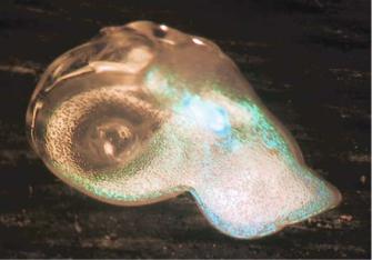
FIGURE 12.2. Poly(lactide) molded from a porous Si photonic crystal template. This polymer contains the drug caffeine. Decay of the optical spectrum (the green color visible in the image) from this fixture provides a surrogate measure of the drug delivery rate.
spectral features are encoded into the material, the fixture conceivably could be probed through the skin or though several millimetres of visibly opaque tissue. This latter concept has been demonstrated with a rugate optical structure made of biodegradable poly-lactide, impregnated with a test drug (Figure 12.2) [56]. Drug release correlates to the decrease in intensity of light reflected by the rugate structure as expected. By placing the spectral feature of the poly-lactide imprint within the low absorbance, near infrared window of human tissue, a drug delivery matrix that could be read through the skin was demonstrated [56].
12.9. OUTLOOK FOR NANOTECHNOLOGY IN PHARMACEUTICAL RESEARCH
The use of porous Si photonic crystals as templates is just one example among many of the application of nanomaterials to the pharmaceutical field. Additional examples include polymer nanoparticles in which the drug is uniformly dispersed, metallic nanoparticles that contain a surface coating of drug, and microporous materials that contain the drug within nanometer-dimension voids. In all of these systems, the high surface to volume ratio of nanoparticles makes them amenable to surface modification for efficient targeting. Advances in nanostructured materials should lead to other approaches for the controlled release of drugs, and they will continue to provide exciting opportunities in medicine.
ACKNOWLEDGEMENTS
The authors gratefully acknowledge helpful discussions with Prof. Sangeeta Bhatia of the Department of Bioengineering at the University of California, San Diego, Dr. Erkki Ruoslahti of the Burnham Institute in La Jolla, California, Dr. Lingyun Cheng and
220 |
´ ´ |
FREDERIQUE CUNIN, YANG YANG LI, AND MICHAEL J. SAILOR |
Dr. William Freeman of the Shiley Eye Center at the University of California, San Diego, and Dr. Jean-Marie Devoisselle and Sylvie Begu of the UMR CNRS/ENSCM in Montpellier, France. The authors thank TREX industries, inc. and the National Cancer Institute of the National Institutes of Health for financial support.
REFERENCES
[1]D.L. Wise. (ed.). Handbook of Pharmaceutical Controlled Release Technology. Marcel Dekker Inc., New York, 2000.
[2]W. Amass, A. Amass, and B. Tighe. A review of biodegradable polymers: uses, current developments in the synthesis and characterization of biodegradable polyesters, blends of biodegradable polymers and recent advances in biodegradation studies. Polym. Int., 47:89–144, 1998.
[3]K.E. Uhrich, S.M. Cannizzaro, R.S. Langer, and K.M. Shakesheff. Polymeric systems for controlled drug release. Chem. Rev., 11:3181–3198, 1999.
[4]K.S. Soppimath, T.M. Aminabhavi, A.R. Kulkarni, and W.E. Rudzinski. Biodegradable polymeric nanoparticles as drug delivery devices. Control. Rel., 70:1–20, 2001.
[5]B. Heurtault, P. Saulnier, B. Pech, J.E. Proust, and J.P Benoit. Biomaterials, 24:4283, 2003.
[6]R.H. Muller, M. Radke, and S.A. Wissing. Solid lipid nanoparticles (SLN) and nanostructured lipid carriers (NLC) in cosmetics and dermatological preparations. Adv. Drug Delivery Rev., 54 (Suppl. 1):S131–S155, 2002.
[7]I. Brigger, C. Dubernet, and P. Couvreur. Nanoparticles in cancer therapy and diagnosis. Adv. Drug Del. Rev., 54:631–651, 2002.
[8]R.R.C. New (ed.). Liposomes a practical approach, Oxford University Press, New York, 1990.
[9]S. B´egu et al. Preparation and characterization of silicious material using liposomes as template. Chem. Commun., 640–641, 2003.
[10]S. B´egu et al. Characterization of a phospholipid bilayer entrapped into non-porous silica nanospheres. J. Mater. Chem., 14:1316–1320, 2004.
[11]E. Ruiz-Hitzky. Functionalizing inorganic solids: towards organic-inorganic nanostructured materials for intelligent and bioinspired systems. The Chem. Rec., 3:88–100, 2003.
[12]C.-Y. Lai et al. A mesoporous silica nanosphere-based carrier system with chemically removable CdS nanoparticle caps for stimuli-responsive controlled release of neurotransmitters and drug molecules. J. Am. Chem. Soc., 125:4451–4459, 2003.
[13]R. Aiello et al. Mesoporous silicate as matrix for drug delivery systems of non-steroidal antiinflamatory drugs. Stud. Surf. Sci. Catal., 14:1165–1172, 2002.
[14]L.T. Canham et al. Derivatized porous silicon mirrors: implantable optical components with slow resorbability. Phys. Stat. Sol. A, 182:521–525, 2000.
[15]U. Cieslaa and F. Schuth¨ . Ordered mesoporous materials. Micropor. Mesopor. Mater., 27:131–149, 1999.
[16]J.S. Beck and J.C. Vartuli Recent advances in the synthesis, characterization and applications of mesoporous molecular sieves. Curr. Opin. Sol. State. Mat. Sci., 1:76–87, 1996.
[17]J.Y. Ying, C.P. Mehnert, and M.S. Wong, Angew. Chem., 38:58, 1999.
[18]N. Husing¨ and U. Schubert. Aerogels—airy materials: chemistry, structure, and properties. Angew. Chem., 37:22–45, 1998.
[19]A. Sayari. Catalysis by crystalline mesoporous molecular sieves. Chem. Mater., 8:1840–1852, 1996.
[20]K. Moller and T. Bein. Inclusion chemistry in periodic mesoporous hosts. Chem. Mater., 10:2950–2963, 1998.
[21]A. Stein, B.J. Melde, and R.C. Schroden. Hybrid inorganic-organic meso-porous silicates-nanoscopic reactors coming of age. Adv. Mater., 12:1403–1419, 2000.
[22]C. Tourn´e-Peteilh et al. The potential of ordered mesoporous for the storage of drugs: the example of a pentapeptide encapsulated in a MSU-Tween 80. Chem. Phys. Chem., 3:2003.
[23]N.K. Mal, M. Fujiwara, and Y. Tanaka. Photocontrolled reversible release of guest molecules from coumarinmodified mesoporous silica. Nature, 421:350–353, 2003.
[24]N.K. Mal, M. Fujiwara, Y. Tanaka, T. Taguchi, and M. Matsukata. Photo-switched storage and release of guest molecules in the pore void of coumarin-modified MCM-41. Chem. Mater., 15:3385–3394, 2003.
NANODESIGNED PORE-CONTAINING SYSTEMS FOR BIOSENSING |
221 |
[25]S.H.C. Anderson, H. Elliott, D.J. Wallis, L.T. Canham, and J.J. Powell. Dissolution of different forms of partially porous silicon wafers under simulated physiological conditions. Phys. Stat. Solidi a-Appl. Res., 197:331–335, 2003.
[26]J.M. Ji, X. Li, L.T. Canham, and J.L. Coffer. Use of microcontact printing methods to direct pattern formation of calcified mesoporous silicon. Adv. Mat., 14:41–43, 2002.
[27]T. Jay, L.T. Canham, K. Heald, C.L. Reeves, and R. Downing. Autoclaving of porous silicon within a hospital environment: potential benefits and problems. Phys. Stat. Solid. A-Appl. Res., 182, 555–560, 2000.
[28]L.T. Canham et al. Derivatized mesoporous silicon with dramatically improved stability in simulated human blood plasma. Adv. Mat., 11:1505-+, 1999.
[29]M. Wainwright, L.T. Canham, K. Al-Wajeeh, and C.L. Reeves. Morphological changes (including filamentation) in Escherichia coli grown under starvation conditions on silicon wafers and other surfaces. Lett. Appl. Microbiol., 29:224–227, 1999.
[30]L.T. Canham et al. Calcium phosphate nucleation on porous silicon: Factors influencing kinetics in acellular simulated body fluids. Thin Solid Films, 297:304–307, 1997.
[31]S.C. Bayliss et al. Phosphate and cell growth on nanostructured semiconductors. J. Mat. Sci. Lett., 16:737– 740, 1997.
[32]S.C. Bayliss, L.D. Buckberry, I. Fletcher, and M.J. Tobin. The culture of neurons on silicon. Sens. Actu. A-Phys., 74:139–142, 1999.
[33]S.C. Bayliss, R. Heald, D.I. Fletcher, and L.D. Buckberry. The culture of mammalian cells on nanostructured silicon. Adv. Mat. 11:318–321, 1999.
[34]S.C. Bayliss, L.D. Buckberry, P.J. Harris, and M Tobin. Nature of the silicon-animal cell interface. J. Por. Mat., 7:191–195, 2000.
[35]A.H. Mayne, S.C. Bayliss, P. Barr, M. Tobin, and L. D. Buckberry. Biologically interfaced porous silicon devices. Phys. Stat. Sol. A, 182:505–513 2000.
[36]J. M. Buriak. Organometallic chemistry on silicon and germanium surfaces. Chem. Rev., 102:1272–1308, 2002.
[37]A.B. Foraker et al. Microfabricated porous silicon particles enhance paracellular delivery of insulin across intestinal Caco-2 cell monolayers. Pharma. Res., 20:110–116, 2003.
[38]R.T. Collins, P.M. Fauchet, and M.A. Tischler. Porous silicon: from luminescence to LEDs. Phys. Today, 50:24–31, 1997.
[39]L. Pavesi and P. Dubos. Random porous silicon multilayers: application to distributed Bragg reflectors and interferential Fabry-Perot filters. Semicon. Sci. Tech., 12:570–575, 1997.
[40]C. Mazzoleni and L. Pavesi. Application to optical components of dielectric porous silicon multilayers. Appl. Phys. Lett., 67:2983–2985, 1995.
[41]V.S. Lin, K. Motesharei, K.S. Dancil, M.J. Sailor, and M.R. Ghadiri. A porous silicon-based optical interferometric biosensor. Science, 278:840–843, 1997.
[42]M.J. Sailor. Properties of Porous Silicon. Canham, L. (ed.), Short Run Press Ltd., London, pp. 364–370, 1997.
[43]K.-P.S. Dancil, D.P. Greiner, and M.J. Sailor. A porous silicon optical biosensor: detection of reversible binding of IgG to a protein A-modified surface. J. Am. Chem. Soc., 121:7925–7930, 1999.
[44]S. Chan, P.M. Fauchet, Y. Li, L.J. Rothberg, and B.L. Miller. Porous silicon microcavities for biosensing applications. Phys. Status Solid A, 182:541–546, 2000.
[45]S. Zangooie, R. Jansson, and H. Arwin. Ellipsometric characterization of anisotropic porous silicon FabryPerot filters and investigation of temperature effects on capillary condensation efficiency. J. Appl. Phys., 86, 850–858, 1999.
[46]A.M Tinsley-Bown et al. Tuning the pore size and surface chemistry of porous silicon for immunoassays.Phys. Status Solid. A, 182:547–553, 2000.
[47]A.G. Nassiopoulos et al. Sub-micrometre luminescent porous silicon structures using lithographically patterned substrates. Thin Sol. Films, 255:329–333, 1995.
[48]M.P. Stewart and J.M. Buriak. Photopatterned hydrosilylation on porous silicon. Angew. Chem. Int. Ed. Engl., 37:3257–3260, 1998.
[49]S. Polarz and M. Antonietti. Porous materials via nanocasting procedures: innovative materials and learning about soft-matter organization. JCS Chem. Commun., 2593–2604, 2002.
[50]M. Wirtz, M. Parker, Y. Kobayashi, and C.R. Martin. Template synthesized nanotubes for chemical separations and analysis. Chem. Eur. J., 16:3572–3578, 2002.
222 |
´ ´ |
FREDERIQUE CUNIN, YANG YANG LI, AND MICHAEL J. SAILOR |
[51]J.C. Hulteen and C.R. Martin. A general template-based method for the preparation of nanomaterials. J. Mater. Chem., 7:1075–1087, 1997.
[52]C.E. Reese, M.E. Baltusavich, J.P. Keim, and S.A. Asher. Development of an intelligent polymerized crystalline colloidal array colorimetric reagent. Anal. Chem., 73:5038–5042, 2001.
[53]X. Xu, S.A. Majetich, and S.A. Asher. Mesoscopic monodisperse ferromagnetic colloids enable magnetically controlled photonic crystals. J. Am. Chem. Soc., 124:13864–13868, 2002.
[54]C. L. Haynes and R. P. Van Duyne. Nanosphere lithography: A versatile nanofabrication tool for studies of size-dependent nanoparticle optics. J. Phys. Chem. B, 105:5599–5611, 2001.
[55]S. Matthias et al. Monodisperse diameter-modulated gold microwires. Adv. Mater., 14:1618–1621, 2002.
[56]Y.Y. Li et al. Polymer replicas of photonic porous silicon for sensing and drug delivery applications.Science, 299:2045–2047, 2003.
[57]V.L.S. Alexeev, C. Anjal A.V. Goponenko, S Das, I.K. Lednev, C.S. Wilcox, D.N. Finegold, and S.A. Asher. High ionic strength glucose-sensing photonic crystal. Anal. Chem., 75:2316–2323, 2003.
[58]G. Mayer and M. Sarikaya. Rigid biological composite materials: structural examples for biomimetic design. Exper. Mech., 42:395–403, 2002.
[59]A.R. Parker, D.R. Mckenzie, and M.C.J Large. Multilayer reflectors in animals using green and gold beetles as contrasting examples. J. Exp. Biol., 201:1307–1313, 1998.
[60]E. Chomski and G.A. Ozin Panoscopic silicon-a material for all length scales. Adv. Mater., 12:1071–1078, 2000.
[61]A.C.J. Sharma, K. Tushar; S. Rasu, V. Lianjun; A. Mohamed, D.N. Finegold, and S.A. Asher. A general photonic crystal sensing motif: creatinine in bodily fluids. J. Am. Chem. Soc., 126:2971–2977, 2004.
[62]P.A. Snow, E.K. Squire, P.S.J. Russell, and L.T. Canham, Vapor sensing using the optical properties of porous silicon Bragg mirrors. J. Appl. Phys., 86:1781–1784, 1999.
[63]S. Chan, S.R. Horner, B.L. Miller, and P.M.Fauchet. Identification of gram negative bacteria using nanoscale silicon microcavities. J. Am. Chem. Soc., 123:11797–11798, 2001.
[64]S. Chan and P.M. Fauchet. Nanoscale microcavities for biomedical sensor applications. Proc. SPIE 3912, 23:2000.
[65]T.A. Schmedake, F. Cunin, J.R. Link, and M.J.S. Sailor. Detection of chemicals using porous silicon “smart dust” particles. Adv. Mater., 14:1270–1272, 2002.
[66]F. Cunin et al. Biomolecular screening with encoded porous silicon photonic crystals. Nat. Mater., 1:39–41 2002.
[67]V. Lehmann, R. Stengl, H. Reisinger, R. Detemple, and W. Theiss. Optical shortpass filters based on macroporous silicon. Appl. Phys. Lett., 78:589–591, 2001.
[68]M. Thonissen and M.G.Berger. Properties of Porous Silicon. Canham, L. (ed.) Short Run Press Ltd., London, pp. 30–37 1997.
[69]G. Vincent Optical properties of porous silicon superlattices. Appl. Phys. Lett., 64:2367–2369, 1994.
[70]M.G. Berger et al. Dielectric filters made of porous silicon: advanced performance by oxidation and new layer structures. Thin Sol. Films, 297:237–240, 1997.
[71]S.O. Meade, M.S. Yoon, K.H. Ahn, and M.J. Sailor. Porous silicon photonic crystals as encoded microcarriers. Adv. Mater., 2004 (in press).
13
Transdermal Drug Delivery using Low-Frequency Sonophoresis
Samir Mitragotri
Department of Chemical Engineering, University of California,
Santa Barbara, CA 93106
13.1. INTRODUCTION
Transdermal drug delivery offers several advantages over traditional drug delivery systems such as oral delivery and injection, especially in regard to protein delivery. These advantages include:
13.1.1. Avoiding Drug Degradation in Gastrointestinal Tract
Orally administered drugs are highly susceptible to gastro-enteric and first pass metabolism. On the other hand, in the case of transdermal drug delivery, the drug diffuses through the skin and is absorbed by the capillary network under the skin. Accordingly, the gastro-intestinal metabolism of the drug is avoided.
13.1.2. Better Patient Compliance
Transdermal drug devices are easier to handle and use than injections. In addition, the pain associated with the injection can also be avoided. These characteristics of transdermal drug delivery offer a significant advantage in cases where frequent drug doses are required, for example, in the case of insulin delivery.