Therapeutic Micro-Nano Technology BioMEMs - Tejlal Desai & Sangeeta Bhatia
.pdf
DIAGNOSTIC AND THERAPEUTIC APPLICATIONS OF METAL NANOSHELLS |
163 |
1.8
1.6
Units) |
1.4 |
|
|
|
|
|
|
|
|
|
|
|
|
|
|
(Arb. |
1.2 |
|
|
|
|
|
|
1 |
|
|
|
|
|
|
|
|
|
|
|
|
|
|
|
Extinction |
0.8 |
|
|
|
|
|
|
0.6 |
|
|
|
|
|
|
|
|
|
|
|
|
|
|
|
|
0.4 |
|
|
|
|
|
|
|
0.2 |
|
|
|
|
|
|
|
0 |
|
|
|
|
|
|
|
400 |
500 |
600 |
700 |
800 |
900 |
1000 |
Wavelength (nm)
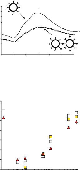
FIGURE 9.4. The principle of the nonoshell immunoasay. Nanoshells conjugated with antibody specific for a particular analyte are well dispersed in an analyte free environment (—), and possess a spectrum with a resonance in the near infrared. In the presence of analyte, however, antibody-antigen interactions induce nanoshell agglutination, a pheonomenon that is easily detected by a reduction in the nanoshell solutions’ original peak resonance (- -).
% Decrease in Ext.
70

 Saline
60 

 Serum
Serum
20% Whole Blood
50
40
30
20
10
0
0.1 |
1 |
10 |
100 |
Log [IgG] (ng/ml)
FIGURE 9.5. Rabbit IgG (analyte) induced aggregation of nanoshells in saline, serum, and blood at 30 min. Successful detection of analyte in serum reveals good sensitivity of the assay, as serum contains a multitude of proteins that could potentially promote nonspecific antibody binding. Likewise, nanoshell performance in whole blood proceeded with only a slight reduction in sensitivity. Whole blood performance demonstrates that nanoshells possess the optical contrast required for detection.

164 |
LEON R. HIRSCH ET AL. |
1 Cm
Swollen
2
Collapsed
3
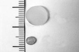
FIGURE 9.6. Nanoshells entrapped within the NIPAAm-co-acrylamide hydrogels absorb near infrared light to drive the phase change of the thermally responsive polymer. Exceeding the LCST caused collapse of the hydrogel material, due to expulsion of water and collapse of the polymer chains.
agent in a pulsatile or staggered fashion. The first goal has been largely addressed by a variety of delivery systems, including osmotically driven pumps and biodegradable matrices (reviewed in [39]). The second goal, controlled modulation of drug delivery, has proved more difficult. Thermally responsive hydrogels and membranes have been extensively evaluated as platforms for the pulsatile delivery of drugs. One of the characteristics of temperature-responsive hydrogels is the presence of a lower critical solution temperature (LCST), a temperature at which the hydrogel material will undergo a dramatic phase change. The driving force for this phase change is based on the interactions between the polymer and the surrounding water [34, 42]. Below the LCST, the most thermodynamically stable configuration is for the water molecules to remain clustered around the polymer chains. Above the LCST, the polymer chains collapse upon each other and minimize interaction with water. Due to this phase change, a macroscopic hydrogel will undergo a drastic change in dimensions, collapsing as the temperature exceeds the LCST, with expulsion of water (and dissolved drug) from the matrix.
N-isopropylacrylamide (NIPAAm) is a commonly used thermoresponsive polymer, and copolymers of NIPAAm and acrylamide form hydrogel materials with LCSTs ranging from 32–60◦C, depending on the composition of the copolymer [29]. Pulsatile delivery of a variety of drugs has been demonstrated from NIPAAm-co-acrylamide hydrogels by subjecting the materials to changes in temperature [29, 47]. While very nice results have been achieved with this system, the practical implementation of the system has been difficult due to issues with inducing temperature changes in implanted materials. In order to develop more easily manipulated implants for modulated drug delivery, composites hydrogels formed from NIPAAm-co-acrylamide and near-infrared absorbing nanoshells have been developed [38]. The composites were fabricated by mixing nanoshells into the monomer mixture. After polymerization, the nanoshells were physically entrapped in the hydrogel matrix. As shown in Figure 9.6, the composite hydrogels undergo a pronounced collapse in response to near infrared light; the nanoshells absorb the light, generating heat within the composite to exceed the LCST of the copolymer.
The collapse of the hydrogel material is completely reversible. When the temperature falls below the LCST, the polymer chains extend and interact with water, causing the

DIAGNOSTIC AND THERAPEUTIC APPLICATIONS OF METAL NANOSHELLS |
165 |
|||||||||||||||
RateRelease min)d.w.g(mg// |
3.5 |
|
|
|
|
|
|
|
|
|
|
|
|
|
||
|
|
|
|
|
|
|
|
|
|
|
|
|||||
3 |
|
|
|
|
|
|
|
|
|
|
|
|
|
|||
|
|
|
|
|
|
|
|
|
|
|
|
|
|
|
||
|
|
2.5 |
|
|
|
|
|
|
|
|
|
|
|
|
|
|
|
|
|
|
|
|
|
|
|
|
|
|
|
|
|||
|
|
2 |
|
|
|
|
|
|
|
|
|
|
|
|
|
|
|
|
1.5 |
|
|
|
|
|
|
|
|
|
|
|
|
|
|
|
|
1 |
|
|
|
|
|
|
|
|
|
|
|
|
|
|
|
|
0.5 |
|
|
|
|
|
|
|
|
|
|
|
|
|
|
|
|
|
|
|
|
|
|
|
|
|
|
|
|
|||
IrradienceLaser |
|
0 |
|
|
|
|
|
|
|
|
|
|
|
|
|
|
|
|
|
|
|
|
|
|
|
|
|
|
|
|
|||
cm(W/ |
1.6 |
|
|
|
|
|
|
|
|
|
|
|
|
|
||
|
|
|
|
|
|
|
|
|
|
|
|
|
||||
|
) |
|
|
|
|
|
|
|
|
|
|
|
|
|
||
|
1.2 |
|
|
|
|
|
|
|
|
|
|
|
|
|
||
|
2 |
|
|
|
|
|
|
|
|
|
|
|
|
|
||
|
|
0.8 |
|
|
|
|
|
|
|
|
|
|
|
|
|
|
|
|
0.4 |
|
|
|
|
|
|
|
|
|
|
|
|
|
|
|
|
|
0 |
|
|
|
|
|
|
|
|
|
|
|
|
|
|
|
|
|
|
|
|
|
|
|
|
|
|
|
|||
|
|
|
-20 |
0 |
20 |
40 |
60 |
80 |
100 |
120 |
|
|||||
|
|
|
|
|
|
|
|
|
Time (min) |
|
|
|
|
|
|
|
FIGURE 9.7. Pulsatile release of protein (top panel) from composite hydrogels was achieved through pulsatile near infrared irradiation (bottom panel).
material to swell. The reversible nature of this phenomenon allows one to partially collapse the material, re-swell it, cycling above and below the LCST repeatedly. If a drug has been incorporated into the hydrogel matrix, each time the hydrogel collapses, a burst of drug will be expelled from the material, as demonstrated in Figure 9.7 [38]. Because of the deep penetration of near infrared light through tissue, the composite hydrogels may be implanted subcutaneously, with the phase change behavior easily manipulated by externally applied light.
9.2.3. Photothermal Ablation
As discussed above for the drug delivery application, nanoshells can be designed to strongly absorb near infrared light and thus generate localized heating, potentially enabling nanoshell-mediated thermal ablation therapies for applications such as cancer treatment. Thermal ablation therapies can provide a minimally invasive alternative to surgical excision of tumors and are particularly attractive for situations where surgery is not possible. Thermal delivery methods under investigation for local tissue ablation include lasers [2, 44], microwave and radio frequency energy [8, 35], magnetic thermal ablation [11], and focused ultrasound [19]. The goal of thermal ablation is to conform a lethal dose of heat to a prescribed tissue volume with as little damage to intervening and surrounding normal tissue as possible, which has been difficult with the majority of techniques currently under investigation. Due to the lack of absorption of near infrared light by tissue components, use of this type of light source, with nanoshells at the desired tissue locations, should minimize collateral tissue damage. In vitro studies with nanoshells bound to breast carcinoma cells have demonstrated effective destruction of the cancerous cells upon exposure to near infrared light [12], with cell damage limited to the laser treatment spot (Figure 9.8).
The efficacy of nanoshell-mediated photothermal ablation has also been assessed in several in vivo studies. Initial studies involved directly injecting nanoshell suspensions into tumor sites and utilizing MRI thermal imaging to monitor temperature profiles during heating [12]. These studies demonstrated rapid heating of nanoshell-laden tissues upon exposure to the near infrared light. Evaluation of the gross pathology and histology demonstrated

166 |
LEON R. HIRSCH ET AL. |
FIGURE 9.8. Breast carcinoma cells treated with nanoshells and near infrared light (821 nm). Cells were stained with the fluorescent viability stain, calcein AM. All cells within the circular laser spot were destroyed. The region of cell death corresponds with the diameter of the laser.
marked tissue damage at the treatment sites, with little or no damage to surrounding tissue. This initial work also provided information about the relationships between nanoshell dosages, light intensity, and duration of illumination with the ultimate thermal profile and resultant tissue damage. However, for the majority of applications, direct injection into the tumor site may not be feasible.
An alternative approach is to inject nanoshells intravenously, allowing them to circulate and accumulate at the tumor site before near infrared treatment. The size of nanoshells is critical to the success of this type of approach. Substantial prior research has investigated the delivery of macromolecules and small particles through the tumor vasculature. These efforts have demonstrated that particles in the 60–400 nm size range will extravasate and accumulate in many tumor types via a passive mechanism referred to as the “enhanced permeability and retention” (EPR) effect [23]. This effect has been attributed to the highly proliferative vasculature within neoplastic tumors. During rapid angiogenesis, defects in the vascular architecture are often present, resulting in leaky vessels. Nanoshells fall within the range applicable for the EPR effect, and thus should accumulate in most tumor types following intravenous injection. The efficacy of photothermal ablation following systemic delivery of nanoshells has been evaluated in a mouse model [27]. Complete regression of tumors was observed following treatment with nanoshells and near infrared light, with no tumor re-growth over at least 60 days. Survival times for mice with the nanoshell treatment in this study were significantly improved compared to untreated mice or those receiving laser treatment alone. Additionally, it is possible to conjugate nanoshells to antibodies to oncoproteins or endothelial markers, which may improve the accumulation of nanoshells in the tumor tissue and further localize nanoshells to targeted cells at the treatment site.
9.2.4. Nanoshells for Molecular Imaging
The drug delivery and thermal ablation applications described in the preceding sections used nanoshells designed to strongly absorb light in the NIR spectral region. By fabricating
DIAGNOSTIC AND THERAPEUTIC APPLICATIONS OF METAL NANOSHELLS |
167 |
nanoshells designed to preferentially scatter rather than absorb light, nanoshells can serve as strong optical contrast agents for a variety of biomedical optical imaging applications. Photonics-based imaging technologies offer the potential for non-invasive, high-resolution in vivo imaging at competitive costs. However, the clinical utility of optical imaging strategies has been significantly constrained both by the limited variety of endogenous chromophores present in tissue and by relatively low levels of optical contrast between normal and diseased tissue. Furthermore, in the case of cancer, where early detection is critical to reducing morbidity and mortality, it is often desirable to image specific molecular biomarkers which are present long before pathologic changes occur at the anatomic level. Imaging biomarkers of interest requires development of targetable optical contrast agents. A recent demonstration of scattering-based molecular imaging used gold colloid conjugated to antibodies to the epidermal growth factor receptor (EGFR) as an optical contrast agent for imaging early cervical precancers [40]. While gold colloid bioconjugates are valuable as contrast agents for detecting superficial epithelial cancers with visible light, a primary challenge in optical contrast agent development has been the need for optical contrast agents at the multiple laser wavelengths within in the NIR spectral region commonly used in optical imaging applications. The facile tunability of nanoshells facilitates their use as NIR contrast agents. In addition, nanoshells offer other advantages relative to conventional imaging agents including more favorable optical scattering properties, enhanced biocompatibility, and reduced susceptibility to chemical/thermal denaturation. Furthermore, as described earlier in this chapter, nanoshells are readily conjugated to antibodies or other targeting moieties of interest enabling molecular specific imaging.
Initial in vitro studies were conducted to demonstrate the potential of nanoshell bioconjugates for molecular imaging applications. These experiments used nanoshell designed to strongly scatter light throughout the NIR “optical window” of 700–1200 nm. To enable molecular targeting, antibodies were conjugated onto nanoshell surfaces using ortho-pyridyl-disulfide-polyethylene-glycol-n-hydroxy-succinimide (OPSS-PEG-NHS) as a linker. Following antibody conjugation, nanoshell surfaces were further modified with PEG-thiol in order to block non-specific adsorption sites. Cells incubated with scattering nanoshell bioconjugates were viewed under darkfield microscopy, a form of microscopy sensitive only to scattered light. Significantly increased optical contrast due to expression of HER2, a clinically relevant cancer biomarker, was observed in HER2-positive breast carnicoma cells targeted with HER2-labeled nanoshells compared to the contrast observed in cells targeted by either nanoshells non-specifically labeled with IgG or control cells which were not exposed to nanoshell conjugates (Figure 9.9). Using a qualitative silver stain capable of detecting the presence of gold on cell surfaces, greater staining intensity was seen in HER2-targeted cells providing additional evidence that the increased contrast seen under darkfield was specifically attributable to nanoshell targeting of the HER2 receptor.
Nanoshell-based molecular contrast agents offer advantages including tunability, size flexibility, and systematic control of optical scattering and absorption properties. While darkfield microscopy is appropriate for in vitro imaging applications, use of nanoshell conjugates in vivo will require more sophisticated imaging techniques. Work currently underway is assessing nanoshell contrast agents in vivo in animal models using scatteringbased optical systems including reflectance confocal microscopy and optical coherence microscopy. Furthermore, the high level of control over nanoshell properties achievable through systematic manipulation of design parameters suggests the potential for biomedical

168 |
LEON R. HIRSCH ET AL. |
FIGURE 9.9. Molecular imaging in living cells using nanoshell bioconjugates. Scattering contrast is evident in HER2+ breast carcinoma cells incubated with HER2 nanoshell bioconjugates (left). Scattering signal is not present when the cells are exposed to nanoshells conjugated to a non-specific Ab (middle) or in control cells not exposed to nanoshells (right).
applications requiring more complex functionalities including integrated imaging and therapy of cancer.
REFERENCES
[1]A.L. Aden and M. Kerker. J. Appl. Phys., 22:1242–1246, 1951.
[2]Z. Amin, W. Thurrell, G.M. Spencer, S.A. Harries,W.E. Grant, S.G. Bown, and W.R. Lees. Invest. Radiol., 28:1148–1154, 1993.
[3]R.D. Averitt, D. Sarkar, and N.J. Halas. Phys. Rev. Lett., 78:4217–4220, 1997.
[4]R.D. Averitt, S.L. Westcott, and N.J. Halas. J. Opt. Soc. Am. B, 16:1824–1832, 1999.
[5]C.F. Bohren and D.R. Huffman. A bsorption and Scattering of Light by Small Particles. Wiley-Interscience, New York, pp. 379, 1983.
[6]K. Eichner. Int. Dent. J., 33:1–10, 1983.
[7]J.G. Fujimoto, M.E. Brezinski, B.J. Tearney, S.A. Boppart, B. Bouma, M.R. Hee, J.F. Southern, and E.A. Swanson. Nat. Med., 1:970–972, 1995.
[8]G.S.Gazelle, S.N. Goldberg, L. Solbiati, and T. Livraghi. Radiology, 217:633–646, 2000.
[9]H. Hagman. Dent. Lab. Rev., 54:28–30, 1979.
[10]G.D. Hale, J.B. Jackson, T.R. Lee, and N.J. Halas. Appl. Phys. Lett., 78:1502–1504, 2000.
[11]I. Hilger, R. Hiergeist, R. Hergt, K. Winnefeld, H. Schubert,and W.A. Kaiser. Invest. Radiol., 37:580–586, 2002.
[12]L.R. Hirsch, J.B. Jackson, A. Lee, N.J. Halas, and J.L. West. Analyt. Chem., 75:2377–2381, 2003.
[13]L.R. Hirsch, R.J. Stafford, J.A. Bankson, S.R. Sershen, B. Rivera, R.E. Price, J.D. Hazle, N.J. Halas,, and J.L. West. Proc. Natl. Acad. Sci. U.S.A., 100:13549–13554, 2003.
[14]M. Ho, M.J. Warrell, D.A. Warrel, D. Bidwell, and A. Voller. Toxicon, 24:211–221, 1986.
[15]N. Holstrum, P. Nilsson, J. Carlsten, and S. Bowland. Biosens. Bioelectron., 13:1287–1295, 1998.
[16]M. Horisberger and J. Rosset. J. Histochem. Cytochem., 25:295–305, 1977.
[17]J.V. Houten, D. Benaron, S. Spilman, and D.K. Stevenson. Ped. Res., 39:470–476, 1996.
[18]D. Huang, E.A. Swanson, C.P. Lin, J.S. Schuman,W.G. Stinson,W. Chang, M.R. Hee, T. Flotte, K. Gregory, C.A. Puliafito, and J.G. Fujimoto. Science, 254:1178–1181, 1991.
[19]F.A. Jolesz and K. Hynynen. Cancer J., S1:100–112, 2002.
[20]B.M. Kapur. Bull. Narcot., 45:116–154, 1993.
[21]E. Kuun, M. Brashaw, and A.d.P. Heyns. Vox Sanguinis, 72:11–15, 1997.
[22]B. Lindholm-Sethson, J.C. Gonzalez, and G. Puu. Langmuir, 14:6705–6708, 1998.
[23]H. Maeda, T. Sawa, and T. Konno, J. Control. Rel., 74:46–61, 2001.
[24]R.G. Nuzzo, F.A. Fusco, and D.L. Allara. J. Am. Chem. Soc., 109:2358–2368, 1987.
[25]S.J. Oldenberg, R.D. Averitt, S.L. Westcott, and N.J. Halas. Chem. Phys. Lett., 28:243–247, 1998.
[26]S.J. Oldenberg, S.L. Westcott, R.D. Averitt, and N.J. Halas. J. Chem. Phys., 111:4729–4735, 1999.
DIAGNOSTIC AND THERAPEUTIC APPLICATIONS OF METAL NANOSHELLS |
169 |
[27]D.P. O’Neal, L.R. Hirsch, N.J. Halas, J.D. Payne, J.L. West. Cancer Lett., (In Press), 2004.
[28]M. Pourbaix. Biomaterials, 5:122–134, 1984.
[29]J.H. Priest, S.L. Murray, R.J. Nelson, and A.S. Hoffman. Revers. Polym. Gels Related Syst., 350:255–264, 1987.
[30]E. Prodan, C. Radloff, N. Halas, and P. Nordlander. Science, 302:419–422, 2003.
[31]M. Quinten. Appl. Phys. B, 73:317–326, 2001.
[32]M. Rajadhyaksha, S. Gonz´alez, J.M. Zavislan, R.R. Anderson, and R.H. Webb. J. Investig. Dermatol., 113:203–303, 1999.
[33]C. Ruan, R. Yang, X. Chen, and J. Deng. J. Electroanalyt. Chem., 455:121–125, 1998.
[34]S. Sasaki, H. Kawasaki, and H. Maeda. Macromolecules, 30:1847–1848.
[35]T. Seki, M. Wakabayashi, T. Nakagawa, M. Imamura, T. Tamai, A. Nishimura, N. Yamashiki, A. Okamura, and K. Inoue. Cancer, 85:1694–1702, 1999.
[36]S. Sershen and J. West. Adv. Drug Del. Rev., 54:1225–1235, 2002.
[37]S.R. Sershen, J.L. Westcott, J.L. West, and N.J. Halas. Appl. Phys. B, 73:379–381, 2001.
[38]S.R. Sershen, S.L. Westcott, N.J. Halas, and J.L. West, J. Biomed. Mater. Res., 51:293–298, 2000.
[39]S.R.Sershen, S.L. Westcott, N.J. Halas, and J.L. West, Appl. Phys. Lett., 80:4609–4611, 2002.
[40]K. Sokolov, M. Follen, I. Pavolva, A. Malpica, R. Lotan, and R. Richards-Kortum. Cancer Res., 63:1999– 2004, 2003.
[41]W. Stober and A. Fink. J. Colloid. Interf. Sci., 26:62–69, 1968.
[42]N. Tanaka, S. Matsukawa, H. Kuroso, and I. Ando. Polymer, 39:4703–4706, 1998.
[43]A. Vogel. Phys. Med. Biol., 42:895–912, 1997.
[44]T.J. Vogl, M.G. Mack, R. Straub, K. Engelmann, S. Zangos, and K. Eichler. Radiologe, 39:764–771, 1999.
[45]R. Weissleder. Nat. Biotech., 19:316–317, 2001.
[46]A. Welch and M. van Gemert (eds.). Optical-Thermal Response of Laser-Irradiated Tissue, Plenum Press, New York, 1995.
[47]R. Yoshida, K. Sakai, T. Okano, and Y. Sakurai. J. Biomat. Sci. Polym. Ed., 6:585–598, 1994.
[48]H.S. Zhou, I. Honma, and H. Komiyama. Phys. Rev. B, 50:12052–12056, 1994
10
Nanoporous Microsystems for Islet Cell Replacement
Tejal A. Desai1, Teri West2, Michael Cohen2, Tony Boiarski2, and
Arfaan Rampersaud2
1Department of Bioengineering and Physiology, University of California, San Francisco, CA 2IMEDD Inc., Columbus, OH 43212
10.1. INTRODUCTION
10.1.1. The Science of Miniaturization (MEMS and BioMEMS)
Micro-Electro-Mechanical Systems technology, commonly known with the acronym MEMS, refers to the fabrication of devices with dimensions on the micrometer scale. For comparison, a human hair is about 80 μm in diameter. The most essential elements of MEMS consist of miniaturized, highly precise, and repeatable structures that can be stationary or moving. These structures are created via fabrication processes and equipment developed for the integrated circuit (IC) industry. The fabrication of MEMS commonly involves bulk or surface machining. Bulk machining defines microstructures by etching directly into the bulk material such as single crystal silicon. The advantage of bulk machining is that it allows the integration of active devices and the use of integrated circuit technology. Surface machining defines the release and movable structure in a polysilicon film or sacrificial layer of silicon dioxide, both deposited on bulk silicon. More complex microchips including multilayer interconnections can be obtained by bonding together and laser drilling of several layers of the components. Typically, microfabrication has a limit of resolution on the order of microns. However, specialized techniques can be used to create features in the nanoscale, as it will be shown later in the fabrication process of the nanoporous membrane. More importantly, the incorporation of new materials and the range of processes now extend far beyond just those found in the IC industry.
172 |
TEJAL A. DESAI ET AL. |
Medicine and biology are among the most promising, although most challenging, fields of application for MEMS. This does not come as a surprise considering that the technology has the capability to fabricate minimally invasive yet highly functional microdevices that match the size range of many structures found in the human body. Examples include pressure sensors that are small enough to fit through 1 mm catheters, but are priced to be disposable, and pacemakers that have incorporated microscale accelerometers to pace the heart in proportion to the patient activity. These tiny devices, also referred to as biomedical microsystems, hold promise for precision surgery with micrometer control, rapid screening of common diseases and genetic predispositions, and autonomous therapeutic management of allergies, pain, and neurodegenerative diseases. The health care implications predicted by successful development of this technology are enormous, including early identification of disease and risk conditions, less trauma and shorter recovery times, and more accessible health care delivery at a lower total cost.
The development of new, affordable, disposable analytic microchips are changing diagnostics. Examples of analytical functions that are benefiting from such developments include blood supply screening, analysis of biopsy samples and body fluids, minimally invasive and noninvasive diagnostic procedures, rapid identification of disease, and early screening. These systems will eventually perform diagnostic procedures in a multiplexed format that incorporates multiple complementary methods. Ultimately these systems will be combined with other devices to create completely integrated analysis and treatment systems.
New drug delivery methods seek to develop tool capable of delivering precise quantities of a drug at the right time and as close as possible to the treatment site. Sustained drug delivery provides the same medicinal effect with higher efficiency, longer duration and less side effects than traditional delivery methods. There are a number of mechanisms to provide timed release of drugs, such as microencapsulation, transdermal patches, and implants. Transdermal release is an attractive alternative for formulations which cannot be effectively delivered using pills and injection because of limitations related to gastrointestinal drug degradation and the inconvenience and pain related to intramuscular and intravenous injections. Implantable devices are preferred for therapies that require several daily injections, such as for diabetes treatment. If the drug level is monitored in real-time, it could also be adapted to metabolic variations. In treatments like chemotherapy, the device can be implanted where the drug is most needed. The current state-of-the art includes systems that are approximately the size of a hockey puck, have a limited battery lifetime of 3–7 years, and rely on the use of power-consumptive electromagnetic dispensing of fixed amounts of medication at programmed intervals, regardless of body need. Among these techniques, implantable pumps have the advantage that the drug therapy can be delivered at the optimal time and concentration to a specific site. MEMS systems combine miniature size, which is amenable to implantability, low power requirements, and the potential to precisely meter fluid samples.
10.1.2. Cellular Delivery and Encapsulation
The immunoisolation of transplanted cells and tissue by size-based semipermeable membranes has emerged as an extremely promising method of treating hormone deficiencies arising from such diseases as Type I diabetes, Alzheimer’s, and hemophilia [1–5]. It has
NANOPOROUS MICROSYSTEMS FOR ISLET CELL REPLACEMENT |
173 |
been demonstrated that cellular transplants, such as isolated pancreatic islets of Langerhans or hepatocytes, respond physiologically both in vitro and in vivo by secreting bioactive substances in response to appropriate stimuli, as long as they are immunoprotected. However, with the exception of autologous cells and tissue, overcoming immunologic rejection of the transplanted cells is still the greatest obstacle. Although approaches involving polymeric microcapsules have yielded promising results for autologous and allogeneic cell transplantation without immunosuppression [15–18], few approaches have been effective for non-immunosuppressed xenogeneic cell encapsulation [19–23], due to mechanical rupture of the membrane, biochemical instability, compatibility with islet cell heterogeneity, and broad pore size distributions [1, 5, 24–26]. While Duvivier et al. showed that size based exclusion is not critical for auto or allografts [64], several groups have found that the protection of porcine islets and xenografts, in general, will be more difficult to protect than allografts, as suggested by studies performed with permeable polymer membranes [30,69]. Their studies also confirmed that cytokines can cross polymeric alginate membranes and damage the islet cells contained inside the capsules. This may not be an issue for allografts but will certainly play a role in xenotransplantation as discussed below.
Immunoisolation of xenogeneic cells requires stringent biological and physical criteria to be met. The immunological response to a xenograft has various features that distinguish it from the alloresponse. The first difference depends on the species crossed, in that the recipient may have natural cytotoxic antibodies against the xenoantigens in the donor tissue. In addition, the major method of recognition of xenoantigens is via the indirect pathway, in which xenoantigen is processed and presented by the host antigen-presenting cells to Th cells. The reaction to xenogeneic cells is very complex and involves not only cells and antibodies, but also complement and a host of cytokines such as tumor necrosis factor, which can inflict cell damage. Thus, the clinical success of encapsulated islet transplantation is still minimal, with less than 30 documented cases of insulin independence occurring from over 250 attempts at clinical islet allo-transplantation since 1983 and no clinically successful cases of xenogeneic islet encapsulation without immunosuppression [27–29]. In light of this, it is critical to turn to new capsule materials, designs, and fabrication approaches, focusing on such fundamental issues as the immunoisolation membrane material, membrane pore parameters, and cellular arrangement within the device. Only then can we begin to develop cell encapsulation devices with long-term therapeutic efficacy and safety.
The immunoisolation membrane should allow permeability of glucose, insulin, oxygen and other metabolically active products, to insure islet functionality and therapeutic effectiveness. It must also prevent the passage of cytotoxic cells, macrophages, antibodies and complement to remain effective. Previous studies indicated that immunoisolation could be attained if C1q and IgG were completely retained, such as the case with membranes having pore diameters between 30 to 50 nm [1]. In fact, Hirotani and Ohgawara measured complement permeability for a membrane with pore size 0.1 to 0.2 microns and reported that complement C3 became inactivated upon the passage through the membrane. Thus, membrane thickness may also be a critical parameter. More recently, it has been elucidated that true immunoisolation also requires the blockage of cytokines such as TNF and cell-secreted antigens. For example, Brauker et al. [33] found that xenografts (CF1 mouse embryonic lung implanted into Lewis rats) were destroyed within 3 weeks even when implanted in devices with intact membranes. The death of the xenogeneic tissues was accompanied by a severe local accumulation of inflammatory cells and a decrease in
