

Therapeutic Micro-Nano Technology BioMEMs - Tejlal Desai & Sangeeta Bhatia
.pdf60 |
YOKO KAMOTANI ET AL. |
on channel surfaces. This technique is collectively known as “laminar flow patterning” and has been found useful in studying cell-cell, cell-surface, cell-medium interactions [22] as well as engineering surface-directed flows [46, 91, 93]. (ii) In the absence of transverse convective fluid motions, mixing between liquid laminar streams occurs only via diffusion at the interface. This characteristic poses both challenges and advantages in microfluidics. It has been demonstrated that the diffusive transport and chemical reaction confined to an area in the vicinity of the interface of two miscible liquids provide novel tools for microelectrode fabrication [43], size-dependent separation of biological molecules [31, 87, 88], and immunoassays [30]. The cross-stream-wise diffusion localized at the interface, however, becomes a rate-limiting factor when it comes to mixing in microfluidics because mixing purely by diffusion is too slow. A rapid and efficient mixing in microfluidics is often achieved by either generation of chaotic convective fluid motions transverse to main laminar flows [76] or simple splitting and recombination of fluid streams to create an increased fluid interface and short diffusion path [39].
4.3.3. Surface Tension
When immiscible fluids (e.g. liquid and air, two immiscible liquids such water and oil) are present, molecules within the liquid are attracted equally from all directions, but those near the interface experience unequal attractions and thus, are drawn towards the center of liquid mass. At the microscale, surface tension originating from this cohesion between liquid molecules at the interface of different fluids becomes a dominant surface force and plays a crucial role in manipulating transport phenomena. Spatial gradients in surface tension that are often created by variations of temperature [14, 15], electric field [32, 47, 59], prepatterned surface energies [46, 92, 93], electrochemical reactions [25] on the walls of microchannels give rise to driving forces for the movement of liquid droplets and thin liquid films or for stable flows of immiscible fluids without any need for mechanical moving parts. In such multiphase microfluidic systems, a Reynolds number that is generally small, itself, does not usually serve as a characteristic system parameter. Instead, geometrical features and ratios of different forces become important in indicating the details of flows. The relative importance between surface tension and other forces is estimated by the following dimensionless parameters.
Ca (capillary number) = |
μL UL |
|
|
|
|
viscous effect |
|
||
σ |
surface tension effect |
|
|
||||||
Bo (Bond number) = |
ρL g L2 |
|
|
|
gravitational effect |
|
|||
σ |
|
surface tension effect |
|||||||
|
|
|
|
||||||
We (Weber number) = |
ρL UL2 L |
|
|
inertial effect |
|||||
σ |
|
surface tension effect |
|||||||
|
|
|
|
||||||
Where μL is liquid viscosity, UL is average velocity of liquid, σ is surface tension, ρL is density of liquid, g is gravitational acceleration, and L is a characteristic length. For example, assuming that there is continuous movement of water (μL = 0.001 kg/ms2, ρL = 1000 kg/m3) droplets or flow of water through air at the average speed of 1 cm/s in a 100 μm- thick polymeric microchannel (σ = 30 dynes/cm), it can be shown that Ca 3.3 × 10⁻⁴, Bo 3.2 × 10⁻³, We 3.3 × 10⁻⁴, implying the dominance of surface tension effects.
AT THE INTERFACE: ADVANCED MICROFLUIDIC ASSAYS FOR STUDY OF CELL FUNCTION |
61 |
Generally, the manipulation of surface tension to change surface wettability in microfluidics is performed in two different formats depending on the reversibility of surface energy changes: (i) In the first case, electric potentials or thermal energies applied to microchannel walls through embedded electrodes enable reversible changes of surface tension. In this arrangement, it has been demonstrated that liquid droplets surrounded by immiscible fluids can be transported quickly to desired locations over two-dimensional arrays of microelectrodes [12, 20, 47]. Such routing of independent droplets and reversible motion of fluids provide novel methods to devise basic building blocks of microfluidic systems such as pumps, valves, and mixers [12, 57]. Also, a precise handling of picoor nanoliter quantities of reagents and analytes isolated in discrete droplets becomes useful for biochemical assays [67]. In addition to droplet-based configurations, the alteration of surface tension by electric potentials has proven useful for reversibly switching flow patterns of continuous air-liquid laminar streams [35] or for transporting thin continuous liquid films [25] as well. (ii) In the second case, surface patterning by laminar flows or UV light can be used to irreversibly generate surface tension gradients. This method is based on the preparation of surfaces patterned with hydrophobic or hydrophilic self-assembled monolayers achieved by laminar flowor UV patterning [46, 92, 93]. The predefined surface free energies facilitate the confinement of liquid samples in hydrophilic regions and assist in guiding continuous liquid streams without requiring external forces to drive the fluid motions. Selective wetting of liquid samples over patterned hydrophilic surfaces enables a nanoscopic patterning of biomolecules such as DNA [70]. Also, this system has been used to realize microfluidic gas-liquid chemical reactions and the microfabrication of thin polymeric membranes [91, 93, 94].
4.4. EXAMPLES OF ADVANCED MICROFLUIDIC CELLULAR BIOASSAYS
Inexpensive rapid microfabrication techniques combined with knowledge of microscale phenomena (e.g. laminar flows and dominance of surface tension) allow development of new advanced microfluidic cellular bioassays. Microfluidic devices for these applications typically have cross-sectional capillary dimensions of 10–500 μm. This size scale is similar to those of biological cells, so that these devices are well suited to the transport, manipulation, and chemical or biochemical treatment of single cells or small number of cells [48]. This section describes several specific systems that take advantage of microfluidics to create controlled microenvironments to study subcellular phenomena, to sort cells by function such as motility, and to analyze cells. These advanced systems allow for the microscale control of interfaces that have not been exploited in traditional systems.
4.4.1. Patterning with Individual Microfluidic Channels
The most straightforward application of microfluidics is the use of different channels to pattern different cells or molecules in different regions on the microscale. The small size of microfluidic channels and the ability to fabricate multiple channels in parallel provides a platform for simultaneously patterning different biomolecules onto a surface.
Delamarche et al. used microfluidic networks for the spatially controlled deposition of biomolecules onto various substrates. The networks are constructed using a PDMS mold
62 |
YOKO KAMOTANI ET AL. |
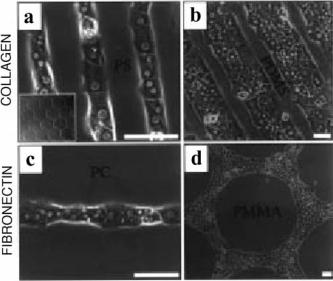
FIGURE 4.2. Phase-contrast optical micrographs of hepatocytes patterned onto various polymers (A: Polystyrene Petri dish, B: PDMS sheet, C: Polycarbonate Petri dish, and D: Poly(methyl methacrylate) disk) 12–24 hours after seeding. Hepatocytes adhere only to regions of collagen (A and B) or fibronectin (C and D). Scale bar is 50 μm. [23] (Reprinted with permission from Folch 1998. Copyright 1998 American Chemical Society)
sealed to a surface to form a conduit. The capillaries are filled with solutions containing the biomolecules of interest, allowed to deposit onto the surface, and the PDMS later peeled off. Using this type of system, immunoglobulins were patterned onto substrates with submicron resolution and used in enzyme-linked immunosorbent assay (ELISA)-type assays [16].
Microfluidics allows manipulation of cellular co-cultures in ways not possible with macroscopic techniques. Toner et al. have selectively delivered different cell suspensions in laminar flows to tissue culture substrates. Another technique uses microchannels saturated with protein solutions that adsorbs onto the surface exposed to the microflow. After microchannels are removed, only the proteins remain and cells attach selectively to these regions. Micropatterned cocultures have been created using patterns of collagen or fibronectin (Figure 4.2) [22].
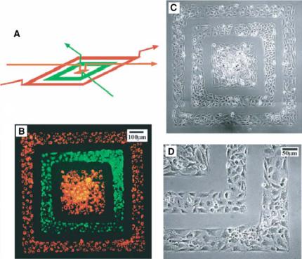
Whitesides et al. have used three-dimensional (3D) microfluidic systems to pattern proteins and cells onto a surface. 3D microfluidic stamps are fabricated using a two-step photolithographic process and sealing two PDMS slabs together. This PDMS structure is then used as a conduit to deliver proteins (bovine serum albumin and fibronectin fluorescently labeled) and cells (bovine capillary endothelial cells (BCEs) and human bladder cancer cells (ECVs) shown in Figure 4.3). This technique allows for the creation of biologically relevant patterns on surfaces with only one additional step of pattern fabrication, alignment, and sealing [10].
Jeon et al. have developed a microfabricated neuronal culture device. The system is created from PDMS and consists of two compartments separated by a physical barrier
AT THE INTERFACE: ADVANCED MICROFLUIDIC ASSAYS FOR STUDY OF CELL FUNCTION |
63 |
FIGURE 4.3. A 3D PDMS stamp shown in (A) is used to deposit two cell types onto a tissue culture dish in a concentric square pattern. Fluorescence (B) and phase-contrast (C and D) pictures show ECVs labeled in green CMFDA and BCEs labeled in a red Dil-conjugated acetylated low density lipoprotein. The cells are cultured with the stamp in place for 24 hours to grow and spread to confluency. The pictures are taken immediately after removing the stamp. (D) is an expanded view of the lower right corner in (C) [10]. (Reprinted with permission from Chiu, Jeon et al. 2000, Copyright 2000 National Academy of Sciences, USA).
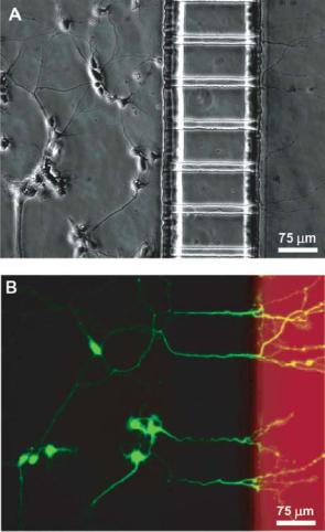
with embedded micron-sized grooves. Neuronal cells are cultured into one compartment and after several days, neurites extend through the grooves and into the second compartment (Figure 4.4). They are also able to micropattern neurites on the surface through microcontact printing to direct neuronal attachment and orientation of the neuronal outgrowth [84].
4.4.2. Multiple Laminar Streams
In the previous section, each microfluidic channel transported one type of molecule or cell. The low Reynolds number flow characteristics in microfluidic channels, however, permits two or more streams of cells and biomolecules to flow next to each other inside of a single microfluidic channel without turbulent mixing. The only mixing at the interface between different streams takes place through diffusion. Researchers have exploited laminar flow in microfluidic networks to pattern proteins, cells, and planar lipid bilayers on substrates with micrometer-scaled resolution [58]. Multiple laminar streams are also
64 |
YOKO KAMOTANI ET AL. |
FIGURE 4.4. Neuronal cell culture inside microfabricated device. Calcein AM and Texas Red dextran are added to the neuritic chamber 1 hour before taking the pictures. (A) Phase micrograph after 4 days in culture of neurons extending to neuritic chamber (on the right). (B) Epifluorescence micrograph of same region with cells stained in green calcein AM, and the neuritic chamber in red dextran [84]. (Reprinted with permission from Taylor, Rhee et al. 2003. Copyright 2003 American Chemical Society)
useful in generating patterns or interfaces and gradients composed of (1) different adhesive regions, (2) different cell types, and (3) different solutions [79].
Multiple laminar streams can also generate and maintain gradients of chemicals. Cells naturally migrate in gradients of soluble molecules called chemoattractants in a process known as chemotaxis. In order to further examine this dynamic behavior, Jeon, Toner, Whitesides et al. have developed a technology that generates a stable, soluble chemoattractant gradient using a device fabricated by soft lithography. The device consists of a network of microfluidic channels with a gradient-generating portion and an observation
AT THE INTERFACE: ADVANCED MICROFLUIDIC ASSAYS FOR STUDY OF CELL FUNCTION |
65 |
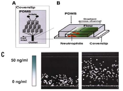
FIGURE 4.5. Schematic diagram of the microfluidic gradient generator. (A) Top view of device with gradient generating and observation regions. (B) 3D view of observation region where cells are exposed to chemoattractant gradients. (C) Micrograph of cells at the beginning of the experiment (0 min, left panel) deposited at the bottom of the field of view, and at the end of the experiment (90 min, right panel). Migration is subjected to a linear increase in IL-8 (0–50 ng/ml). Bar, 200 μm [40]. (Reproduced by permission from Jeon, Baskaran et al. 2002. Copyright 2002 Nature Publishing Group www.nature.com)
portion. (Figure 4.5A) The gradient-generating portion consists of a pyramidal branched array of channels that split, combine, and mix fluid streams. The channels recombine in the main channel to form a well-defined concentration gradient that spanned the width of the channel. (Figure 4.5B).
A gradient of interleukin-8 (IL-8) was formed in the device and the migration of neutrophils was observed. Figure 4.5C shows the results of one experiment where cells are initially positioned at the side of lower IL-8 concentration and subjected to a linear increase in IL-8 (1–50 ng/ml). The cells move towards the higher concentration. The ability to generate and maintain stable linear gradients allowed straightforward determination of chemotaxis coefficients demonstrating the power of microfluidics for quantitative cell biology. Comparison of cell migration in stable cliff-shaped and hill-shaped gradient profiles also allowed observation of complex migratory behaviors of the neutrophils previously unappreciated [40]. Whitesides et al. have also created substrate-bound gradients of proteins using laminar flows in microchannels. Linear gradients are created through streams that flow through long serpentine channels where mixing occurs. All streams converge into one channel where the gradient is established. Gradients of laminin and bovine serum albumin are demonstrated and neuronal axon orientation studied [18].

66 |
YOKO KAMOTANI ET AL. |
4.4.3. PARTCELL
Cellular transduction—the process of translating extracellular stimuli from the environment into responses such as growth, migration, or differentiation—occurs through subcellular components, including the cytoskeleton and various scaffolding proteins. These structural elements are distributed heterogeneously, but not randomly, throughout the cell with micron scale spatial variations. It has been proposed that structural order of these components is necessary for the propagation and processing of cellular signals. In order to study the role of such microscale organization, a method to perturb or manipulate specific domains within a cell with high molecular sensitivity and micron-scale spatial accuracy is needed [7]. Microfluidics can provide a solution. A technique, called PARTCELL (partial treatment of cells using laminar flows), positions the interface between multiple laminar flows over a single cell to deliver reagents with subcellular spatial selectivity.
In a typical set up, a microsystem is made from PDMS embossed with microchannel features sealed onto a glass surface. Streams containing different liquids are made to flow into three inlets and combine in a main channel to form parallel streams. (Figure 4.6a) The width of the streams and the position of the interface are adjusted by altering the relative amounts of fluid flowing in from the inlets. This setup allows for the patterned delivery of chemicals, colloids, and other reagents to a portion of a cell. (Figure 4.6b) Advantages to PARTCELL include: the facile generation of laminar flows which makes the procedure experimentally simple, the ability to pattern over delicate structures such as the cell surfaces, and the use of PDMS which makes fabrication easy and cell culture and analysis convenient. PARTCELL can be applied to a variety of studies including ones of cell dynamics, cell polarity, spatially regulated signaling, drug screening, and other cellular phenomena.
When cell permeable reagents are delivered to subcellular regions of surface-attached cells, a stable chemical concentration gradient is established and maintained within the cell. Such subcellular “positioning” of rapidly diffusing small molecules to a part of a
FIGURE 4.6. Schematic drawing of PARTCELL setup. (a) Three inlets combine to form parallel streams in main channel. (b) Close up view of cell placed at the interface between laminar streams.
AT THE INTERFACE: ADVANCED MICROFLUIDIC ASSAYS FOR STUDY OF CELL FUNCTION |
67 |
cell interior is counter intuitive considering the small size of cells. PARTCELL makes this possible because the continuous fluid flows impose rapid influx of reagent to one cell region and efflux from another region [82]. This has been demonstrated in a living bovine capillary endothelial (BCE) cell by labeling two different mitochondria subpopulations within the cell with a green and red fluorescent dye respectively. In a similar procedure actin filaments were disrupted in selective regions of the actin microstructure by treatment with a membrane-permeable molecule that binds to actin monomers [83].
PARTCELL can also be used to manipulate the cell-substrate interactions. For example, trypsin can be delivered selectively to a portion of a cell resulting in selective trypsinmediated detachment of the exposed region of the cell from the surface [82]. The method may be useful in obtaining insights into cell migration processes which often involve various matrix proteinases. PARTCELL also allows for the localized delivery of chemicals to only a portion of the cell surface. For example, flow of fluorescently labeled, acetylated, lowdensity lipoprotein (Ac-LDL) to only a portion of the surface led to spatially selective endocytosis of Ac-LDL [82].
PARTCELL uses microfluidics to perform subcellular manipulations that are difficult to perform otherwise. Limitations to PARTCELL include the need to seed the cells inside the channels and only being able to deliver the subcellular treatment in parallel laminar flows (as opposed to delivery to a point). Bradke et al. have developed a related technique for subcellular treatment and demonstrated the use of two pipettes to partially treat a neurite outgrowth with a small-molecule drug [6]. This method has the advantage of not requiring seeding of cells inside microfluidic channels, however, this method requires precise handling of micromanipulators and the open fluidic system needed makes the fluid flow more susceptible to disturbances and variations.
The ability to stimulate parts of single cells has led to discovery of new states and functions of cells not observed in traditional in vitro cultures. Bastiaens et al. first demonstrated that local stimulation of the epidermal growth factor receptor (EGFR) using epidermal growth factor (EGF) attached to microbeads leads to global phosphorylation of EGFR in the plasma membrane of MCF7 breast adenocarcinoma cells, in a ligand independent manner. Although microbeads served as a convenient tool for subcellular EGF delivery, the positioning of the microbeads on single cells was random in these studies. Furthermore, immobilization of EGF molecules on a comparatively huge microsphere alters cellular dynamics such as endocytosis of EGF. To overcome these limitations, Miyawaki et al used PARTCELL to perform subcellular stimulation of COS cells with a laminar flow stream of rhodamine-labelled EGF [56]. The PARTCELL experiments, which used special fluorescent indicators to visualize activation of Ras family G proteins [56] and tyrosine phosphorylation [45], were able to clarify the role of EGFR endocytosis as well as receptor density on the ligand independent lateral propagation (LILP) of EGF signals (Figure 4.7). Whereas many believed that LILP was a universal phenomena in cells, the PARTCELL experiments were able to show that endocytosis of activated EGFR could suppress LILP. Furthermore, it was shown that whether LILP occurs or not in a cell depends more generally on the amount of EGF stimulation, and receptor density [66]. Together with results from subsequent studies on the effect of tyrosine phosphatase inhibition on LILP and a mathematical analysis of EGF signaling [60], a picture is now emerging of how a reaction network of positive and negative regulators determines when and if LILP will occur within a cell (Tisher & Bastien, 2003). Although the physiological roles of LILP are still speculative, it may play a role in

68 |
YOKO KAMOTANI ET AL. |
FIGURE 4.7. Local stimulation of normal COS cells with Rhodamine-EGF to visualize Ras activation.
(A) Fluorescence image before stimulation. Violet region is exposed to rhodamine-EGF. Four ROIs are assigned across the laminar flows. (B) Rhodamine-EGF fluorescence after 10 minutes of stimulation. Cell is outlined with a dotted line. (C) Superimposed outlines of cell at 0 (black) and 20 (red) minutes. (D) Dual emission ratio images presented in intensity-modified display (IMD) mode. Right hand corner shows time in minutes after stimulation [66]. (Reproduced by permission from Sawano et al. 2002. Copyright 2002 Cell Press)
invasion and proliferation of cancer cells as EGFR is overexpressed in many carcinomas [66].
4.4.4. Microscale Integrated Sperm Sorter (MISS)
Laminar flows, which eliminates turbulent mixing, and the small dimensions of microfluidic systems, which allows rapid diffusion of small particles, has been used to separate particles based on their size. For example, Yager et al. have developed an H-filter in which two different liquid streams—one stream containing a mixture of different size particles such as cells and proteins, and another stream just composed of liquid—are introduced into a common channel. The two fluid streams flow parallel to one another, the large particles following the original streamline, while small particles diffuse rapidly between the streams and separate from the original solution. At the end of the main channel is a splitting junction which separates different regions of the main channel for downstream processing and analysis [30].
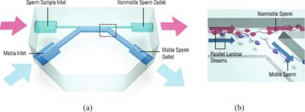
Such microfluidic interfaces can also be used for function-based cell sorting. Cho et al. designed a microfluidic system called the microscale integrated sperm sorter (MISS) to isolate motile sperm from nonmotile sperm and cellular debris [11]. This device also takes advantage of laminar flows in microfluidic channels, in a manner similar to H-filters, but applies the concept to sorting of cells. Motile sperm are isolated based on their ability to cross streamlines in a laminar fluid stream. The device can be used in clinical settings where small amounts of sperm need to be sorted. It can be used to select the most viable sperm for in vitro fertilization procedures and as disposable, at-home diagnostic tests for male infertility.
This simple, disposable device is made from poly(dimethylsiloxane) (PDMS) that is cast onto a silicon wafer with the desired microchannel features. After curing, the PDMS is peeled off and plasma oxidized to irreversibly seal it onto a glass surface. The design
AT THE INTERFACE: ADVANCED MICROFLUIDIC ASSAYS FOR STUDY OF CELL FUNCTION |
69 |
FIGURE 4.8. Schematic drawing of sperm sorting device. (a) Top view of device with two inlets and two outlet reservoirs. (b) Close up view of outlet channels where motile sperm are separated from nonmotile sperm.
contains two inlets (one for the sperm sample and one for the media) and two outlets (one for nonmotile sperm and one for motile sperm) shown in Figure 4.8a. Nonmotile sperm and debris flow along their initial streamlines and exit out one outlet, while the motile sperm swim into a parallel stream and exit out a separate outlet. (Figure 4.8b) The fluid flow is driven by a passively driven pumping system which uses horizontally oriented reservoirs for gravity-driven pumping. Gravity, surface tension, and channel resistance contribute to give a steady flow rate of sperm.
MISS is a self-contained, readily fabricated, functional microdevice that can manipulate living cells using microfluidics, avoiding the need for electronics or external power sources. It can be used in clinical settings and in the field of assisted reproductive technology. The device isolates sperm based on their motility and can be extended to the sorting of any other microorganisms that can swim efficiently. Other potential future uses include that for the preliminary screening of male infertility, as an indicator of toxicity, multiple connections or parallel integration of multiple MISS systems, and sorting on more subtle differences in motility [11].
4.4.5. Air-Sheath Flow Cytometry
Flow cytometry is a powerful tool to rapidly measure physicochemical characteristics of cells in liquid suspension using fluorescent probes that bind to specific cell-associated molecules (e.g. fluorochrome-labeled antibodies, nucleic acid probes, cell function probes, green fluorescent proteins). As labeled cells injected into the center of a fast stream of sheath liquid flow in single-file past a focused laser beam, they generate light scattering and fluorescence emission that are directed to detectors and converted into electric pulses. The measured amount of scattered light and fluorescence is used to enumerate cells at high rates ( 25,000 cells/sec) and is correlated with the properties of cells such as shape, size [71]. The high-speed analytical capabilities of conventional flow cytometers have gained widespread use in routine laboratories and also, there have been many efforts to implement such functionalities into miniaturized bioanalytical systems. As a core component of flow cytometry, rapid delivery of cells suspended in narrow liquid streams is crucial for reliable analyses and has been an important theme in developing microfluidic systems that can be integrated with miniaturized optics to enable high-speed detection and enumeration of