

Therapeutic Micro-Nano Technology BioMEMs - Tejlal Desai & Sangeeta Bhatia
.pdf224 |
SAMIR MITRAGOTRI |
13.1.3. Sustained Release of the Drug can be Obtained
Transdermal drug delivery can provide sustained release of drugs over a sufficiently long time (up to a week). Hence, it is possible to maintain a steady drug concentration in the blood. This is especially important for drugs with a narrow therapeutic window.
Transdermal drug delivery, however, suffers from the severe limitation that the permeability of the skin is very low. Therefore, it is difficult to deliver drugs across the skin at a therapeutically relevant rate. This, in fact, is the main reason why only a handful of low-molecular weight drugs are clinically administered by this route today.
13.2. ULTRASOUND IN MEDICAL APPLICATIONS
Ultrasound is used in various medical therapies including lithotripsy [1], hyperthermia [2], thrombolysis [3], lipoplasty [4], wound healing [5], fracture healing [6], and drug delivery (sonophoresis [7–9], sonoporation [10–13], triggered drug release [14–17], and targeted drug delivery [18, 19]). A majority of these applications have come about in the last decade. Ultrasound has also been used to enhance transdermal transport of various drugs including macromolecules [7–9, 20–69]. This type of enhancement is termed sonophoresis, indicating the enhanced transport of molecules under the influence of ultrasound. Ultrasound at various frequencies in the range of 20 kHz–16 MHz has been used to enhance skin permeability. However, transdermal transport enhancement induced by low-frequency ultrasound ( f < 100 kHz) has been found to be more significant than that induced by high frequency ultrasound [7, 8, 70].
13.3. SONOPHORESIS: ULTRASOUND-MEDIATED TRANSDERMAL TRANSPORT
The first published report on sonophoresis dates back to 1950’s. Fellinger and Schmidt [71] reported successful treatment of polyarthritis of the hand’s digital joints using hydrocortisone ointment with sonophoresis. It was subsequently shown that hydrocortisone injection combined with ultrasound “massage” yielded better outcome compared to simple hydrocortisone injections for bursitis treatment [72]. Cameroy [26] reported success using carbocaine sonophoresis for closed Colle’s fractures. In a series of publications Griffin et al. showed improved treatment of elbow epicondylitis, bicipital tendonitis, shoulder osteoarthritis, shoulder bursitis and knee osteoarthritis by combined application of hydrocortisone and ultrasound [29–31, 73]. Improved dermal penetration using ultrasound was also reported for local anesthetics [20, 74, 75].
Studies demonstrated that ultrasound enhanced the percutaneous absorption of methyl and ethyl nicotinate by disordering the structured lipids in the stratum corneum. Similar conclusions were reached by Hofman and Moll [76] who studied the percutaneous absorption of benzyl nicotinate. While several investigators reported positive effect of ultrasound on drug permeation, lack of an effect of ultrasound on skin permeation was also reported in certain cases. For example, Williams reported no detectable effect of ultrasound on the rate of penetration of three anesthetic preparations through human skin [63].
TRANSDERMAL DRUG DELIVERY USING LOW-FREQUENCY SONOPHORESIS |
225 |
Levy et al. [77] showed that 3–5 minutes of ultrasound exposure (1 MHz, 1.5 W/cm2) increased transdermal permeation of mannitol and physostigmine across hairless rat skin in vivo by up to 15-fold. They also reported that the lag time typically associated with transdermal drug delivery was nearly-completely eliminated after exposure to ultrasound. Mitragotri et al. reported in vitro permeation enhncement of several low-molecular weight drugs under the same ultrasound conditions [41].
Bommanan et al. [23, 24] hypothesized that since the absorption coefficient of the skin varies directly with the ultrasound frequency, high frequency ultrasound energy would concentrate more in the epidermis, thus leading to higher enhancements. In order to assess this hypothesis, they studied the effect of high-frequency ultrasound (2 MHz-16 MHz) on permeability of salicylic acid (dissolved in a gel) through hairless guinea pig skin in vivo. They found that a 20 minute application of ultrasound (0.2 W/cm2) at a frequency of 2 MHz did not significantly enhance the amount of salicylic acid penetrating the skin. However, 10 MHz ultrasound under otherwise same conditions resulted in a 4-fold increase and 16 MHz ultrasound resulted in about a 2.5-fold increase in transdermal salicylic acid transport [23, 24].
13.4. LOW-FREQUENCY SONOPHORESIS
Low-frequency sonophoresis has been a topic of extensive research only in the last 10 years. Tachibana et al. [60–62] reported that application of low-frequency ultrasound (48 kHz) enhanced transdermal transport of lidocaine and insulin across hairless rat skin in vivo. They found that the blood glucose level of a hairless rat immersed in a beaker filled with insulin solution (20 U/ml) and placed in an ultrasound bath (48 kHz, 5000 Pa) decreased by 50 % in 240 minutes [62]. They also showed that application of ultrasound under similar conditions prolonged the anesthetic effect of transdermally administrated lidocaine in hairless rats [61] and enhanced transdermal insulin transport in rabbits. Mitragotri et al. [8, 43] showed that application of ultrasound at even lower frequencies (20 kHz) enhances transdermal transport of various low-molecular weight drugs including corticosterone and high-molecular weight proteins such as insulin, γ-interferon, and erythropoeitin across the human skin in vitro. Quantitatively, Mitragotri et al. compared the enhancement ratios (ratio of the sonophoretic and passive permeabilities measured in vitro across human cadaver skin) induced by therapeutic ultrasound (1 MHz) and low-frequency ultrasound (20 kHz) for four permeants, butanol, corticosterone, salicylic acid, and sucrose. They found that the enhancement induced by low-frequency ultrasound is up to 1000-fold higher than that induced by therapeutic ultrasound [43].
Low-frequency sonophoresis can be classified into two categories; simultaneous sonophoresis and pretreatment sonophoresis. Simultaneous sonophoresis corresponds to a simultaneous application of drug and ultrasound to the skin. This was the first mode in which low-frequency sonophoresis was shown to be effective. This method enhances transdermal transport in two ways: i) enhanced diffusion through structural alterations of the skin and ii) convection induced by ultrasound. Transdermal transport enhancement induced by this type of sonophoresis decreases after ultrasound is turned off [49]. Although this method can be used to achieve a temporal control over skin permeability, it requires that the
226 |
SAMIR MITRAGOTRI |
patients use a wearable ultrasound device for drug delivery. In pretreatment sonophoresis, a short application of ultrasound is used to permeabilize skin prior to drug delivery. The skin remains in a state of high permeability for several hours. Drugs can be delivered through permeabilized skin during this period. In this approach, the patient does not need to wear the ultrasound device.
13.5. LOW-FREQUENCY SONOPHORESIS: CHOICE OF PARAMETERS
The enhancement induced by low-frequency sonophoresis is determined by four main ultrasound parameters, frequency, intensity, duty cycle, and application time. A detailed investigation of the dependence of permeability enhancement on frequency and intensity in the low-frequency regime (20 kHz < f < 100 kHz) has been reported by Tezel et al. [7]. At each frequency, there exists an intensity below which no detectable enhancement is observed. This intensity is referred to as the threshold intensity. Once the intensity exceeds this threshold, the enhancement increases strongly with the intensity until another threshold intensity, referred to as the decoupling intensity is reached. Beyond this intensity, the enhancement does not increase with further increase in the intensity due to acoustic decoupling. The threshold intensity for porcine skin increased from about 0.11 W/cm2 at 19.6 kHz to more than 2 W/cm2 at 93.4 kHz. At a given intensity, the enhancement decreased with increasing ultrasound frequency.
The dependence of enhancement on intensity, duty cycle, and application time can be combined into a single parameter, total energy density delivered from the transducer, E = It where I is the ultrasound intensity (W/cm2), t is the net exposure time (seconds). As a general trend, no significant enhancement is observed until a threshold energy dose is reached. The threshold energy doses for various frequencies were found to be 10 J/cm2 for 19.6 kHz, 63 J/cm2 at 36.9 kHz, 103 J/cm2 at 58.9 kHz, 304 J/cm2 for 76.6 kHz, and 1305 J/cm2 at 93.4 kHz. Thus, the threshold energy dose increased by about 130-fold as the frequency increased from 19.6 kHz to 93.4 kHz. The dependence of enhancement on energy density after the threshold is different for different frequencies. For extremely highenergy doses (say 104 J/cm2), the enhancement induced by all the frequencies is comparable. However, for lower energy doses, the differences between various different frequencies are significant and the choice of frequency may affect the effectiveness of sonophoresis.
In addition to frequency and energy density, sonophoretic enhancement also depends on additional parameters including the distance between the transducer and the skin, gas concentration in the coupling medium, and the transducer geometry. Detailed dependence of enhancement on these parameters has not been yet studied.
13.6. MACROMOLECULAR DELIVERY
13.6.1. Peptides and Proteins
Low-frequency sonophoresis has been shown to deliver several macromolecular drugs (Figure 13.1). Tachibana and Tachibana demonstrated that a 5 minute exposure to ultrasound (40 kHz 3000–5000 Pa) induced a significant reduction of blood glucose levels in rats
TRANSDERMAL DRUG DELIVERY USING LOW-FREQUENCY SONOPHORESIS |
227 |
|
120 |
2.5 |
|
Blood Glucose Level (mg/dl)
100 |
|
|
|
|
|
(U/ml) |
80 |
|
|
|
|
|
|
|
|
|
|
|
Blood |
|
60 |
|
|
|
|
|
|
|
|
|
|
|
in |
|
|
|
|
|
|
|
|
40 |
|
|
|
|
|
LevelaXa |
|
|
|
|
|
|
|
20 |
|
|
|
|
|
|
0 |
|
|
|
|
|
|
0 |
20 |
40 |
60 |
80 |
100 |
120 |
Time (Minutes)
2
1.5
1
US
0.5
0
0 |
100 |
200 |
300 |
400 |
500 |
Time (Minutes)
A B
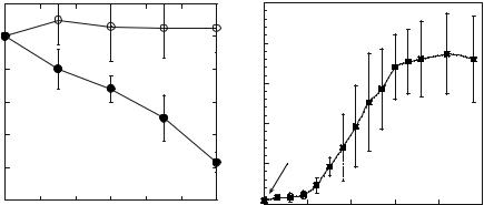
FIGURE 13.1. (A) Transdermal insulin delivery using low-frequency sonophoresis (closed circles). Open circles show controls (insulin placed on untreated skin. Skin was permeabilized by a short application of ultrasound ( 60-fold enhancement of skin conductivity). Insulin (500 U/ml) was placed on permeabilized skin. (B) Transdermal delivery of low-molecular weight heparin (LMWH) with low-frequency sonophoresis. Skin was pretreated with ultrasound for 2 minutes and skin conductance was increased. A solution of LMWH was placed on sonophoretically permeabilized skin. Delivery of LMWH was assessed by measuring plasma aXa activity. Application of LMWH on non-sonicated skin did not increase plasma aXa activity.
exposed to insulin [62]. Specifically, the glucose level decreased to 34% of the initial value at lower pressures and to 22% of the initial value at higher acoustic pressures. Comparable results were obtained in rabbits at somewhat higher frequencies (150 kHz). Mitragotri et al. performed in vitro and in vivo evaluation of the effect of low-frequency ultrasound on transdermal delivery of proteins [8]. Application of low-frequency ultrasound (20 kHz, 125 mW/cm2, 100 msec pulses applied every second) enhanced transdermal transport of proteins including insulin, γ-interferon, and erythropoeitin across human cadaver skin in vitro [8]. Ultrasound under the same conditions delivered therapeutic doses of insulin across hairless rat skin in vivo from a chamber glued on the rat’s back and filled with an insulin solution (100 U/ml) [8]. A simultaneous application of insulin and ultrasound (20 kHz, 225 mW/cm2, 100 msec pulses applied every second) reduced the blood glucose level of diabetic hairless rats from about 400 mg/dL to 200 mg/dL in 30 minutes. A corresponding change in plasma insulin levels was observed during sonophoresis. Boucaud et al. also demonstrated a dose-dependent hypoglycemia in hairless rats exposed to ultrasound and insulin [70]. At an energy dose of 900 J/cm2 75% reduction in glucose levels was reported. Pretreatment of skin by low-frequency ultrasound (20 kHz, 7 W/cm2) has also been shown to enhance skin permeability to insulin [78].
13.6.2. Low-molecular Weight Heparin
Low-frequency ultrasound has also been shown to deliver low molecular weight heparin (LMWH) across the skin [48]. Transdermal LMWH delivery was measured by monitoring
228 |
SAMIR MITRAGOTRI |
aXa activity in blood. No significant aXa activity was observed when LMWH was placed on non-treated skin. However, significant amount of LMWH was transported transdermally after ultrasound pretreatment. aXa activity in the blood increased slowly for about 2 hours, after which, it increased rapidly before achieving a steady state after 4 hours at a value of about 2U/ml [48]. Effect of transdermally delivered LMWH was observed well beyond 6 hours in contrast to intravenous or subcutaneous injections, which resulted only in transient biological activity.
13.6.3. Oligonucleotides
Low-frequency ultrasound has also been shown to enhance dermal penetration of oligonucleotides (ODN). A 10-minute application of ultrasound (20 kHz and 2.4 W/cm2) increased skin ODN permeability to 4.5 × 10⁻⁵ cm/hr compared to nearly undetectable values of non-treated skin. A significant amount of ODN was also localized in the skin. Greater enhancements of ODN delivery were obtained by simultaneous application of ultrasound and ODN. Experiments performed with FITC-labeled ODN revealed that ODN is largely localized in the superficial layers of the skin. An estimate of local concentration of ODN in the skin was performed. Assuming a depth of penetration of 100–1000 μm the estimated concentration of ODN in the skin at the end of ultrasound application was about 0.53–5.3 % of the donor concentration. ODN penetration into skin due to LFS was heterogeneous. Heterogeneity of dermal penetration was visualized by monitoring penetration of a dye, sulforhodamine B (SRB) that was incorporated in the coupling medium. SRB penetration clearly indicated 4–5 intensely stained spots ( 1 mm in diameter), which were termed as Localized Transport Pathways, LTPs. To further ensure that ODN penetrated into skin without losing integrity, skin exposed to ISIS 13920 in the presence of ultrasound was assessed using immunohistochemistry. No visible staining was observed in case of passive delivery, however the skin treated with LFS was heavily stained suggesting penetration of oligonucleotide delivery. ODN was localized in the epidermis as well as dermis. Furthermore microscopy studies suggested that ODN penetrated into epidermal cells. This is a particularly appealing feature since viable epidermal cells are an attractive target for ODN delivery.
13.6.4. Vaccines
Recently, low-frequency sonophoresis has also been used to deliver vaccines across the skin (Unpublished data). Transcutaneous immunization promises to be a potent novel vaccination technique since topical immunization elicits both systemic and mucosal immunity [79]. The latter is of great importance, since a significant number of pathogens invade the host via mucosal surfaces [80]. TCI is based on the premise that systemic and mucosal immune responses can be initiated by stimulation of the LCs in the skin.
Ultrasonic delivery of TTx generated a strong IgG response in animals. Delivery of 1.3 μg of TTx generated an immune response comparable to that induced by 10 μg subcutaneous injection. Studies have shown that an IgG antibody response generated by only 5 μg subcutaneous injection is sufficient for protection against a lethal dose of tetanus toxin [81]. Ultrasonic delivery of TTx also generated a strong mucosal immune response. A large number of TT specific plasma cells were found in the intestine. In addition, significant
TRANSDERMAL DRUG DELIVERY USING LOW-FREQUENCY SONOPHORESIS |
229 |
presence of anti-TT antibodies was observed throughout the intestine (unpublished data). Secretory IgA produced by local antibody secreting cells (ASC), i.e. B-lymphocytes, is an important line of defense for mucosal surfaces such as the respiratory or intestinal tract. The polymeric structure of the secretory IgA results in effective cross-linking of large antigens, which are then easily trapped in mucus and eliminated by the action of cilia in the respiratory tract or peristalsis in the gut [80, 82]. Secretory IgA also inhibits infection and colonization by directly preventing the adhesion of bacteria and viruses to mucosal epithelial cells [82, 83].
Two possible mechanisms were proposed to explain why pretreatment of skin with lowfrequency ultrasound prior to contact with the antigen vaccine may enhance the immune response. One possible mechanism is that ultrasound pretreatment results in increased delivery of the vaccine compared to control, thus enabling sufficient amount of vaccine to enter the skin in order to activate the skins immune response. However, a comparison of the response obtained by TCI and subcutaneous immunization shows that IgG immune response elicited by TCI is almost 10 -fold more effective per dose compared to subcutaneous injections. The second mechanism involves the involvement of Langerhans and immune cells of the skin that effectively capture the antigen and present it to the immune system. Clear activation of LCs was observed after ultrasonic TTx delivery. LC activation is partly induced by the entry of the antigen and partly by the direct effect of ultrasound on skin. Mechanisms responsible for ultrasound-induced activation of LCs are not clear, although barrier disruption or release of pro-inflammatory signals by the keratinocytes are possible candidates.
13.7. TRANSDERMAL GLUCOSE EXTRACTION USING SONOPHORESIS
Low-frequency ultrasound skin pretreatment has also been used to extract glucose and other analytes from the skin (transport in the opposite direction; from the interstitial compartment through the skin into a reservoir filled with water placed on the top of the pretreated skin). Kost et al. demonstrated that ultrasound pretreatment followed by vacuum application extracts sufficient amounts of interstitial fluid to perform continuous glucose monitoring [37]. The measured glucose flux after application of vacuum (10 in Hg) on ultrasound-pretreated skin was about 52 ± 30 μg/cm2/hr when the average serum glucose concentration of the rat was 183 mg/dL, a flux about 100 time higher compared to passive flux across non-treated skin [84]. This flux corresponds to interstitial fluid (ISF) extraction rate of 25.6 μl/cm2/hr.
Correlation between sonophoretically extracted glucose and blood glucose values was assessed in rats [85]. In these experiments, rat skin was exposed to ultrasound. Multiple extractions were performed using vacuum (10 in Hg for 5 minutes applied every 20 minutes) over a period of 2 hours. The first transdermal flux was used to calculate the calibration factor. Blood glucose levels of rats were varied by infusing insulin intravenously at a rate of 10 mU/min for 2 hours. Transdermally extracted glucose flux correlated well with the changes in the blood glucose level in the hypoand hyperglycemic range. The relationship between the predicted and measured glucose values was linear (r = 0.97). Similar results were reported by Kost et al. in the tests performed in human volunteers [37]. Specifically, ultrasound was used to permeabilize skin of human volunteers. A short application of
230 |
SAMIR MITRAGOTRI |
ultrasound permeabilized skin for about 15 hours. During this period, interstitial fluid was extracted every 30 minutes. Concentration of glucose in the extracted fluid was measured and compared with blood glucose values. The results showed good correlation between glucose in the interstitial fluid and in the blood. Furthermore, patients reported no pain upon ultrasound application. More recently, Kost et al. reported clinical studies performed on diabetic volunteers where ultrasound was used to permeabilize skin and glucose was collected by diffusion instead of vacuum through permeabilized skin. Glucose flux through sonicated site averaged 11 nmol/cm2/hours [86].
Safety of low-frequency sonophoresis has been evaluated in several studies. Histological studies performed on rat and pig skin indicated no structural changes in the skin on a length scale of μm [43]. Accordingly, the structural changes in the stratum corneum appear to occur at a sub-micron scale. Singer et al. performed a toxicological analysis of low-frequency sonophoresis. They found a dose-dependent effect of ultrasound on skin. They concluded that low-frequency ultrasound at low intensities appears safe for enhancing the topical delivery of medications, producing only minimal urticarial reactions. Higher-intensity ultrasound produced significant thermal effects. [87] Boucaud et al. also performed a microstructural analysis of skin samples exposed to ultrasound. They reported no detectable changes in the skin structure of human skin at an intensity of 2.5 W/cm2. Hairless rat skin exposed to the same intensity showed slight and transient erythema and dermal necrosis at 24 hours [88]. Tolerance of low-frequency ultrasound by patients has been reported in a number of studies. Kost et al. reported that low-frequency ultrasound was well-tolerated by patients [37]. More recently, a clinical study on the use of low-frequency ultrasound for lidocaine delivery has also been reported [89]. However, it must be realized that ultrasound, like any other energy source, is likely to exhibit a window of parameters within which safe application can be practiced. Accordingly, a careful selection of parameters must be performed in sonophoresis studies.
13.8. MECHANISMS OF LOW-FREQUENCY SONOPHORESIS
Significant attention has been devoted to understand the mechanisms of low-frequency sonophoresis [23, 41, 64, 90, 91]. A consensus has been reached that acoustic cavitation, the formation and collapse of gaseous cavities, is responsible for low-frequency sonophoresis [9, 41, 57, 91]. Below, we summarize the current conclusions of the mechanistic investigations of sonophoresis.
During low-frequency sonophoresis, cavitation is predominantly induced in the coupling medium (the liquid present between the ultrasound transducer and the skin [57]). The maximum radius of the cavitation bubbles is related to the frequency and acoustic pressure amplitude. Under the conditions used for low-frequency sonophoresis ( f 20–100 kHz and pressure amplitudes 1–2.4 bar) the maximum bubble radius is estimated to be between 10–100 μm. Owing to the large bubble size, cavitation is unlikely to occur within the 15 μm thick SC during low-frequency sonophoresis. Accordingly, cavitation in the coupling medium is of primary interest during low-frequency sonophoresis.
Two types of cavitation, stable or inertial have been evaluated for their role in sonophoresis. Stable cavitation corresponds to periodic growth and oscillations of bubbles while inertial cavitation corresponds to violent growth and collapse of cavitation bubbles [92]. Using
TRANSDERMAL DRUG DELIVERY USING LOW-FREQUENCY SONOPHORESIS |
231 |
A B C
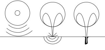
FIGURE 13.2. Three possible modes through which inertial cavitation may enhance SC permeability. (A) Spherical collapse near the SC surface emits shock waves, which can potentially disrupt the SC lipid bilayers. (B) Impact of an acoustic microjet on the SC surface. The microjet possessing a radius about one tenth of the maximum bubble diameter impacts the SC surface without penetrating into it. The impact pressure of the microjet may enhance SC permeability by disrupting SC lipid bilayers. (C) Microjets may physically penetrate into the SC and enhance the SC permeability.
acoustic spectroscopy, stable as well as inertial cavitation has been quantified [9, 57]. The overall dependence of inertial cavitation on ultrasound intensity was found to be similar to that of conductivity enhancement [9, 57]. Specifically, ultrasound intensity above threshold intensity is required before inception of inertial cavitation is observed. This threshold corresponds to minimum pressure amplitude required to induce rapid growth and collapse of cavitation nuclei. Beyond this threshold, white noise (indicator of inertial cavitation) increased linearly with ultrasound intensity, although at any given intensity, inertial cavitation activity decreased rapidly with ultrasound frequency [57]. The threshold intensity for the occurrence of inertial cavitation increased with increasing ultrasound frequency. This dependence reflects the fact that growth of cavitation bubbles becomes increasingly difficult with increasing ultrasound frequency. Tezel et al. showed that regardless of the intensity and frequency, skin conductivity enhancement correlated universally with the cavitation energy density [57]. These data suggested a strong role played by inertial cavitation in low-frequency sonophoresis.
Inertial cavitation occurs in the bulk coupling medium as well as near the skin surface. Inertial cavitation at both locations may potentially be responsible for conductivity enhancement. Three mechanisms by which inertial cavitation events might enhance SC permeability were proposed [93] (Figure 13.2). These include bubbles that collapse symmetrically and emit a shock wave, which can disrupt the SC lipid bilayers and acoustic microjets that might impact the SC without penetration. Impact of microjets may also be responsible for SC lipid bilayer disruption. Microjets resulting from collapsing bubbles near the SC surface may also potentially penetrate into the SC and disrupt the structure.
Inertial cavitation in the vicinity of a surface is fundamentally different from that away from the surface. Specifically, collapse of spherical cavitation bubbles in the bulk solution is symmetric and results in the formation of a shock wave. This shock wave can potentially disrupt the structure of the lipid bilayers. However, the amplitude of the shock wave decreases rapidly with the distance. Collapse of cavitation bubbles near boundaries
232 |
SAMIR MITRAGOTRI |
(especially rigid ones) has been extensively studied in the literature [94]. Specifically, Naude and Ellis showed that cavitation bubbles travel under the influence of ultrasound field towards the boundary and collapse near the boundary depending on its proximity to the surface [95]. The collapse of cavitation bubbles near the boundary is asymmetric due to the difference in the surrounding condition on either side of the bubble. Specifically, the asymmetry in the surroundings leads to the generation of asymmetry in the pressure, which ultimately leads to the formation of a liquid microjet directed towards the surface. The diameter of the microjet is much smaller than that of the maximum bubble radius. There have been several estimates of the speed of the liquid microjet when it strikes the surface (between 50 and 180 m/s [96–98]).
In a recent study, Tezel et al. evaluated the effect of spherical collapses as well as microjets on skin permeability enhancement [91]. They concluded that both types of cavitation events may be responsible for sonophoresis. Regardless of the precise mode of collapse, about 10 collapses/second/cm2 in the form of spherical collapses or microjets near the surface of the stratum corneum were suggested to explain experimentally observed conductivity enhancements. They also reported that bubble collapses only close to the stratum corneum surface ( 50 μm) contribute to sonophoresis.
Disruption of SC lipid bilayers due to bubble-induced shock waves or microjet impact may enhance skin permeability by at least two mechanisms. First, a moderate level of disruption decreases the structural order of lipid bilayers and increases solute diffusion coefficient [99]. At a higher level of disruption, lipid bilayers may loose structural integrity and facilitate penetration of the coupling medium into the SC. Since many sonophoresis experiments reported in the literature are performed using coupling media comprising aqueous solutions of surfactants, disruption of SC lipid bilayers enhances incorporation of surfactants into lipid bilayers. Incorporation of excessive water and surfactants further promotes bilayer disruption, thereby opening pathways for solute permeation [100, 101]. Recently, Alvarez-Roman reported that lipid extraction also plays a role in low-frequency sonophoresis [64]. They reported that about 30% of the stratum corneum lipids were removed during low-frequency sonophoresis.
13.9. CONCLUSIONS
Low-frequency sonophoresis has been shown to increase skin permeability to a variety of lowas well as high-molecular weight drugs including insulin and low-molecular weight heparin. Ultrasound-mediated enhancement of transdermal transport is mediated by inertial cavitation. Collapse of cavitation bubbles near the stratum corneum is hypothesized to disrupt its structure due to cavitation-generated shock waves or microjets.
REFERENCES
[1]A.J. Coleman and J.E. Saunders. A review of the physical properties and biological effects of the high amplitude acoustic field used in extracorporeal lithotripsy. Ultrasonics, 31:75–89, 1993.
[2]C.J. Diederich and K. Hynnen. Ultrasound technology for hyperthemia. Ultrasound Med. Biol., 25:871–887, 1999.
TRANSDERMAL DRUG DELIVERY USING LOW-FREQUENCY SONOPHORESIS |
233 |
[3]A.V. Alexandrov. Ultrasound-enhanced thrombolysis for stroke: clinical significance. Eur. J. Ultrasound, 16:131–140, 2002.
[4]J.C. Goes and A. Landecker. Ultrasound-induced lipoplasty (UAL) in breast surgery. Aesthetic Plast. Surg., 26:1–9, 2002.
[5]C.A. Speed. Therapeutic ultrasound in soft tissue lesions. Rheumatology, 40:1331–1336, 2001.
[6]M. Hadjiargyrou, K. McLeod, J.P. Ryaby, and C. Rubin.Enhancement of fracture healing by low intensity Ultrasound. Clin. Orthop., 355(Suppl):S216–S229, 1998.
[7]A. Tezel, A. Sens, J. Tuscherer, and S. Mitragotri. Frequency dependece of sonophoresis. Pharm. Res., 18:1694–1700, 2001.
[8]S. Mitragotri, D. Blankschtein, and R. Langer, Ultrasound-mediated transdermal protein delivery. Science, 269:850–853, 1995.
[9]H. Tang, D. Blankschtein, and R. Langer. An investigation of the role of cavitation in low-ferquency ultrasound-mediated transdermal drug transport. Pharm. Res., 19:1160–1169, 2002.
[10]H.R. Guzman, D.X. Nguyen, S. Khan, and M.R. Prausnitz. Ultrasound-mediated disruption of cell membranes. I. Quantification of molecular uptake and viability. J. Acoust. Soc. Am., 110:588–596, 2001.
[11]M.B. Sundaram J. and Mitragotri S. An experimental analysis of ultrasound-induced permeabilization.
Biophy. J., 2002.
[12]D. Miller and J. Quddus. Sonoporation of monolayer cells by diagnostic ultrasound activation of contrastagent gas bodies. Ultrasound Med. Biol., 26:661–667, 2000.
[13]J.Wu, J.P. Ross and J.-F. Chiu. Reparable sonoporation generated by microstreaming. J. Acoust. Soc. Amer., 111:1460–1464, 2002.
[14]J.L. Nelson, B.L. Roeder, J.C. Carmen, F. Roloff, and W.G. Pitt. Ultrasonically activated chemotherapeutic drug delivery in a rat model. Cancer Res., 62:7280–7283, 2002.
[15]R.J. Price and S. Kaul. Contrast ultrasound targeted drug and gene delivery: an update on a new therapeutic modality. J. Cardiovasc. Pharmacol. Ther., 7:171–180, 2002.
[16]C.S. Kwok, P.D. Mourad, L.A. Crum, and B.D. Ratner. Self-assembled molecular structures as ultrasonically-responsive barrier membranes for pulastile delivery. J. Biomed. Mater. Res., 57:151–164, 2001.
[17]J. Kost, K. Leong, and R. Langer. Ultrasound-enhanced polymer degradation and release of incorporated substances. Proc. Natl. Acad. Sci., 86:7663–7666, 1989.
[18]J.R. Linder. Evolving applications of contrast ultrasound. Am. J. Cardiol., 90:72J–80J, 2002.
[19]E.C. Unger, E. Hersh, M. Vannan, T.O. Matsunaga, and T. McCreery. Local drug and genen delivery through microbubbles. Prog. Cardiovasc., 44:45–54, 2001.
[20]H.A.E. Benson, J.C. McElnay, and R. Harland. Phonophoresis of lingocaine and prilocaine from Emla cream. Int. J. Pharm., 44:65–69, 1988.
[21]H.A.E. Benson, J.C. McElnay, and R. Harland. Use of ultrasound to enhance percutaneous absorption of benzydamine. Phys. Ther., 69:113–118, 1989.
[22]H.A.E. Benson, J.C. McElnay, and J. Hadgraft. Influence of ultrasound on the percutaneous absorption of nicotinate esters. Pharm. Res., 9:1279–1283, 1991.
[23]D. Bommannan, G.K. Menon, H. Okuyama, P.M. Elias, and R.H. Guy. Sonophoresis. II. examination of the mechanism(s) of ultrasound-enhanced transdermal drug delivery. Pharm. Res., 9:1043–1047, 1992.
[24]D. Bommannan, H. Okuyama, P. Stauffer, and R.H. Guy. Sonophoresis. I. The use of high-frequency ultrasounnd to enhance transdermal drug delivery. Pharm. Res., 9:559–564, 1992.
[25]N.N. Byl, A. McKenzie, B. Halliday, T. Wong, and J. O’nConell. The effects of phonophoresis with corticosteroids: a controlled pilot study. J. Orth. Sports Phys. Ther., 18:590–600, 1993.
[26]B.M. Cameroy. Ultrasound enhanced local anesthesia. Am. J. Orthoped., 8:47, 1966.
[27]C.D. Ciccone, B.Q. Leggin, and J.J. Callamaro. Effects of ultrasound and trolamine salicylate phonophoresis on delayed-onset muscle soreness. Phys. Ther., 71:666–678, 1991.
[28]J.E. Griffin and J. Touchstone. Ultrasonic movement of cortisol in to pig tissue. Am. J. Phys. Med., 44:20–25.
[29]J.E. Griffin, J.L. Echternach, R.E. Proce, and J.C. Touchstone. Patients treated with ultrasonic driven hydrocortisone and with ultrasound alone. Phys. Ther., 47:600–601, 1967.
[30]J.E. Griffin and J.C. Touchstone. Low-intensity phonophoresis of cortisol in swine. Phys. Ther., 48:1136– 1344, 1968.
[31]J. Griffin, E. and J.C. Touchstone. Effects of ultrasonic frequency on phonophoresis of cortisol into swine tissues. Am. J. Phys. Med., 51:62–78, 1972.