

Therapeutic Micro-Nano Technology BioMEMs - Tejlal Desai & Sangeeta Bhatia
.pdfVASCULAR ZIP CODES AND NANOPARTICLE TARGETING |
133 |
targeted with homing peptides. The RGD integrin recognition sequence can direct adenovirus vectors to cells expressing the αvβ3 integrin, such as angiogenic endothelial cells [21, 40]. Chemical mimetics of the RGD sequence have been used to target tumor vasculature with lipid-based nanoparticles designed for imaging purposes or as gene therapy vectors [23, 31, 41].
The targeted nanoparticles used so far can seek out and concentrate at the intended target tissue. However, having reached their target, they are only capable of providing a signal or passively releasing a drug or gene payload. For the full potential of nanotechnology to be realized in medicine, it will be necessary to build more functions into such particles. One potentially important advance would be a nanoparticle in which the binding to the target acts as a switch that triggers a response such as a signal or release of a drug. Better still, the nanoparticle could incorporate a switch that can be controlled from outside, so that the functions of the particle can be activated at will and at an optimal time. Finally, the device should be capable of recognizing changes caused by the treatment and report back on the results.
The sensor aspect has essentially been solved. The homing peptides described here, as well as proteins, antibodies, and nucleic acid molecules that recognize suitable targets, can serve as sensors. Using sensors that detect vascular markers offers the considerable advantage that the endothelial marker molecule recognized by the sensor is readily available to a circulating particle. Moreover, endothelial binding is frequently followed by transendothelial transport into the tissue. In addition, some of our newest peptides recognize markers that are shared by the tumor endothelium and tumor cells [28, 35]. The dual recognition allows these peptides to deliver a payload both to tumor vessels and the tumor parenchyma. These peptides and their payload are taken up and internalized by the target cells; in some cases, the result is a tremendous concentration of the payload in tumors [29]. These peptides can also direct exquisitely specific homing of intravenously injected quantum dots into target tissues. For example, we have been able to label tumor blood vessels with quantum dots in one color and the lymphatics in another in the same tumor [2]. With the sensors in hand, the next challenge in the field is to couple the sensors to molecular switches that convert the sensor signal to a response.
7.8. FUTURE DIRECTIONS
Our homing peptide studies indicate that the endothelial cells (and possibly mural cells as well) in each vascular bed express a unique complement of cell surface molecules. Only a few of the molecules that act as receptors for homing peptides are known, but their diversity (proteases, integrins, growth factor receptors, and proteoglycans; Ruoslahti, 2002) suggests that they represent a heterogeneous group of proteins with a variety of functions. The function of any given marker molecule is likely to be important for the vascular bed that expresses it. Such molecules can also function as tissue-specific receptors for metastatic tumor cells [7]. It will be important to accelerate the discovery of homing peptide receptors; we are working on a method that allows simultaneous identification of a homing peptide and its receptor.
The homing peptides that recognize tumor vasculature fall into two categories: (1) those that bind to angiogenesis-associated molecules and recognize the vasculature of all tumors
134 |
ERKKI RUOSLAHTI |
(and other angiogenic lesions) and (2) those that recognize the vasculature of some, but not all tumors. The latter kind would be potentially advantageous in therapeutic applications because sites of beneficial angiogenesis would not be affected. Our current screening efforts are directed at discovering peptides that would broadly recognize the vasculature of a given tumor type.
Homing peptide work shows that tumor lymphatics, like tumor blood vessels, differ from their normal counterpart vessels. This offers a possibility of attacking tumors from two directions, through the blood vessels and through the lymphatics, a strategy that remains to be explored.
We have described homing peptides that are internalized by the target cells and can take a payload into the cell nucleus. We are currently testing drug conjugates to utilize these properties of the peptides. We are also taking advantage of the sensor capabilities of homing peptides in designing nanodevices that can become internalized in tumor endothelial calls and tumor cells.
Being able to sense disease-associated changes in the vasculature may make it possible to develop nanodevices that serve a sentinels reporting back on early signs of a disease, such as pre-malignant lesions. We have already shown that it is possible to identify peptides that specifically recognize pre-malignant lesions by the vascular signature [22, 26]. The sentinel nanodevices would be altered upon an encounter with a specific target and would return to the general circulation to report back on their survey of the vasculature. Ingestible nanoparticles could similarly patrol the gut, and other designs could be developed for premalignant lesions of the urinary bladder, and so on. We are in the process of isolating peptides that recognize premalignant epithelial cells in these tissues to provide the sensors for such diagnostic nanodevices. An encounter with the target by a nanosensor for premalignant lesions could also induce release of a drug from the device, making it into both a diagnostic and preventative tool.
Adding responsiveness to an external signal, such a magnetic field, would allow external control of nanodevices that have identified their target in the body to effect a therapy in a highly focused manner. Finally, another set of sensors operating the same way as the homing signals, but designed to detect apoptosis, cellular distress, cell proliferation, or inflammation, could monitor the state of the tissue of interest for the physician. The design of nanodevices capable of performing these tasks will require collaboration among a diverse group of scientists with specialties ranging from tumor biology to materials science. Success in endeavors such as these is likely to bring nanomedicine to the forefront of medical practice.
ACKNOWLEDGEMENT
The author’s work was supported by the NIH grants CA 82713 and Cancer Center Support Grant CA 30199, and the DAMD 17-02-1-0315 Innovator Award from the Department of Defense.
REFERENCES
[1]K. Alitalo and P. Carmeliet. Molecular mechanisms of lymphangiogenesis in health and disease. Cancer Cell, 1:219–227, 2002.
VASCULAR ZIP CODES AND NANOPARTICLE TARGETING |
135 |
[2]M.E. Akerman, W.C. Chan, P. Laakkonen, S.N. Bhatia, and E. Ruoslahti. Nanocrystal targeting in vivo. Proc. Natl. Acad. Sci. U.S.A., 99:12617–12621, 2002.
[3]W. Arap, W. Haedicke, M. Bernasconi,, R. Kain, D. Rajotte, S. Krajewski, H.M. Ellerby, D.E. Bredesen, R. Pasqualini, and E. Ruoslahti. Targeting the prostate for destruction through a vascular address. Proc. Natl. Acad. Sci. U.S.A., 99:1527–1531, 2002.
[4]W. Arap, R. Pasqualini, and E. Ruoslahti. Cancer treatment by targeted drug delivery to tumor vasculature in a mouse model. Science, 279:377–380, 1998.
[5]P.J. Bates, J.B. Kahlon, S.D. Thomas, Trent, and D.M. Millet. Antiproliferative activity of G-rich Oligonucleotides correlates with protein binding. J.Biol.Chem., 274:26369–26377, 1999.
[6]D.M. Brown, M. Pellecchia, and E. Ruoslahti. Drug identification through in vivo screening of chemical libraries. ChemBioChemistry, 5:871–875.
[7]D. Brown, and E. Ruoslahti. Metadherin, a novel cell-surface protein in breast tumors that mediates lung metastasis. Cancer Cell, 5:365–374, 2004.
[8]S. Christian, J. Pilch, K. Porkka, P. Laakkonen, and E. Ruoslahti. Nucleoilin expressed at the cell surface is a marker of endothelial cells in tumor blood vessels. J. Cell. Biol., 163:871–878, 2003.
[9]F. Curnis, A. Sacchi, L. Borgna, F. Magni, A. Gasparri, and A. Corti. Enhancement of tumor necrosis factor antitumor immunotherapeutic properties by targeted delivery to aminopeptidase N (CD13). Nature Biotech., 18:1185–1190, 2000.
[10]F. Curnis, G. Arrigoni, A. Sacchi, L. Fischetti, W. Arap, R. Pasqualini, and A. Corti. Differential binding of drugs containing the NGR motif to CD13 isoforms in tumor vessels, epithelia, and myeloid cells. Cancer Res., 62:867–874, 2002.
[11]A.M. Derfus, W.C. Chan, and S.N. Bhatia. Probing the cytotoxicity of semiconductor quantum dots. Nano Letters, 4:11–18, 2004.
[12]A.M. Derfus, W.C. Chan, and S.N. Bhatia, Intracellular delivery of quantum dots for live cell labeling and organelle tracking. Adv. Mat. published online 5/19/04, 2004.
[13]B.S. Ding, Y.J. Zhou, X. Y. Chen, J. Zhang, P.X. Zhang, Z. Y. Sun, X. Y. Tan, and J. N. Liu. Lung endothelium targeting for pulmonary embolism thrombolysis. Circulation, 108:2892–2898, 2003.
[14]Ellerby, W. Arap, L.M. Ellerby, R. Kain, R. Andrusiak, G. D. Rio, S. Krajewski, C. R. Lombardo, R., Rao,
E.Ruoslahti. et al. Anti-cancer activity of targeted pro-apoptotic peptides. Nat. Med., 5:1032–1038, 1999.
[15]B.P. Eliceiri and D.A. Cheresh. The role of alphav integrins during angiogenesis: insights into potential mechanisms of action and clinical development. J. Clin. Invest., 103:1227–1230, 1999.
[16]B.P. Eliceiri and D.A. Cheresh. Adhesion events in angiogenesis. Curr. Opini. Cell Biol., 13:563–568, 2001.
[17]M. Essler and E. Ruoslahti. Molecular specialization of breast vasculature: a breast-homing phagedisplayed peptide binds to aminopeptidase P in breast vasculature. Proc. Natl. Acad. Sci. U.S.A., 99:2252–2257, 2002.
[18]M.N. Fukuda, C. Ohyama, K. Lowitz, O. Matsuo, R. Pasqualini, E., Ruoslahti, and M. Fukuda. Apeptide mimic of E-selectin ligand inhibits sialyl Lewis X-dependent lung colonization of tumor cells. Cancer Res., 60:450–456, 2000.
[19]D.M. Gerlag, E. Borges, P.P. Tak, H.M. Ellerby, D.E., Bredesen, R. Pasqualini, E. Ruoslahti, and G.S. Firestein. Suppression of murine collagen-induced arthritis by targeted apoptosis of synovial neovasculature. Arth. Res., 3:357–361, 2001.
[20]C. Halin, S. Rondini, F. Nilsson, A. Berndt, H. Kosmehl, L. Zardi, and D. Neri. Enhancement of the antitumor activity of interleukin-12 by targeted delivery to neovasculature. Nat. Biotechnol., 20:264–269, 2002.
[21]Y.S. Haviv, J.L. Blackwell, A. Kanerva, P. Nagi,V. Krasnykh, I. Dmitriev, M. Wang, S. Naito, X. Lei,
A.Hemminki, et al. Adenoviral gene therapy for renal cancer requires retargeting to alternative cellular receptors. Cancer Res., 62:4273–4281, 2002.
[22]J.A. Hoffman, E. Giraudo M. Singh, M. Inoue, K. Porkka, D. Hanahan, and E. Ruoslahti. Progressive vascular changes in a transgenic mouse model of squamous cell carcinoma. Cancer Cell, 4:383–391, 2003.
[23]J. D. Hood, M. Bednarski, R. Frausto, S. Guccione, R. A. Reisfeld, R. Xiang, and D. A. Cheresh. Tumor regression by targeted gene delivery to the neovasculature. Science, 296:2404–2407, 2002.
[24]P. Houston, J. Goodman, A. Lewis, C. J. Campbell, and M. Braddock. Homing markers for atherosclerosis: applications for drug delivery, gene delivery and vascular imaging. FEBS Lett., 492:73–77, 2001.
[25]R.C. Johnson, D. Zhu, H.G. Augustin-Voss, and B.U. Pauli. Lung endothelial dipeptidyl peptidase IV is an adhesion molecule for lung metastatic rat breast and prostate carcinoma cells. J. Cell Biol., 121:1423–1432, 1993.
136 |
ERKKI RUOSLAHTI |
[26]J.A. Joyce, P. Laakkonen, M. Bernasconi, G. Bergers, E. Ruoslahti, and D. Hanahan. Stage-specific vascular markers revealed by phage display in a mouse model of pancreatic islet tumorigenesis. Cancer Cell, 4:393– 403, 2003.
[27]S. Kim, K. Bell, S.A. Mousa, and J.A. Varner. A regulation of angiogenesis in vivo by ligation of integrin α5β1 with the central cell-binding domain of fibronectin. Am. J. Pathol., 156:1345–1362, 2000.
[28]P. Laakkonen, K. Porkka, J.A. Hoffman, and E. Ruoslahti. A tumor-homing peptide with a lymphatic vesselrelated targeting specificity. Nat. Med., 8:743–751, 2002.
[29]P. Laakkonen, M.E. Akerman, H. Biliran, M. Yang, F. Ferrer, T. Karpanen, R.M. Hoffman, and E. Ruoslathi. Antitumor activity of a homing peptide that targets tumor lymphatics and tumor cells. Proc. Natl. Acad. Sci.
U.S.A., 2004.
¨
[30] U. Langel. Cell-Penetrating Peptides: Processes and Applications. Boca Raton, Florida, CRC Press, 2003. [31] G.M. Lanza and S.A. Wickline. Targeted ultrasonic contrast agents for molecular imaging and therapy. Curr.
Prob. Cardiol., 28:625–653, 2003.
[32] D.P. McIntosh, X.Y. Tan, P. Oh, and J. E. Schnitzer. Targeting endothelium and its dynamic caveolae for tissue-specific transcytosis in vivo: a pathway to overcome cell barriers to drug and gene delivery. Proc. Natl. Acad. Sci. U.S.A., 99:1996–2001, 2002.
[33] R. Pasqualini and E. Ruoslahti. Organ targeting in vivo using phage display peptide libraries. Nature, 380:364– 366, 1996.
[34] R. Pasqualini, E. Koivunen, R. Kain, J. Lahdenranta, M. Sakamoto, A. Stryhn, R. A. Ashmun, L. H. Shapiro, W. Arap, and E. Ruoslahti. Aminopeptidase N is a receptor for tumor-homing peptides and a target for inhibiting angiogenesis. Cancer Res., 60:722–727, 2000.
[35] K. Porkka, P. Laakkonen, J.A. Hoffman, M. Bernasconi, and E. Ruoslahti. Targeting of peptides to the nuclei of tumor cells and tumor endothelial cells in vivo. Proc. Natl. Acad. Sci. U.S.A., 99:7444–7449, 2002.
[36] D. Rajotte and E. Ruoslahti. Membrane dipeptidase is the receptor for a lung-targeting peptide identified by in vivo phage display. J. Biol. Chem., 274:11593–11598, 1999.
[37] E. Ruoslahti. Specialization of tumour vasculature. Nat. Rev. Cancer, 2:83–90, 2002.
[38] E. Ruoslahti, RGD story: A personal account. A landmark essay. Matrix Biol., 22:459–465, 2003.
[39] E. Ruoslahti and D. Rajotte. An address system in the vasculature of normal tissues and tumors. Annu. Rev. Immunol., 18:813–827, 2000.
[40] T.J. Wickham. Targeting adenovirus. Gene Ther., 7:110–114, 2000.
[41] P.M. Winter, S.D. Caruthers, A. Kassner, T.D. Harris, L.K. Chinen, J.S. Allen, E.K. Lacy, H. Zhang, J.D. Robertson, S.A. Wickline, and G.M. Lanza. Molecular imaging of angiogenesis in nascent Vx-2 rabbit tumors using a novel alpha(nu)beta3-targeted nanoparticle and 1.5 tesla magnetic resonance imaging. Cancer Res., 63:5838–5843, 2003.
[42] L. Zhang, J.A. Hoffman, and E. Ruoslahti. Molecular profiling of heart endothelial cell. Circulation, (In press) 2005.
8
Engineering Biocompatible Quantum Dots for Ultrasensitive, Real-Time Biological
Imaging and Detection
Wen Jiang , Anupam Singhal , Hans Fischer †, Sawitri Mardyani , and Warren C. W. Chan †
Institute of Biomaterials and Biomedical Engineering, 4 Taddle Creek Rd., 408, Toronto, ON, M5S 3G9, Canada
†Department of Materials Science & Engineering, 4 Taddle Creek Rd., 408, Toronto, ON, M5S 3G9, Canada
8.1. INTRODUCTION
Advances in the design of optical probes have played a central role in the emergence of photon-based microscopy techniques for biological imaging and detection [1–6]. These advances have led to the elucidation of the biological function and activity of many proteins, nucleic acids, and other molecules in living cells, tissues, and animals. Currently, the molecular architecture of greater than 70% of all optical probes consists of an “optical emitter” attached to a “targeting molecule” [4]. The targeting molecule directs the optical emitter to specific biological sites where the optical emitter can then be used to detect the activities of biomolecules. The most popular optical probes have been traditionally designed from organic-based molecules; for instance, probes for the imaging of cellular cytoskeleton are based on the conjugation of red-fluorescent molecule Texas Red to the small targeting organic molecule phalloidin (for labeling actin fibers) and green-fluorescent Alexa Fluor 488 to a recognition antibody (for labeling microtubules) [4]. Hundreds of different types of organic-based fluorescent probes are commercially available. These probes can be used
138 |
WEN JIANG ET AL. |
in numerous applications, including the staining of DNA and proteins, detection of subtle differences in the ionic content in living cells, or detection of protein structures [4, 7–10]. Due to their complex molecular structures, however, organic fluorophores often exhibit unfavorable absorption and emission properties, such as photobleaching, environmental quenching, broad and asymmetric emission spectra, and the inability to excite multiple fluorophores of more than 2–3 colors at a single wavelength [10, 11].
Recent developments in the field of nanotechnology have led to the fabrication of a new generation of inorganic probes that overcomes many of the problems associated with organic-based probes [6, 12–14]. Furthermore, these nanometer-sized probes provide novel and unique properties for biological applications that are not available with organicbased fluorophores. At the forefront of this new class of inorganic probes are semiconductor nanocrystals, also known as quantum dots (“QDs”). The physical properties and applications of quantum dots have been heavily investigated in many physics and engineering labs since the early 1980s [15–17]. QDs are defined as particles with physical dimensions smaller than the exciton Bohr radius; this gives rise to a unique phenomenon known as quantumconfinement. Quantum confinement, which refers to the confinement of charge carriers (i.e. electrons and holes) within a material, embarks QDs with unique optical and electronic properties that are unavailable to semiconductors in bulk solids. Although initial interest in QDs focused in physical applicatons (e.g,. making computer chips and light-emitting diodes), recent work by Nie, Alivisatos, and their coworkers have highlighted the great promise of QDs in biological applications [18, 19]. As biological probes, QDs are extremely bright (1 QD ≈ 10 to 20 organic fluorophores), have high resistance to photobleaching, have narrow spectral linewidths, and have sizeand materials-tunable emission that can be excited using a single wavelength [18, 19]. Some of these properties are illustrated in figure 8.1.
When used in biological research and applications, these optical properties will lead to improved detection sensitivity for analysis, and to simplification in experimental and instrumental design. In this book chapter, we describe the development of inorganic semiconductor QD probes for biological and medical applications.
8.2. SYNTHESIS AND SURFACE CHEMISTRY
8.2.1. Synthesis of QDs that are Soluble in Organic Solvents
Breakthroughs in the synthesis of quantum dots by Bawendi [20, 21], Guyot-Sionnest [22], Alivisatos [23] and their coworkers have paved the way for using QDs in biological applications. Prior to their work, a broad range of strategies were attempted to synthesize QDs with high optical quality (i.e., non-aggregated QDs with bright emission and stable against photodegradation). These methods include synthesis of QDs in aqueous buffers with thioglycerol as a stabilizing agent, synthesis of QDs in yeast cells with expressed proteins containing glutathione as stabilizing agents, or synthesis of QDs inside micelle systems [15, 24–27]. However, all of these approaches produced QDs with broad size distributions (relative standard deviation > 15%) and low quantum yields (< 20%). These synthetic systems produced QDs with poor optical and size properties because the reaction conditions did not provide a proper environment for rapid and even nucleation (this leads
ENGINEERING BIOCOMPATIBLE QUANTUM DOTS FOR ULTRASENSITIVE |
139 |
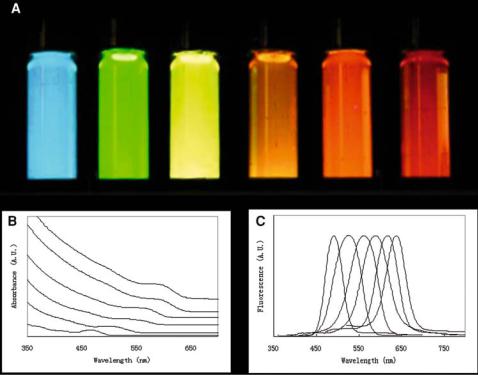
FIGURE 8.1. Size-tunable optical properties of QDs (left, 2.0 nm to right, 6.0 nm—diameter of QDs). (A) The unique emission colours of different sized QDs are observed under UV-excitation by a handheld lamp. (B) Absorbance and (C) fluorescence spectra of different-sized QDs. For the fluorescence measurement, an excitation wavelength of 400 nm is used. QDs = quantum dots.
to QDs with broad size distribution) and for careful growth of QDs (this leads to QDs with large amounts of defect sites).
In the 1990s, research by Bawendi, Guyot-Sionnest, Alivisatos and their coworkers demonstrated the key to prepare high-quality QDs is to use an organometallic reaction scheme in non-aqueous solvents. Furthermore, the reaction must take place at high temperatures to achieve rapid and even nucleation [21–23]. In one common approach high-quality QDs are synthesized via the high-temperature (e.g., 350◦C) pyrolysis of organometallic and chalcogenide reagents in the solvent tri-n-octylphosphine oxide (TOPO). After rapid injection, the TOPO solution appears light yellow. As QDs grow bigger, the solution color evolves from a light yellow to a dark ruby red. At the desired size, the QDs can be isolated using solvent precipitation and centrifugation. Currently, the most established protocols for the synthesis of QDs are those from groups II-VI (e.g., CdSe, CdTe, CdS, and ZnSe) and group III-V (e.g., InP and InAs) of the periodic table [2, 19, 28–30].
La Mer and Dinegar developed theoretical model systems to explain the nucleation and growth of colloidal particles in the 1950s. Their theory has recently been adapted toward explaining QD nucleation and growth [31]. Based on their theory, the nucleation of QDs
140 |
WEN JIANG ET AL. |
occurs when the concentration of precursor molecules are at a high enough level that they exceed the so-called “nucleation threshold”. As QDs grow over time, the concentrations of precursor molecules fall below the threshold level and, ultimately, the Ostwald Ripening process replaces the nucleation process [32]. Ostwald Ripening refers to the breakdown of smaller QDs to provide the needed precursor atoms to build larger QDs. The Ostwald Ripening process is a thermal dependent process, where the growth of larger QDs requires high temperature—the breakdown of QDs in the Ostwald Ripening process is related to the temperature (i.e., larger QDs require higher temperatures). As a result, careful control of growth temperatures can be used to accurately control the average size and size distribution of QDs. During the synthesis, the size and size distributions are monitored by absorbance and fluorescence spectrophotometry. When the desired optical properties are attained, the temperature is reduced to prevent further QD growth or dissolution.
Numerous groups have developed variations to the organometallic approach for the mass-scale synthesis of QDs [30, 33]. In particular, Peng and coworkers have demonstrated that organometallic precursors (e.g. dimethyl cadmium) can be replaced with nonpyrophoric and less costly “greener” reagents (e.g. cadmium oxide, CdO, or cadmium acetate, Cd(Ac)2) [34, 35]. These “alternative routes” to the synthesis of QDs in organic media can be used to reproducibly prepare high-quality CdS, CdSe, and CdTe QDs. Since QDs formed with using greener reagents exhibit slower reaction kinetics (e.g. slower nucleation), extended nucleation periods allow increased quantities of “greener” precursors to be injected at the start of the reaction; with more nucleation sites, more QDs per reaction can be synthesized. Using this synthesis technique, QDs have been successfully synthesized in quantities greater than 1 gram.
In addition to development techniques for the synthesis of high quality QDs, Bawendi, Guyot-Sionnest, Alivisatos and their coworkers demonstrated that the fluorescence efficiency of QDs could be enhanced by the formation of a secondary surface inorganic shell on top of the QD core structure [21–23]. Improvements in the fluorescence efficiency could be attributed to the removal of surface defect sites that traps excited or mobile electrons (see optical properties section below for details). The semiconductor capping material must have larger bandgap energy than the core QD and must have a bond length that is similar to that of the core. For CdSe QDs, CdS and ZnS are excellent shells. For example, the growth of a ZnS layer on CdSe at a temperature below the CdSe nucleating temperature ( 100 to 310◦C) has produced CdSe/ZnS core/shell QDs that exhibit luminescence yields up to 85% [21–23].
There have been great efforts recently to develop better approaches to systematically control the nucleation and growth of semiconductor QDs. A major goal in QD synthesis is to prepare QDs with a discrete size (< 0.1% in size distribution) or shape (e.g., spherical vs. rod-shaped) since the size, shape, and morphology of QDs are closely related to their optical and electronic properties [36, 37]. Although the commonly used flask synthesis has led to the synthesis of high quality QDs, there is great difficulty in controlling the kinetics and thermodynamic factors in the reaction vessel. To manipulate these factors, microfluidic technology has been used as a platform for improving the synthesis of QDs [38]. In a microfuidic chip, micrometer-sized reservoirs, channels, and flow control systems are used to regulate the amount and concentration of precursor solvents. Furthermore, temperature systems can be incorporated onto the chip; this can provide greater control and manipulation of reaction temperature. In effect, the synthesis of QDs may be better controlled and their dimensional properties dramatically improved. Chan et al. demonstrated the controlled
ENGINEERING BIOCOMPATIBLE QUANTUM DOTS FOR ULTRASENSITIVE |
141 |
synthesis of CdSe QDs using a microfluidic chip [38]. The synthesis of a series of CdSe QDs with average diameters of 2.44, 2.54, 2.64, and 2.69 +/ − 0.06 nm in the microfluidics chip was achieved. Although early in development, research by Chan et al. show feasibility in using microfluidic technology for manipulating the synthetic conditions of QDs; this technology holds promise for improving QD synthesis.
Other emerging areas of research in QD synthesis are the design of QDs with various shapes (e.g., rod-shaped), structures (e.g., tetrapod-branched QDs), and composition (e.g., doping CdSe QDs with Te or Mn). All of these changes can influence the optical and electronic properties of the QDs or embark new properties (e.g, magnetism) into the QDs. To date, these QDs have not found applications in biology; however, we expect this to change in the near future. We refer the interested reader to some research developments in this area of QD research [36, 37, 39].
8.2.2. Modification of Surface Chemistry of QDs for Biological Applications
Since the surfaces chemistry of both the core and core/shell QDs are typically coated with hydrophobic ligands, great efforts have been made to modify the surface chemistry of QDs in order to render them biocompatible [13, 18, 19, 40–43]. One approach to achieving QD biocompatibility is the use of displacement chemistry techniques, where the TOPO molecules on the QD surface are replaced with bifunctional molecules (e.g. mercaptoacetic acid and phospho-alcohols). One end of the bifunctional molecule contains a functional group (e.g., –SH or –P) that can interact with metal atoms on the surface of the QDs and out-compete the phosphine oxide from the TOPO molecules for binding to the metal atoms on the QD surface. The other end of the bifunctional molecules must contain a hydrophilic functional group (e.g., alcohol or carboxylic acid functional groups). Since the hydrophilic functional groups protrude from the QD surface, the QDs become charged and extremely polar. The resulting QDs are thus water-soluble and biocompatible. Furthermore, these QDs can be conjugated to biomolecules by reacting the surface alcohol and carboxylic groups with proteins, peptides, and oligonucleotides through various reaction routes.
A second approach to interface QDs with biological systems is to design amphiphilic molecules that interact with the TOPO-molecules on the QD surface. These molecules (e.g., phospholipids) typically contain both hydrophobic and hydrophilic regions. The hydrophobic end interacts with the TOPO molecule through hydrophobic-hydrophobic interactions, while the hydrophilic end, containing carboxylic acid or alcohol functional groups, protrudes from the QD surface. In this scheme, the hydrophobic stabilizing ligand TOPO do not interact with the aqueous solvent and the QDs become biocompatible because of the QD surface hydrophilic functional groups.
In either the displacement or amphiphilic techniques, the QDs may aggregate out of solution. With the displacement technique, the bifunctional stabilizing ligand on the QD surface desorbs off—this leads to the loss of surface charges that cause particle-to-particle repulsion; while in the second technique, the desorption of the amphilphile molecule exposes the hydrophobic TOPO to aqueous solventsthis leads to aggregation via hydrophobic- to-hydrophobic interactions. To alleviate this problem, cross-linking schemes to lock the organic shell from desorbing the surface of the QDs has been developed. One typical approach is the use of the amino acid lysine or short-chain of poly-lysine to cross-link the carboxylic acids on the surface of QDs via carbodiimide catalyst [41, 44, 45]. This will
142
O
QD |
S CH2 COH + H2N |
Biomolecule |
EDAC |
|
QD |
Streptavidin + Biotin |
Biomolecule |
|
WEN JIANG ET AL. |
|
O |
QD |
S CH2 C N Biomolecule |
|
H |
QD |
Streptavidin – Biotin Biomolecule |
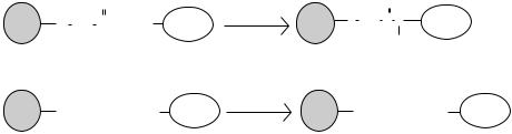
FIGURE 8.2. Methods to coat QD surface with biorecognition molecules. (A) QDs containing the carboxylic acid surface functional groups can be reacted to biomolecules containing primary amino groups through carbodiimide catalysis. EDAC (which is 1-ethyl-3-[3-(dimethylamino)propyl]carbodiimide) is a commonly used water-soluble carbodiimide. The end result of this reaction is an amide bond that links the QDs to the biomolecule. (B) QDs coated with streptavidin have also been a used for linking biotinylated biomolecules. The protein streptavidin has an extremely high binding affinity to the small organic molecule biotin.
lock the stabilizing organic ligand in place and prevent the organic ligands from desorbing the QD surface. With the organic coating in place and the appropriate surface charges, QDs will have long-term stability against aggregation.
Functional groups on QD surface provide sites to conjugate QDs to biomolecules (e.g., proteins, peptides, oligonucleotides) in order to form optical probes (figure 8.2).
One approach to bioconjugating QDs employs conventional carbodiimide chemistry [18, 46]. The addition of carbodiimide molecules to carboxylic acid-coated QDs yields an acylisourea intermediate group, which can be readily attacked by primary amines on biomolecules to form an amide linkage. Electrostatic interactions can also be used to link biomolecules onto the surface of QDs. Mattoussi and coworkers engineered a protein with a positive-charged leucine zipper and demonstrated the adsorption of such a protein onto the surface of a negatively-charged QD [13, 47, 48]. Another popular approach for linking QDs to biomolecules is through a streptavidin-biotin interaction [49, 50]. QDs are initially coated with the protein streptavidin and incubated with biotinylated biomolecules (such as biotin-conjugated antibodies). Streptavidin and biotin has a high binding affinity to each other (Kd 10−14 M, figure 8.2) [51]. A complex of QD-streptavidin-biotin-antibody is formed; when purified from uncomplexed biotin-antibody, they are ready for use as an optical probe.
8.3. OPTICAL PROPERTIES
QDs have garnered broad interests from biological and medical research communities because of their unique optical and electronic properties. Unlike organic-based fluorophores, the properties of QDs can be manipulated by simply changing their size, shape, or composition; basic semiconductor quantum chemistry and physics have been utilized to explain these properties (figure 8.3).
Bulk semiconductor materials contain two energy bands—conduction and valence band. The energy difference between the two bands is called the bandgap energy (Ev). In bulk semiconductor, the Ev is generally a fixed value that is dependent upon the composition of the material. When the semiconductor is in its normal “unexcited” state, the valence