

Therapeutic Micro-Nano Technology BioMEMs - Tejlal Desai & Sangeeta Bhatia
.pdf20 |
ANJANA JAIN AND RAVI V. BELLAMKONDA |
[31]A. Jain, S.M. Brady-Kalnay, and R.V. Bellamkonda. Modulation of Rho GTPase activity alleviates chondroitin sulfate proteoglycan-dependent inhibition of neurite extension. J. Neurosci. Res., 77(2):299–307, 2004.
[32]K. Kataoka, Y. Suzuki, M. Kitada, T. Hashimoto, H. Chou, H. Bai, M. Ohta, S. Wu, K. Suzuki, and C. Ide. Alginate enhances elongation of early regenerating axons in spinal cord of young rats. Tissue. Eng., 10(3-4):493–504, 2004.
[33]T. Khan, M. Dauzvardis, and S. Sayers. Carbon filament implants promote axonal growth across the transected rat spinal cord. Brain. Res., 541(1):139–145, 1991.
[34]D.H. Kim and T.A. Jahng. Continuous brain-derived neurotrophic factor (BDNF) infusion after methylprednisolone treatment in severe spinal cord injury. J. Korean. Med. Sci., 19(1):113–122, 2004.
[35]V. Kottis, P. Thibault, D. Mikol, Z.C. Xiao, R. Zhang, P. Dergham, P.E. Braun. Oligodendrocyte-myelin glycoprotein (OMgp) is an inhibitor of neurite outgrowth. J. Neurochem., 82(6):1566–1569, 2002.
[36]C.E. Krewson, M.L. Klarman, and W.M. Saltzman. Distribution of nerve growth factor following direct delivery to brain interstitium. Brain. Res., 680(1–2):196–206, 1995.
[37]A. Lakatos, S.C. Barnett, R.J. Franklin. Olfactory ensheathing cells induce less host astrocyte response and chondroitin sulphate proteoglycan expression than Schwann cells following transplantation into adult CNS white matter. Exp. Neurol., 184(1):237–246, 2003.
[38]A.C. Lee, V.M. Yu, J.B. Lowe, 3rd, M.J. Brenner, D.A. Hunter, S.E. Mackinnon, S.E. Sakiyama-Elbert. Controlled release of nerve growth factor enhances sciatic nerve regeneration. Exp. Neurol., 184(1):295– 303, 2003.
[39]L.S. Liu, T. Khan, S.T. Sayers, M.F. Dauzvardis, and C.L. Trausch. Electrophysiological improvement after co-implantation of carbon filaments and fetal tissue in the contused rat spinal cord. Neurosci. Lett., 200(3):199–202, 1995.
[40]Y. Luo and M.S. Shoichet. A photolabile hydrogel for guided three-dimensional cell growth and migration. Nat. Mater., 3(4):249–253, 2004.
[41]M.P. Mattson, A. Taylor-Hunter, and S.B. Kater. Neurite outgrowth in individual neurons of a neuronal population is differentially regulated by calcium and cyclic AMP. J. Neurosci., 8(5):1704–1711, 1988.
[42]R.J. McKeon, A. Hoke, and J. Silver. Injury-induced proteoglycans inhibit the potential for laminin-mediated axon growth on astrocytic scars. Exp. Neurol., 136(1):32–43, 1995.
[43]L. McKerracher, S. David, D.L. Jackson, V. Kottis, R.J. Dunn, and P.E. Braun. Identification of myelinassociated glycoprotein as a major myelin-derived inhibitor of neurite growth. Neuron, 13(4):805–811, 1994.
[44]N.J. Meilander, X. Yu, N.P. Ziats, and R.V. Bellamkonda. Lipid-based microtubular drug delivery vehicles. J. Control. Release., 71(1):141–152, 2001.
[45]P.P. Monnier, A. Sierra, J.M. Schwab, S. Henke-Fahle, and B.K. Mueller. The Rho/ROCK pathway mediates neurite growth-inhibitory activity associated with the chondroitin sulfate proteoglycans of the CNS glial scar. Mol. Cell. Neurosci., 22(3):319–330, 2003.
[46]D.A. Morgenstern, R.A. Asher, and J.W. Fawcett. Chondroitin sulphate proteoglycans in the CNS injury response. Prog. Brain. Res., 137:313–332, 2002.
[47]A. Mosahebi, M. Wiberg, and G. Terenghi. Addition of fibronectin to alginate matrix improves peripheral nerve regeneration in tissue-engineered conduits. Tissue. Eng., 9(2):209–218, 2003.
[48]G. Mukhopadhyay, P. Doherty, F.S. Walsh, P.R. Crocker, and M.T. Filbin. A novel role for myelin-associated glycoprotein as an inhibitor of axonal regeneration. Neuron, 13(3):757–767, 1994.
[49]T.T. Ngo, P.J. Waggoner, A.A. Romero, K.D. Nelson, R.C. Eberhart, and G.M. Smith. Poly(L-Lactide) microfilaments enhance peripheral nerve regeneration across extended nerve lesions. J. Neurosci. Res., 72(2):227–238, 2003.
[50]B. Niederost, T. Oertle, J. Fritsche, R.A. McKinney, and C.E. Bandtlow. Nogo-A and myelin-associated glycoprotein mediate neurite growth inhibition by antagonistic regulation of RhoA and Rac1. J. Neurosci., 22(23):10368–10376, 2002.
[51]C.D. Nobes and A. Hall. Rho, rac, and cdc42 GTPases regulate the assembly of multimolecular focal complexes associated with actin stress fibers, lamellipodia, and filopodia. Cell, 81(1):53–62, 1995.
[52]L.N. Novikova, L.N. Novikov, and J.O. Kellerth. Differential effects of neurotrophins on neuronal survival and axonal regeneration after spinal cord injury in adult rats. J. Comp. Neurol., 452(3):255–263, 2002.
NANOAND MICRO-TECHNOLOGY |
21 |
[53]D.D. Pearse, F.C. Pereira, A.E. Marcillo, M.L. Bates, Y.A. Berrocal, M.T. Filbin, and M.B. Bunge. cAMP and Schwann cells promote axonal growth and functional recovery after spinal cord injury. Nat. Med., 10(6):610–616, 2004.
[54]J. Qiu, D. Cai, H. Dai, M. McAtee, P.N. Hoffman, B.S. Bregman, and M.T. Filbin. Spinal axon regeneration induced by elevation of cyclic AMP. Neuron, 34(6):895–903, 2002.
[55]N. Rangappa, A. Romero, K.D. Nelson, R.C. Eberhart, and G.M. Smith. Laminin-coated poly(L-lactide) filaments induce robust neurite growth while providing directional orientation. J. Biomed. Mater. Res., 51(4):625–634, 2000.
[56]S.E. Sakiyama-Elbert and J.A. Hubbell. Controlled release of nerve growth factor from a heparin-containing fibrin-based cell ingrowth matrix. J. Control. Release, 69(1):149–158, 2000a.
[57]S.E. Sakiyama-Elbert and J.A. Hubbell. Development of fibrin derivatives for controlled release of heparinbinding growth factors. J. Control Release, 65(3):389–402, 2000b.
[58]F.F. Santos-Benito and A. Ramon-Cueto. Olfactory ensheathing glia transplantation: a therapy to promote repair in the mammalian central nervous system. Anat. Rec., 271B(1):77–85, 2003.
[59]C.E. Schmidt and J.B. Leach. Neural tissue engineering: strategies for repair and regeneration. Annu. Rev. Biomed. Eng., 5:293–347, 2003.
[60]D. Shaw and M.S. Shoichet. Toward spinal cord injury repair strategies: peptide surface modification of expanded poly(tetrafluoroethylene) fibers for guided neurite outgrowth in vitro. J. Craniofac. Surg. 14(3):308– 316, 2003.
[61]V.R. Sinha and A. Trehan. Biodegradable microspheres for protein delivery. J. Control. Release, 90(3):261– 280, 2003.
[62]W. Sufan, Y. Suzuki, M. Tanihara, K. Ohnishi, K. Suzuki, K. Endo, and Y. Nishimura. Sciatic nerve regeneration through alginate with tubulation or nontubulation repair in cat. J. Neurotrauma., 18(3):329–338, 2001.
[63]S. Sunderland, Sir. Nerve Injuries and Their Repair: A Critical Appraisal. Edinburgh: Churchill Livingstone, 1991.
[64]T. Takami, M. Oudega, M.L. Bates, P.M. Wood, N. Kleitman, and M.B. Bunge. Schwann cell but not olfactory ensheathing glia transplants improve hindlimb locomotor performance in the moderately contused adult rat thoracic spinal cord. J. Neurosci., 22(15):6670–6681, 2002.
[65]R.F. Valentini, P. Aebischer, S.R. Winn, P.M. Galletti. Collagenand laminin-containing gels impede peripheral nerve regeneration through semipermeable nerve guidance channels. Exp. Neurol., 98(2):350–356, 1987.
[66]E. Verdu, R.O. Labrador, F.J. Rodriguez, D. Ceballos, J. Fores, and X. Navarro. Alignment of collagen and laminin-containing gels improve nerve regeneration within silicone tubes. Restor. Neurol. Neurosci., 20(5):169–179, 2002.
[67]N. Weidner, A. Blesch, R.J. Grill, and M.H. Tuszynski. Nerve growth factor-hypersecreting Schwann cell grafts augment and guide spinal cord axonal growth and remyelinate central nervous system axons in a phenotypically appropriate manner that correlates with expression of L1. J. Comp. Neurol., 413(4):495–506, 1999.
[68]M.R. Wells, K. Kraus, D.K. Batter, D.G. Blunt, J. Weremowitz, S.E. Lynch, H.N. Antoniades, and
H.A. Hansson. Gel matrix vehicles for growth factor application in nerve gap injuries repaired with tubes: a comparison of biomatrix, collagen, and methylcellulose. Exp. Neurol., 146(2):395–402, 1997.
[69]W.D. Whetstone, J.Y. Hsu, M. Eisenberg, Z. Werb, and L.J. Noble-Haeusslein. Blood-spinal cord barrier after spinal cord injury: relation to revascularization and wound healing. J. Neurosci. Res., 74(2):227–239, 2003.
[70]M.J. Winton, C.I. Dubreuil, D. Lasko, N. Leclerc, and L. McKerracher. Characterization of new cell permeable C3-like proteins that inactivate Rho and stimulate neurite outgrowth on inhibitory substrates. J. Biol. Chem., 277(36):32820–32829, 2002.
[71]S. Woerly, V.D. Doan, F. Evans-Martin, C.G. Paramore, and J.D. Peduzzi. Spinal cord reconstruction using NeuroGel implants and functional recovery after chronic injury. J. Neurosci. Res., 66(6):1187–1197, 2001.
[72]S. Woerly, V.D. Doan, N. Sosa, J. de Vellis, A. Espinosa-Jeffrey. Prevention of gliotic scar formation by NeuroGel allows partial endogenous repair of transected cat spinal cord. J. Neurosci. Res., 75(2):262–272, 2004.
22 |
ANJANA JAIN AND RAVI V. BELLAMKONDA |
[73]X. Xu, W.C. Yee, P.Y. Hwang, H. Yu, A.C. Wan, S. Gao, K.L. Boon, H.Q. Mao, K.W. Leong, and S. Wang. Peripheral nerve regeneration with sustained release of poly(phosphoester) microencapsulated nerve growth factor within nerve guide conduits. Biomaterials, 24(13):2405–2412, 2003.
[74]X.M. Xu, A. Chen, V. Guenard, N. Kleitman, and M.B. Bunge. Bridging Schwann cell transplants promote axonal regeneration from both the rostral and caudal stumps of transected adult rat spinal cord. J. Neurocytol., 26(1):1–16, 1997.
[75]X.M. Xu, S.X. Zhang, H. Li, P. Aebischer, and M.B. Bunge. Regrowth of axons into the distal spinal cord through a Schwann-cell-seeded mini-channel implanted into hemisected adult rat spinal cord. Eur. J. Neurosci., 11(5):1723–1740, 1999.
[76]L.W. Yick, P.T. Cheung, K.F. So, and W. Wu. Axonal regeneration of Clarke’s neurons beyond the spinal cord injury scar after treatment with chondroitinase ABC. Exp. Neurol., 182(1):160–168, 2003.
[77]S. Yoshii and M. Oka. Collagen filaments as a scaffold for nerve regeneration. J. Biomed. Mater. Res., 56(3):400–405, 2001.
[78]S. Yoshii, M. Oka, M. Shima, A. Taniguchi, and M. Akagi. Bridging a 30-mm nerve defect using collagen filaments. J. Biomed. Mater. Res., 67A(2):467–474, 2003.
[79]X. Yu and R.V. Bellamkonda. Tissue-engineered scaffolds are effective alternatives to autografts for bridging peripheral nerve gaps. Tissue Eng., 9(3):421–430, 2003.
[80]X. Yu, G.P. Dillon, and R.B. Bellamkonda. A laminin and nerve growth factor-laden three-dimensional scaffold for enhanced neurite extension. Tissue Eng., 5(4):291–304, 1999.
[81]D.W. Zochodne. The microenvironment of injured and regenerating peripheral nerves. Muscle Nerve Suppl., 9:S33–38, 2000.
[82]J. Zuo, D. Neubauer, K. Dyess, T.A. Ferguson, and D. Muir. Degradation of chondroitin sulfate proteoglycan enhances the neurite-promoting potential of spinal cord tissue. Exp. Neurol., 154(2):654–662, 1998.
[83]J. Zuo, D. Neubauer, J. Graham, C.A. Krekoski, T.A. Ferguson, and D. Muir. Regeneration of axons after nerve transection repair is enhanced by degradation of chondroitin sulfate proteoglycan. Exp. Neurol., 176(1):221– 228, 2002.
2
3-D Fabrication Technology for Tissue Engineering
Alice A. Chen, Valerie Liu Tsang, Dirk R. Albrecht, and Sangeeta N. Bhatia
HarvardMIT Division of Health Sciences and Technology (HST), Electrical Engineering and Computer Science, Massachusetts Institute of Technology; Department of Medicine, Brigham & Women’s Hospital
2.1. INTRODUCTION
Tissue engineering typically involves the combination of cells and biomaterials to form tissues with the goal of replacing or restoring physiological functions lost in diseased organs. The biomaterial scaffolds are designed to provide mechanical support for the cells; however, in practice, the simple addition of cells to porous scaffolds often does not recapitulate sufficient tissue function. Scaffold design previously focused on the incorporation of macroscale features such as interconnected pores for nutrient transport and tissue remodeling. One strategy to further augment the function of tissue-engineered constructs is to mimic the in vivo tissue microarchitecture and cellular microenvirnment. Tissues in the body are divided into repeating functional units (e.g., nephron, islet) [1], whose 3-D architecture coordinates the processes of multiple types of specialized cells. Further, the local environment of these cells presents biochemical and physical stimuli that specifically modulate both cellular functions, e.g. biosynthesis and metabolism, and cellular fate processes such as differentiation, proliferation, apoptosis and migration. Thus, the fabrication of functional 3-D tissue constructs that incorporate both microscale features for appropriate cell functions and macroscale mechanical and transport properties demands control over chemistry and architecture over multiple length scales.
Tissue engineering scaffolds that mimic the complex architecture of native tissues have been more difficult to produce than conventional porous polymer scaffolds that support undirected cell adhesion and spreading within homogeneous and relatively large
24 |
ALICE A. CHEN ET AL. |
(millimeter scale) constructs [2]. Recently, computer-controlled rapid-prototyping technologies have been adapted toward the fabrication of 3-D scaffolds with precise geometric control at the macroand micro-scale. These 3-D fabrication approaches offer numerous opportunities with great potential for tissue engineering. For example, the function of complex tissue units is expected to rely on the independent control of macroand micro-scale features. The incorporation of vascular beds would allow for larger constructs than could be supported by nutrient diffusion alone. In addition, the combination of clinical imaging data with CAD-based freeform techniques allows the fabrication of replacement tissues that are customized to the shape of a particular defect. Finally, the large-scale production of identical functional tissue units may find use in cell-based assays for drug discovery or for fundamental biological studies. Recently, microfabrication tools have been applied to the study of cell-cell and cell-matrix interactions within a two-dimensional cell culture context [1]. Extending these studies to three dimensional cellular control may provide further insight on cellular interactions and structure/function relationships within a tissue.
In this review, we describe various three-dimensional technologies used in tissue design and fabrication and compare their modes of assembly, spatial resolution, development stage, and feasibility for tissue engineering. Specifically, our discussion focuses on three general approaches (Fig. 2.1): acellular polymer scaffold fabrication, cellular assembly techniques, and hybrid cell/scaffold strategies.
2.2. FABRICATION OF ACELLULAR CONSTRUCTS
Traditional scaffold fabrication methods, including solvent-casting/particulate- leaching, gas foaming, fiber bonding, phase separation, and emulsion freeze drying, allow for limited control of pore size and shape but lack the sensitivity to precisely determine scaffold architecture [3]. In contrast, CAD-based rapid prototyping methods provide excellent spatial control over polymer architecture and have recently been applied to the fabrication of 3-D tissue engineering scaffolds. In Figure 2.1, various methods for creating acellular scaffolds are categorized according to their modes of fabrication, using heat, light, adhesives, or molding. These techniques are presented below, along with recent applications and advances.
2.2.1. Heat-Mediated 3D Fabrication
Fabrication by heat energy combines pre-fabricated polymer layers into simple threedimensional structures by raising the polymer above its glass transition temperature and fusing the softened layers together with applied pressure [4]. In sheet lamination fabrication, laser-cut polymer sheets are sequentially bonded by the application of heat and pressure. Currently, scaffolds created with this method have very low void volume and are generally too dense for the construction of tissues with high cellularity.
Lamination techniques can also be used to fabricate more intricate scaffolds that contain small, well-defined pores to increase void volume. For example, biodegradable polyester polymers such as poly(DL-lactic-co-glycolic) acid (PLGA), have been micropatterned by various techniques and laminated into three-dimensional structures. Borenstein and colleagues constructed thin biodegradable films containing small trenches by casting

3-D FABRICATION TECHNOLOGY FOR TISSUE ENGINEERING |
25 |
FIGURE 2.1. Summary of 3-D Scaffold Fabrication Methods. Acellular scaffolds can be fabricated using various techniques, such as heat (FDM), chemicals (3-DP), light (SLA), and molding. Cells themselves can be incorporated in the fabrication process by cellular addition or by photopatterning of hydrogels.
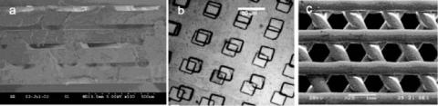
PLGA onto microfabricated silicon masters. When laminated together, these patterned films formed a vascular tissue engineering scaffold with 20 μm diameter channels between layers (Fig. 2.2a) [5]. Researchers later developed similar scaffolds with soft lithography techniques that utilize inexpensive elastomeric polydimethylsiloxane (PDMS) molds cast from silicon masters [6]. By introducing a PLGA solution into the mold and heating, Bhatia and colleagues created polymer layers that exhibited microstructures similar in shape and resolution (20–30 μm) to those on the silicon master and could be fused together (Fig. 2.2b) [7]. To further increase the scaffold surface area for cell attachment and proliferation,
26 |
ALICE A. CHEN ET AL. |
FIGURE 2.2. Fabrication using Heat. (a–b) Molded Lamination. Membranes of the biodegradable polymer PLGA are cast from silicon (a) or PDMS (b) molds and then laminated to create 3-D scaffolds. In (a), layers of PLGA are fused together to form microfluidic channels for vascular tissue engineering (c) Fused Deposition Molding. Molten biomaterials are extruded through a nozzle to build 3-D scaffolds layer by layer. (Photo courtesy of Jeff Borenstein and Kevin King, Draper Laboratory).
micropores can be incorporated into the patterned PLGA membranes by solvent casting and particulate leaching strategies.
Selective laser sintering (SLS) is a heat-based fabrication technique that uses laser energy to combine powdered polymeric materials into defined shapes. A laser beam directed across a powder bed locally increases polymer temperature to fuse with the surrounding material and form a layer of patterned structures [4]. Three-dimensional SLS scaffolds are created sequentially with fresh powder deposited over each patterned layer. Unfused powder released from the scaffold yields high porosity and surface area while retaining mechanical integrity. The pattern resolution of SLS is limited by the diameter of the laser beam diameter to about 400 um [4], and maximum pore size is about 50 um due to the powder particle size [8]. Lee and Barlow first utilized SLS with polymer-coated calcium phosphate powders to fabricate oral implants and demonstrated extensive bone tissue ingrowth in dog models [9]. Since then, Leong and others have broadened SLS utilization for various biopolymer applications [8].
Fused deposition modeling (FDM) combines heat and extrusion techniques to create 3-D scaffolds layer by layer. A nozzle directs a stream of molten plastic or ceramic onto a previously deposited layer of material. By altering the direction of material deposition with each layer, scaffolds with complex internal organization can be formed (Fig. 2.2c). Zein and Hutmacher used this method to produce biodegradable poly(ε-caprolactone) (PCL) scaffolds exhibiting various honeycomb geometries with finely tuned pore and channel dimensions of 250–700 μm [10]. Primary human fibroblasts cultured in these scaffolds proliferated and produced extracellular matrix [11], and scaffolds composed of other biocompatible polymers and composites have demonstrated utility for various tissue engineering applications [12–14]. While FDM exhibits high pattern resolution in the xy-plane, it is limited in the z-direction by the diameter of the extruded polymer filament that defines layer thickness and corresponding pore height. Further, high processing temperatures limit the biomaterials that are compatible with the method. However, FDM capabilities are expanding with new developments such as multi-phase jet solidification (MJS), a technique that allows simultaneous extrusion of multiple melted materials [15].
3-D plotting is a similar heat-based extrusion technology that is not limited to synthetic polymers that must withstand high temperatures while retaining their desired properties such as degradation and biocompatibility. Instead, fabrication is based on a sol-gel phase
3-D FABRICATION TECHNOLOGY FOR TISSUE ENGINEERING |
27 |
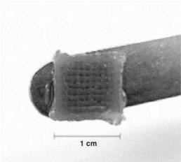
FIGURE 2.3. 3-D Plotting. Heated liquid agar solidifies into a 3-D hydrogel scaffold when deposited into a cooled medium. (from [16], reprinted with permission of Elsevier).
transition that occurs at lower temperatures. This strategy has been demonstrated with natural hydrogel biomaterials that are substantially more versatile for tissue engineering applications. For example, Mulhaupt and coworkers deposited agar and gelatin solutions heated to 90◦C into a cooled plotting medium, resulting in a 3-D hydrogel scaffold (Figure 2.3) [16]. Similarly, Ang and colleagues used robotic dispensing to form chitosan and chitosanhydroxyapatite scaffolds [17]. Following fibrin treatment, these scaffolds supported the adhesion of human osteosarcoma cells or mouse fibroblasts.
2.2.2. Light-Mediated Fabrication
Light energy can also be used to fabricate structured 3-D polymer scaffolds. Photopolymerization uses light to initiate a chain reaction that solidifies a liquid polymer solution. Stereolithography (SLA) is a photopolymerization method that utilizes a deflected UV laser beam to irradiate and solidify exposed polymer regions at the surface of a vat of photosensitive polymer (Fig. 2.4). Multiple layers are formed sequentially by lowering the stage and repeating the laser illumination. While SLA machines are traditionally used to build prototypes and molds for implants, Cooke et al. fabricated biodegradable 3-D polymer scaffolds for bony tissue consisting of diethyl fumarate, poly(propylene fumarate) and the photoinitiator bisacylphosphine oxide [18]. Similarly, a photocurable ceramic acrylate suspension formed cancellous bone [19] and hydroxyapatite bone tissue scaffolds [20], with overall dimensions suitable for healing critical-sized (4-mm thickness, 50-mm diameter) bone defects. As with SLS, stereolithography is limited in resolution by laser beam diameter to approximately 250 μm, although small-spot laser systems have demonstrated the production of smaller (70 μm) features [4].
Light energy can also be used to photopolymerize hydrogel polymer scaffolds that are less rigid than conventional stereolithography materials. Hydrogels are crosslinked networks of insoluble hydrophilic polymers that swell with water. Their increasing popularity as tissue

28 |
ALICE A. CHEN ET AL. |
FIGURE 2.4. Stereolithography. (a) UV light is used to crosslink the material in specific regions of a layer. The elevator is then lowered to reveal a new layer of polymer, and the process is repeated to create the desired shape.
(b) A prototype scaffold designed using SLA (from [18], reprinted by permission of John Wiley & Sons, Inc.).
engineering biomaterials reflects mechanical properties and high water content analogous to those of natural tissue. Yu and colleagues demonstrated a photolithographic method of patterning layers of dried 2-hydroxyethyl methacrylate that were subsequently rehydrated and seeded with cells [21]. However, the resolution of hydrogel scaffold fabrication may be compromised during rehydration of the polymer. Instead, Matsuda et al. later created scaffolds with improved strength and limited swelling using combinations of vinylated polysaccharides and diacrylated polyethylene glycol [22]. Additionally, the photopatterning of hydrogels has recently been extended to incorporate living cells into hybrid constructs, as discussed in a later section.
2.2.3. Adhesive-Mediated Fabrication
Scaffolds fabricated by binding polymers with solvents or adhesives, rather than by heat or light, circumvent biomaterial limitations for thermostable polymers or for biocompatible photoinitiators. Three-dimensional printing (3-DP), for instance, utilizes an ink jet printer to deposit a binder solution onto a polymer powder bed. Multiple layers can be fabricated and stacked with dimensions on the scale of polymer particle size (approximately 200–300 μm)
3-D FABRICATION TECHNOLOGY FOR TISSUE ENGINEERING |
29 |
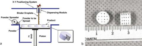
FIGURE 2.5. 3-D printing. Ink jet technology is used to print a binder solution onto a bed of polymer powder. An additional layer of powder is then deposited, and the process is repeated to form 3-D scaffolds (a) from Therics, website, with permission; (b) from [24], reprinted with permission of Leppincott Williams & Wilkins.
(Fig. 2.5) [23]. Scaffolds composed of natural biopolymers such as starch, dextran, and gelatin can be formed using aqueous solvents, and further can incorporate micropores by particle leaching. Griffith at al. explored porous scaffolds of PLGA for liver tissue engineering and demonstrated rat hepatocyte attachment [24]. Others have extended this technique to examine the effects of pore size on the attachment, growth, and matrix deposition of different cell types [25].
Pressure assisted microsyringe (PAM) fabrication is another adhesion-based technique that uses a solvent to bind polymers in a layer by layer format. A stage controlled microsyringe delivery system deposits a stream of polymer dissolved in solvent through a 10–20 μm glass capillary needle [7]. The polymer stream thickness can be modified by varying the solution viscosity, syringe-tip diameter, syringe pressure, and stage motor speed, to generate structures that range in dimension from 5 μm to 600 μm. This method is similar to FDM scaffold fabrication, but is capable of high resolution features and does not require heat. However, the limited size of the syringe-needle system prohibits the use of particulate leaching to increase microporosity and scaffold surface area.
2.2.4. Indirect Fabrication by Molding
In addition to the methods described above that directly fabricate 3-D scaffolds, scaffolds can also be cast from microstructured molds formed using the same methods. This indirect fabrication strategy is advantageous for sensitive biomaterials that are incompatible with fabrication conditions, since only the mold itself is subjected to the processing environment. Further, the resulting scaffold represents an inverse of the mold, thereby extending the 3-D design possibilities. For example, Orton et al. casted a hydroxyapatite/acrylate suspension onto a negative epoxy mandible mold made by stereolithography (Fig. 2.6a) [26]. After heat-curing the polymer, the mold and acrylate binder were incinerated. The resulting hydroxyapatite scaffolds contained different internal channel architectures and resulted in bone ingrowth in minipigs up to nine weeks post-implantation [27]. Others have created molds for indirect scaffolds using 3-DP by depositing wax or other low melting point compounds that can be later removed with elevated temperature or solvents. This method has been combined with particulate leaching to indirectly fabricate porous scaffolds composed of hypoxyapatite, poly(L)lactide, and polyglycolide [28]. Sachlos et al. similarly used ink