

Therapeutic Micro-Nano Technology BioMEMs - Tejlal Desai & Sangeeta Bhatia
.pdf10 |
ANJANA JAIN AND RAVI V. BELLAMKONDA |
1.2.3. Other Hydrogel Scaffolds
There are other hydrogels that can be used as scaffolds besides the three main ones discussed above. Some of the other hydrogels are Matrigel, NeuroGelTM, and Biomatrix. Matrigel is made out of a mixture of ECM proteins, such as LN and collagen. In vivo studies have shown that Matrigel alone is not adequate scaffold to promote axonal outgrowth [25, 65]. However, when the Matrigel is used in conjunction with Schwann Cells, axonal outgrowth is significantly noticeable. NeuroGelTM is a crosslinked copolymer hydrogel made of N-2-(hydroxypropyl) methacrylamide. When this hydrogel was inserted into the thoracic region of the spinal cord after a contusion injury, it was observed that the rats that had implanted NeuroGelTM in the lesioned cavity had an improved locomotion according to the BBB test and there was evidence of axonal fibers infiltrating the hydrogel, thereby crossing the tissue-implant interface [71]. NeuroGelTM also demonstrated the capability to hinder glial scar formation when it was implanted in the lesion of spinal cords in adult rats [72]. Biomatrix is a hydrogel, similar to Matrigel, made of ECM proteins, such as LN. However, Biomatrix does not appear to have as adequate regenerative capabilities as collagen and other hydrogels [68].
1.2.4. Spatial Control: Contact Guidance as a Strategy to Promote Regeneration
It was previously mentioned that besides the use of hydrogels as a scaffold, fibers could also be utilized to direct axonal growth from the proximal to distal ends of the nerve. This is another strategic technique to gain spatial control of proteins using a substrate. Tubes are inserted between the nerve gaps and then the nerve ends are sutured to the tubes with the fibers placed through the length of the tube (Fig. 1.1D). Due to the fibers being oriented longitudinally through the tube, it provides the orientation for the axons to grow from the proximal to distal end of the gap. Fibers are used to encourage the occurrence of two events in order to obtain successful myelinated axonal regeneration in the PNS. The first event is the formation of the fibrin matrix, which will have the same orientation as the filaments. The second event that needs to occur is the infiltration of Schwann cells. The goal is to have the Schwann cells adhere to the filaments and travel along the entire length of the filaments, which is the length of the nerve gap. This would then provide an environment which would encourage axonal outgrowth. Poly (L-Lactide) (PLLA) is another material that is commonly used to make filaments. In an in vitro study, it was demonstrated that if the PLLA was coated with LN, then the neurite outgrowth was significantly greater than neurite outgrowth on uncoated PLLA surface or the poly-L-lysine coated filaments [55]. Tubes are generally used to encapsulate the filaments and provide an environment for axonal growth along the filaments. However, in a study conducted in the PNS, collagen filaments were sutured to the proximal and distal ends of the nerve without the aid of tubes in vivo. The study showed that the number of myelinated axons that regenerated was greater to that found in the group that received the autograft, although it was not significantly greater [77]. This is the only study that did not use a conduit for the filaments or any neurotrophic factors, however, the regeneration was abundant and demonstrated that perhaps these two components are not completely necessary if the proper conditions are provided for axonal growth. Another variable that needs to be considered in the application of fibers is the number of fibers that should be inserted between the nerve ends. In studies conducted by Yoshii et al., collagen
NANOAND MICRO-TECHNOLOGY |
11 |
filaments were sutured to the sciatic nerve ends without the aid of a tube, two thousand filaments were connected at the ends to keep them joined over a 20 mm and 30 mm gap [77, 78]. The myelinated axon regeneration was comparable to the results observed with autografts for the 20 mm gap [77]. However, in the case of the 30 mm gap, the axonal regeneration was significantly less. These studies suggest that a large number of filaments would aid in axonal outgrowth. However, in another study, which inserted PLLA filaments inside silicone tubes, demonstrated that a lower packing density of filaments elicited the greatest number of myelinated axons [49].
Although filaments are predominantly used in the PNS, studies have been performed where filaments were inserted in CNS to promote axonal outgrowth. Carbon filaments were implanted in the lesion of a fully transected rat spinal cord. The carbon filaments allowed a scaffold for axons to advance through the lesion [33]. This study was taken further, where 10,000 carbon filaments were cultured with fetal tissue and implanted into the spinal cord lesion. This condition exhibited an improvement in electrical conduction through the injured axons [39]. A study conducted by the same group who inserted 2000 filaments into a nerve gap in the PNS, utilized the collagen filaments to encourage axonal regeneration in the CNS after spinal cord injury (SCI) [78]. Four thousand collagen fibers were inserted between the two nerve ends parallel to the spinal cord. It was demonstrated that the collagen fibers provided an adequate scaffold to bridge the nerve ends and allow axons to extend across the gap.
It was previously mentioned that proteins and oligopeptides could be coupled to hydrogels. A similar method was used to couple peptides to fibers that could potentially be implanted as a scaffold in the CNS. Two laminin peptides, YIGSR and IKVAV, were coupled to poly(tetrafluoroethylene) (PTFE) fibers and DRGs were cultured to observe neurite extension [60]. The peptide surface modified fibers encouraged neurite outgrowth; however, the neurites could not extend along unmodified PTFE fibers. To have successful axonal regeneration using fibers as the scaffold, it is important to either use a biomaterial that encourages fibrin matrix formation and Schwann cell infiltration or to coat the fibers with a protein that does those things. Current research has demonstrated that fibers made out of collagen, coated with proteins, such as collagen or laminin, or oligopeptides have produced the most significant axonal regeneration. Controlling proteins spatially through fiber scaffolds allows a surface for axons to adhere, as well as orient the direction of growth.
1.2.5. Spatial Control: Nerve Guide Conduits Provide an Environment for Axonal Regeneration
The use of nerve guide conduits has greatly influenced axonal regeneration. They aid in providing a scaffold to promote axonal regeneration and have the potential to both spatially and temporally control the protein environment at the site of injury. Importantly, the conduit serves as a physical barrier to prevent proteins and other molecules from inhibiting axonal regeneration. When NGCs were first being used, it was believed that the best material for the tube was silicone due to its mechanical properties. However, silicone NGCs are non-absorbable, non-semipermeable and require a second surgery to remove the conduit, otherwise it could cause chronic tissue response, such as scar formation, as well as nerve compression [18]. Most NGCs in use today are semi-permeable and even biodegradable. However, as NGCs have been extensively reviewed elsewhere [8, 18, 30, 59], we choose
12 |
ANJANA JAIN AND RAVI V. BELLAMKONDA |
to concentrate this chapter on approaches where the NGCs are used as carriers for other bioactive agents to enhance their functionality.
1.2.6. Spatial Control: Cell-scaffold Constructs as a Way of Combining Permissive Substrates with Stimuli for Regeneration
Cell transplantation techniques are an elegant way to combine two promising strategies to elicit regeneration: permissive substrates and spatio-temporally controlled delivery of trophic factors at the site of injury. This strategy has been explored both in the CNS and the PNS and is described below. Typically, NGCs are used as carriers for the delivery of these cells to the site of injury in the PNS or the CNS (Fig. 1.1E).
Schwann cells and OEG are two cell types commonly used to promote regeneration in the CNS, while Schwann cells are typically the cells of choice in the PNS. These cells provide both trophic cues, as well as physical, contact guidance type cues in promoting regeneration as described below. The use of cells, such as these glia, utilizes the strategy that modulates intrinsic mechanisms to promote axonal outgrowth. The transplantation of Schwann cells and OEG allows for spatial control of growth factors and other proteins, which are secreted by the cells.
Schwann cells have been shown to enhance peripheral nerve regeneration. It was mentioned previously that infiltration by endogenous Schwann cells increased axonal regeneration [24]. Schwann cells were embedded in a scaffold, such as Matrigel, and transplanted into an NGC implanted between two nerve ends, myelinated and unmyelinated axons are regenerated [25]. It was believed that by implanting Schwann cells already present throughout the conduit, the pace of regeneration could be increased. Schwann cells align along the tube and arrange themselves so that they are end to end, which is called Bungner bands. It was demonstrated that syngeneic Schwann cells elicited a better axonal regeneration than heterologous Schwann cells, which elicited an immune response [25]. It was also shown that as the Schwann cell density increased in the NGC, the axonal regeneration improved along similar lines to nerve autografts. Schwann cells myelinate peripheral nerves and it has been established that transplantation of these cells encourages the outgrowth of myelinated and unmyelinated axons.
Schwann cells have also shown to promote regeneration in the CNS. In studies that transected rat spinal cords and then implanted grafts containing Schwann cells and Matrigel, it was demonstrated that the number of myelinated and unmyelinated axons was greater compared to grafts containing only Matrigel and the myelinated axons formed fascicles through the conduit [74, 75]. In another study that transplanted Schwann cells into the spinal cord, it was shown that Schwann cells that released increased amounts of NGF had significantly more axons growing into the graft compared to Schwann cells that were not modified to release increased amounts of NGF [67]. It was also demonstrated that these Schwann cells expressed the same phenotype and myelinated axons in the CNS as in the PNS. The combination of NGF and Schwann cells allows for the outgrowth of axons into the grafts due to the presence of NGF and then the Schwann cells provides direction for axonal growth due to the Bunger bands [67]. It was mentioned previously, cAMP has been investigated to promote axonal regeneration. In a study cAMP and Schwann cells were both inserted into the spinal cord to observe whether there was a synergistic effect [53]. The results demonstrated that by implanting Schwann cells and elevating cAMP, the
NANOAND MICRO-TECHNOLOGY |
13 |
number of myelinated axons increased and functional recovery was observed compared to the transplantation of only Schwann cells.
Unlike Schwann cells, which can be transplanted in both the PNS and CNS, OEG is primarily transplanted in the CNS to promote axonal regeneration. OEG ensheath olfactory axons and shield the axons from inhibitory molecules exposed in the environment, thus allowing the axons to regenerate throughout adult life [58]. OEG demonstrates a promising method to ensheath the axons in other areas of the CNS that are injured and aid in regeneration. The olfactory bulb is the main supplier for OEG and one of the main benefits of using this source for OEG is because the glia can migrate into other regions of the CNS and integrate with other CNS glia [58].
Comparisons have been made between Schwann cells and OEG for their effectiveness in promoting axonal regeneration in the CNS. In a study that was comparing the response of astrocytes and CSPG expression after OEG or Schwann cell transplantation in the CNS, it was demonstrated that OEG elicited less of an astrocytic response and lower expression of CSPG compared to Schwann cells [37]. Although OEG do not induce as severe a response as Schwann cells do, Schwann cells have shown more promising results in improving locomotor performance compared to OEG after adult rats have suffered from contused thoracic SCI [64].
It was mentioned earlier that astrocytes can also be used as a substrate for axonal outgrowth. These studies were performed in vitro. It was demonstrated that uniformly orienting the astrocytes and organizing the ECM and cell adhesion molecules in order to culture neurons on the astrocytes lead to the enhancement of neurites extending in a direction parallel to the astrocytes [9]. The use of glial cells, such as astrocytes, as a substrate can be combined with a biomatrix to enhance neurite extension in a specific direction [20]. Glial cells were cultured on the biodegradable poly(D,L)-lactide matrices to orient the cells in a specific direction. Although this substrate did not enhance either the number of extended neurites or the length of the neurites, the cultured cortical neurons extended neurites along the orientation of the glial cells/biomatrix substrate.
1.3. TEMPORALLY CONTROLLING THE RELEASE OF PROTEINS
As important as it is to control the proteins spatially, it is equally imperative to control the amount of protein delivered over a period of time. Regeneration over long nerve gaps requires several months. Therefore, for axonal outgrowth to occur during this time period, the microenvironment must be actively supportive over this time scale. If proteins, such as Rho GTPases and neurotrophic factors, are only administered as a single dose at the time of implantation of the scaffold, then some of the protein will be taken up intracellularly, diffuse into the surrounding tissue, and degrade. Then there will not be a therapeutic level of protein to promote axonal outgrowth over the time necessary to have complete regeneration. For example, it was concluded that after local administration of NGF into the brain, the half-life of NGF was 30 minutes [36]. Once the effective concentration for the proteins is known, then it can be delivered and sustained. Sustaining the presence of proteins at the effective concentration can be achieved through a controlled slow release delivery system. There are currently four main techniques that are being investigated for controlling protein concentration at the site of injury over time: (1) osmotic pumps, (2) embedded
14 ANJANA JAIN AND RAVI V. BELLAMKONDA
Osmotic |
|
||
protein |
pump |
for |
|
release |
|||
|
|
||
 Lesion site
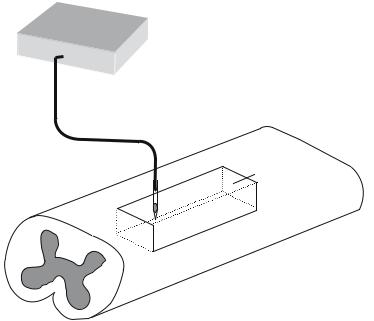
FIGURE 1.2. Osmotic Pumps for Temporal Control of Proteins. An external osmotic pump provides a reservoir of protein that is delivered via a catheter implanted near or at the lesion area.
microspheres, (3) microtubules and (4) enzyme dependent demand-driven trophic factor release.
1.3.1. Temporal Control: Osmotic Pumps Release Protein to Encourage Axonal Outgrowth
Osmotic pumps can be used to deliver proteins, such as neurotrophic factors, to promote axonal regeneration. Osmotic pumps are mostly utilized to deliver the proteins in the CNS. There are two parts to this delivery system, one component is the infusion pump that is usually implanted under the skin on the back of the animal, and other component is the catheter that is inserted in the lesion of the nerve (Fig. 1.2).
1.3.1.1. Temporal Control: Using Osmotic Pumps to Stimulate Process Extension by Sustained, Local Trophic Factor Delivery Several studies have investigated the benefits of continuous infusion of the neurotrophic factors BDNF and NT-3 after SCI. Typically, after SCI, methylprednisolone (MP) is administered to the patient. It has been demonstrated that the levels of BDNF and NT-3 decrease after the administration of MP. In a study, after treatment of MP, it was concluded that if BDNF was continuously delivered, then the rats locomotor function improved [34]. In a study that delivered both BDNF and NT-3 over a short time period (2 weeks) and a longer time period (8 weeks), it was shown that only the rats treated with BDNF and NT-3 over the 8 week time period allowed for the survival of the rubrospinal neurons [52]. However, rubrospinal axonal regeneration was not observed.
NANOAND MICRO-TECHNOLOGY |
15 |
In another study that delivered either NT-3 or BDNF for 4 weeks into the spinal cord after it was crushed, the rats treated with BDNF did not exhibit any axonal regeneration. However, fiber sprouting was observed into and through the lesion in the rats that had NT-3 administered to the spinal cord lesion [11]. In a study that infused only BDNF for two weeks into the rat motor cortex after SCI, sprouting of corticospinal fibers was observed; however, axonal regeneration did not occur into the peripheral nerve transplant that was placed in the lesion [27]. The constant release of neurotrophic factors using the osmotic pump appears to exhibit therapeutic results. The site of administration seems to affect the response of axonal regeneration and fiber sprouting. The only disadvantage of utilizing the osmotic pump is the different locations of its components.
1.3.1.2. Temporal Control: Alleviation of Inhibitory Environments by Using Osmotic Pumps It was mentioned above that osmotic pumps can be used to deliver neurotrophic factors to the CNS to modulate intra-neuronal mechanisms. Osmotic pumps have also been utilized to infuse IN-1 antibody that neutralizes NOGO-A, an isoform of NOGO that is one of the main inhibitory molecules located in the glial scar [13]. It was observed that after 2 weeks of IN-1 delivery, regenerating fibers were observed through the lesion in the thoracic region into the lumbar region of the spinal cord. Therefore, the use of osmotic pumps can also be used to deliver proteins that can neutralize the inhibitory environment of the glial scar.
Other than the use of osmotic pumps to deliver proteins, Gelfoam, an insoluble gelatin sponge, was used to deliver chondroitinase ABC into the spinal cord lesion. The animals treated with chondroitinase ABC filled Gelfoam displayed axonal regeneration of the Clarke’s neurons through the lesion area and it was exhibited that CSPG was digested by the chondroitinase ABC [76].
1.3.2. Temporal Control: Slow Release of Trophic Factors Using Microspheres
Microspheres, used in drug delivery applications, are being investigated to deliver protein to the PNS and CNS in order to encourage axonal outgrowth (Fig. 1.3). Microspheres have an advantage over osmotic pumps because a single administration is needed to release the protein over time. The size of the microspheres depends upon the application. The size of the microparticles in the studies that use microspheres to promote axonal outgrowth is around 12-16 μm. The materials that are used to make the microsphere are typically biodegradable polymers. The use of copolymers and altering the ratio of the polymers can affect the biodegradation profiles because the polymeric characteristics, such as glass transition temperature and hydrophilicities, change [61]. The polymeric materials mostly used for the microspheres are poly(lactic acid) (PLA), the copolymer poly(lactic-co-glycolic acid) (PLGA) and polyphosoesters. When investigating a specific polymer or another biomaterial, it is important to make sure that when the material degrades it does not denature the protein due to the possible immunogeneic response it can cause, thus altering the release profile and bioactivity [61].
1.3.2.1. Temporal Control: Use of Microspheres to Stimulate Process Extension in the PNS and CNS Most of the research, currently, focuses on delivering NGF loaded microspheres to regenerate nerves in the PNS. In a study performed by Xu et al., NGF
16 |
ANJANA JAIN AND RAVI V. BELLAMKONDA |
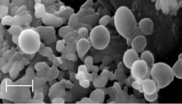
1μm
FIGURE 1.3. SEM Image of Microspheres. Microspheres can be used to encapsulate protein that will be slowly released as the microsphere degrades. Scale bar = 1μm. Figure courtesy of YT Kim and RV Bellamkonda, Department of Biomedical Engineering, Georgia Institute of Technology.
was loaded into poly(phosphoester) (PPE) microspheres. First, in in vitro studies, it was determined that the microspheres released bioactive NGF up to 10 weeks. The NGF loaded PPE microspheres in a saline solution were loaded into PPE NGCs. When these constructs were implanted into rat sciatic nerves, it was observed that treatment with NGF loaded microspheres in the NGC had a cable that bridged the entire 10 mm gap between the nerve ends. Also, compared to the controls, there were more myelinated axons, higher fiber density, and thicker myelin sheath [73].
In the CNS, one of the first studies conducted using microspheres to deliver protein to the CNS was by Camarata et al. In order to combat neurodegenerative disease, they inserted microspheres loaded with NGF that could be released in vivo for 4 to 5 weeks [14]. In another in vitro study, the number of days NGF was released was increased to 91 days. Various ratio of PLGA were tested to determine the release characteristics, as well as poly(ε-caparolactone) (PCL) [15]. The surface morphology of the microspheres that are loaded versus unloaded ones is different. The surface of protein loaded microspheres is rougher, whereas the unloaded microspheres have a smoother surface. The smaller the microsphere, the greater the surface area, thus increasing the degradation rate of the microsphere and release of the protein.
1.3.3. Temporal Control: Lipid Microtubules for Sustained Release of Stimulatory Trophic Factors
Another method to slowly release protein in the CNS and PNS is the use of lipid microtubules, also referred to as microcylinders (Fig. 1.4). These microtubules are hollow cylinders with a diameter of 0.5 μm [44]. The length of the microtubules varies based on the time period in which the protein, DNA, or other desired molecule needs to be released. The molecule is released at the ends of the microtubules, which is the reason why the length of the microcylinders controls the release profile of the protein. In a study previously mentioned, to aid axonal regeneration in the PNS, a two-step slow release system was developed. The first step was NGF loaded microtubules, which had a length of 40 μm, and
NANOAND MICRO-TECHNOLOGY |
17 |
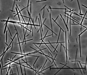
50 μm
FIGURE 1.4. Micrograph Image of Lipid Microtubules. The image depicts the lipid microtubules being on average 40 μm in length. The microtubules release the protein from the ends. Scale bar = 50 μm.
the second step was the loaded microtubules embedded in agarose hydrogel [79]. The two step release system was thus, first the diffusion of the NGF from the microtubules into the agarose and then the release of the NGF from the agarose into the gap between the two nerve ends. This slow release system allows the NGF to last longer in the nerve gap and prevents degradation or dilution by macrophages and other fluids. Two months post-implantation, a cable formed, the number of myelinated axons was statistically similar to the autograft condition, and the density of myelinated axons was similar to that of the autograft and a normal sciatic nerve.
1.3.4. Temporal Control: Demand Driven Release of Trophic Factors
Another form of controlled release of a protein is the fibrin matrix, which was initially developed for wound healing. Cells that migrate to the area degrade the matrix through proteolysis, thereby releasing the contained protein (Fig. 1.5) [57]. A fibrin matrix covalently coupled to heparin that interacted with neurotrophins, NGF, BDNF, and NT-3 was developed. It was demonstrated in vitro that the neurite outgrowth was enhanced when the neurotrophins were released using this delivery system compared when soluble neurotrophins were added to the fibrin matrix [56]. When the heparin immobilized fibrin matrix was implanted in a nerve gap in the PNS, fiber sprouting was observed through the conduit to the distal end [38].
1.4. CONCLUSION
The advancement in CNS and PNS regeneration has been due to the utilization of nanoand micro-technologies. Most of the technology that has been developed has been geared
18 |
ANJANA JAIN AND RAVI V. BELLAMKONDA |
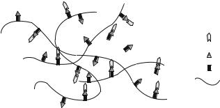
Growth factor binding to heparin
Bound heparin
Peptide coupling heparin to matrix
Fibrin matrix
FIGURE 1.5. Schematic of Fibrin Matrix Releasing Protein. The peptides bind the heparin to the fibrin matrix. The growth factor or protein is then able to bind the heparin, thus attaching the growth factor to the matrix. The cells migrating to the area will then degrade the matrix releasing the growth factor. Figure adapted from Ref. [57].
towards controlling proteins spatially and temporally. There are three main strategies used to elicit axonal outgrowth after injury, which allows spatial and temporal control of proteins. The three strategies mentioned are to 1) provide permissive bioactive substrates for the axonal outgrowth; (2) use trophic factors to stimulate growth; and (3) alleviate signaling due to the inhibitory entities present in the extracellular environment to allow axons to regenerate between the proximal and distal ends.
This chapter briefly describes studies that have incorporated various nanoand microtechnologies using biomaterials based design. While, for analytical convenience we divide this chapter into sections with various strategies, it is becoming evident that a coordinated, multiple component strategy may be required for successful regeneration. For example, one approach is to design a substrate that is coupled to proteins, contains either Schwann cells or OEG, and has a delivery vehicle slowly releasing proteins. The key combination remains elusive and is the focus of active, ongoing investigation.
REFERENCES
[1]P. Aebischer, A.N. Salessiotis, and S.R. Winn. Basic fibroblast growth factor released from synthetic guidance channels facilitates peripheral nerve regeneration across long nerve gaps. J. Neurosci. Res., 23(3):282–289, 1989.
[2]A.M. Avellino, D. Hart, A.T. Dailey, M. MacKinnon, D. Ellegala, and M. Kliot. Differential macrophage responses in the peripheral and central nervous system during wallerian degeneration of axons. Exp. Neurol., 136(2):183–198, 1995.
[3]A. Baird, P.A. Walicke. Fibroblast growth factors. Br. Med. Bull., 45(2):438–452, 1989.
[4]A.P. Balgude, X. Yu, A. Szymanski, and R.V. Bellamkonda. Agarose gel stiffness determines rate of DRG neurite extension in 3D cultures. Biomaterials, 22(10):1077–1084, 2001.
[5]C.E. Bandtlow. Regeneration in the central nervous system. Exp. Gerontol., 38(1–2):79–86, 2003.
[6]R. Bellamkonda, J.P. Ranieri, and P. Aebischer. Laminin oligopeptide derivatized agarose gels allow threedimensional neurite extension in vitro. J. Neurosci. Res., 41(4):501–509, 1995a.
[7]R. Bellamkonda, J.P. Ranieri, N. Bouche, and P. Aebischer. Hydrogel-based three-dimensional matrix for neural cells. J. Biomed. Mater. Res., 29(5):663–671, 1995b.
[8]R. Bellamkonda and P. Aebischer. Review: Tissue Engineering in the Nervous System. Biotech. Bioeng., 43:543–1994, 1993.
NANOAND MICRO-TECHNOLOGY |
19 |
[9]R. Biran, M.D. Noble, and P.A. Tresco. Directed nerve outgrowth is enhanced by engineered glial substrates. Exp. Neurol., 184(1):141–152, 2003.
[10]M. Borkenhagen, J.F. Clemence, H. Sigrist, and P. Aebischer. Three-dimensional extracellular matrix engineering in the nervous system. J. Biomed. Mater. Res., 40(3):392–400, 1998.
[11]E.J. Bradbury, S. Khemani, R. Von King, J.V. Priestley, and S.B. McMahon. NT-3 promotes growth of lesioned adult rat sensory axons ascending in the dorsal columns of the spinal cord. Eur. J. Neurosci., 11(11):3873–3883, 1999.
[12]E.J. Bradbury, L.D. Moon, R.J. Popat, V.R. King, G.S. Bennett, P.N. Patel, J.W. Fawcett, and S.B. McMahon. Chondroitinase ABC promotes functional recovery after spinal cord injury. Nature, 416(6881):636–640, 2002.
[13]C. Brosamle, A.B. Huber, M. Fiedler, A. Skerra, and M.E. Schwab. Regeneration of lesioned corticospinal tract fibers in the adult rat induced by a recombinant, humanized IN-1 antibody fragment. J. Neurosci.,
20(21):8061–8068, 2000.
[14] P.J. Camarata, R. Suryanarayanan, D.A. Turner, R.G. Parker, and T.J. Ebner. Sustained release of nerve growth factor from biodegradable polymer microspheres. Neurosurgery, 30(3):313–319, 1992.
[15]X. Cao and M.S. Schoichet. Delivering neuroactive molecules from biodegradable microspheres for application in central nervous system disorders. Biomaterials, 20(4):329–339, 1999.
[16]D. Ceballos, X. Navarro, N. Dubey, G. Wendelschafer-Crabb, W.R. Kennedy, and R.T. Tranquillo. Magnetically aligned collagen gel filling a collagen nerve guide improves peripheral nerve regeneration. Exp. Neurol., 158(2):290–300, 1999.
[17]L.J. Chamberlain, I.V. Yannas, H.P. Hsu, G. Strichartz, and M. Spector. Collagen-GAG substrate enhances the quality of nerve regeneration through collagen tubes up to level of autograft. Exp. Neurol., 154(2):315–329, 1998.
[18]L.B. Dahlin and G. Lundborg. Use of tubes in peripheral nerve repair. Neurosurg. Clin. N. Am., 12(2):341– 352, 2001.
[19]S. David and S. Lacroix. Molecular Approaches to Spinal Cord Repair. Annu. Rev. Neurosci., 2003.
[20]R. Deumens, G.C. Koopmans, C.G. Den Bakker, V. Maquet, S. Blacher, W.M. Honig, R. Jerome, J.P. Pirard, H.W. Steinbusch, and E.A. Joosten. Alignment of glial cells stimulates directional neurite growth of CNS neurons in vitro. Neuroscience, 125(3):591–604, 2004.
[21]C.I. Dubreuil, M.J. Winton, and L. McKerracher. Rho activation patterns after spinal cord injury and the role of activated Rho in apoptosis in the central nervous system. J. Cell. Biol., 162(2):233–243, 2003.
[22]G.R. Evans. Peripheral nerve injury: a review and approach to tissue engineered constructs. Anat. Rec., 263(4):396–404, 2001.
[23]J.W. Fawcett and R.A. Asher. The glial scar and central nervous system repair. Brain. Res. Bull., 49(6):377– 391, 1999.
[24]S.P. Frostick, Q. Yin, and G.J. Kemp. Schwann cells, neurotrophic factors, and peripheral nerve regeneration. Microsurgery, 18(7):397–405, 1998.
[25]V. Guenard, N. Kleitman, T.K. Morrissey, R.P. Bunge, and P. Aebischer. Syngeneic Schwann cells derived from adult nerves seeded in semipermeable guidance channels enhance peripheral nerve regeneration. J. Neurosci., 12(9):3310–3320, 1992.
[26]T. Hashimoto, Y. Suzuki, M. Kitada, K. Kataoka, S. Wu, K. Suzuki, K. Endo, Y. Nishimura, and C. Ide. Peripheral nerve regeneration through alginate gel: analysis of early outgrowth and late increase in diameter of regenerating axons. Exp. Brain. Res., 146(3):356–368, 2002.
[27]G.W. Hiebert, K. Khodarahmi, J. McGraw, J.D. Steeves, and W. Tetzlaff. Brain-derived neurotrophic factor applied to the motor cortex promotes sprouting of corticospinal fibers but not regeneration into a peripheral nerve transplant. J. Neurosci. Res., 69(2):160–168, 2002.
[28]A. Hoke and J. Silver. Proteoglycans and other repulsive molecules in glial boundaries during development and regeneration of the nervous system. Prog. Brain. Res., 108:149–163, 1996.
[29]D.A. Houweling, A.J. Lankhorst, W.H. Gispen, P.R. Bar, and E.A. Joosten. Collagen containing neurotrophin- 3 (NT-3) attracts regrowing injured corticospinal axons in the adult rat spinal cord and promotes partial functional recovery. Exp. Neurol., 153(1):49–59, 1998.
[30]T.W. Hudson, G.R. Evans, and C.E. Schmidt. Engineering strategies for peripheral nerve repair. Orthop. Clin. North. Am., 31(3):485–498, 2000.