

Therapeutic Micro-Nano Technology BioMEMs - Tejlal Desai & Sangeeta Bhatia
.pdf90 |
|
|
|
LAURA J. ITLE, WON-GUN KOH, AND MICHAEL V. PISHKO |
|||||||||||||||
|
|
|
|
|
|
|
|
||||||||||||
Assay/Phenotype |
Hepatocytes |
Fibroblasts |
HUVEC |
Myocyte |
Neuroblastoma |
Macrophage |
|
||||||||||||
Viability A |
|
|
|
|
|
|
|
|
|
|
|
|
|
|
|
|
|
|
|
|
|
|
|
|
|
|
|
|
|
|
|
|
|
|
|
|
|
|
|
|
|
|
|
|
|
|
|
|
|
|
|
|
|
|
|
|
|
|
|
Viability B |
|
|
|
|
|
|
|
|
|
|
|
|
|
|
|
|
|
|
|
|
|
|
|
|
|
|
|
|
|
|
|
|
|
|
|
|
|
|
|
|
|
|
|
|
|
|
|
|
|
|
|
|
|
|
|
|
|
|
|
Apoptosis A |
|
|
|
|
|
|
|
|
|
|
|
|
|
|
|
|
|
|
|
|
|
|
|
|
|
|
|
|
|
|
|
|
|
|
|
|
|
|
|
Apoptosis B |
|
|
|
|
|
|
|
|
|
|
|
|
|
|
|
|
|
|
|
|
|
|
|
|
|
|
|
|
|
|
|
|
|
|
|
|
|
|
|
Caspase Activity |
|
|
|
|
|
|
|
|
|
|
|
|
|
|
|
|
|
|
|
|
|
|
|
|
|
|
|
|
|
|
|
|
|
|
|
|
|
|
|
|
|
|
|
|
|
|
|
|
|
|
|
|
|
|
|
|
|
|
|
Endocytosis |
|
|
|
|
|
|
|
|
|
|
|
|
|
|
|
|
|
|
|
|
|
|
|
|
|
|
|
|
|
|
|
|
|
|
|
|
|
|
|
|
|
|
|
|
|
|
|
|
|
|
|
|
|
|
|
|
|
|
|
Exocytosis |
|
|
|
|
|
|
|
|
|
|
|
|
|
|
|
|
|
|
|
|
|
|
|
|
|
|
|
|
|
|
|
|
|
|
|
|
|
|
|
|
|
|
|
|
|
|
|
|
|
|
|
|
|
|
|
|
|
|
|
Nitric Oxide |
|
|
|
|
|
|
|
|
|
|
|
|
|
|
|
|
|
|
|
|
|
|
|
|
|
|
|
|
|
|
|
|
|
|
|
|
|
|
|
|
|
|
|
|
|
|
|
|
|
|
|
|
|
|
|
|
|
|
|
L-type Ca2+ channels |
|
|
|
|
|
|
|
|
|
|
|
|
|
|
|
|
|
|
|
|
|
|
|
|
|
|
|
|
|
|
|
|
|
|
|
|
|
|
|
|
|
|
|
|
|
|
|
|
|
|
|
|
|
|
|
|
|
|
|
Cell-matrix adhesion |
|
|
|
|
|
|
|
|
|
|
|
|
|
|
|
|
|
|
|
|
|
|
|
|
|
|
|
|
|
|
|
|
|
|
|
|
|
|
|
|
|
|
|
|
|
|
|
|
|
|
|
|
|
|
|
|
|
|
|
Cell-cell adhesion |
|
|
|
|
|
|
|
|
|
|
|
|
|
|
|
|
|
|
|
|
|
|
|
|
|
|
|
|
|
|
|
|
|
|
|
|
|
|
|
|
|
|
|
|
|
|
|
|
|
|
|
|
|
|
|
|
|
|
|
Chemotaxis |
|
|
|
|
|
|
|
|
|
|
|
|
|
|
|
|
|
|
|
|
|
|
|
|
|
|
|
|
|
|
|
|
|
|
|
|
|
|
|
|
|
|
|
|
|
|
|
|
|
|
|
|
|
|
|
|
|
|
|
|
|
|
|
|
|
|
|
|
|
|
|
|
|
|
|
|
|
|
|
FIGURE 5.5. Schematic of a potential multi-phenotypic biosensor. Six different cell lines are isolated in hydrogel arrays in six different microfluidic channels. Several cellular assays can be performed using one array.
Multi-phenotypic arrays offer the advantage of providing more information about cellular responses than existing arrays and lend themselves to miniaturization, which could lead to high degrees of multiplexing. Multi-phenotypic cell based biosensors show promise in the area of high-throughput drug screening, the detection of biochemical warfare agents, and the detection of environmental toxins. While the use of nucleic acid and protein arrays is well established, the uses of multi-phenotypic cellular arrays are in the infancy of their development, creating an exciting and dynamic area of research.
REFERENCES
[1]B.M. Paddle. Biosens. Bioelectron., 11:1079–1113, 1996.
[2]L. Bousse. Sens. Actu. B, 34:270–275, 1996.
[3]C. Ziegler. Fresenius J. Anal. Chem., 366:552–559, 2000.
[4]J. Hodgson. Nat. Biotechnol., 19:722–726, 2001.
[5]J. Knight. Nature, 418:474–475, 2002.
[6]T. Chovan and A. Guttman. Trends Biotechnol., 20:116–122, 2002.
[7]C. Ziegler and W. Gopel. Curr. Opin. Chem. Biol. 2:585–591, 1998.
[8]M. Matsuzawa, R.S. Potember, D.A. Stenger, V. Krauthamer, J. Neurosci. Methods. 50:253–260, 1993.
[9]B.J. Spargo, M.A. Testoff, T.B. Nielsen, D.A. Stenger, J.J. Hickman, and A.S. Rudolph. Proc. Natl. Acad. Sci. U.S.A., 91:11070–11074, 1994.
[10]M. Matsuzawa, P. Liesi, and W. Knoll. J. Neurosci. Methods, 69:189–196, 1996.
[11]W. Ma, Q.Y. Liu, D. Jung, P. Manos, J.J. Pancrazio, A.E. Schaffner, J.L. Barker, and D.A. Stenger. Brain Res. Dev. Brain Res., 111:231–243, 1998.
[12]V. Brisson and R.D. Tilton. Biotechnol. Bioeng., 77:290–295, 2002.
[13]J.F. Clemence, J.P. Ranieri, P. Aebischer, and H. Sigrist. Bioconjug. Chem., 6:411–417, 1995.
[14]G. Chen, Y. Ito, Y. Imanishi, A. Magnani, S. Lamponi, and R. Barbucci. Bioconjug. Chem., 8:730–734, 1997.
[15]S. Rohr, R. Fluckiger-Labrada, and J.P. Kucera. Pflugers Arch., 446:125–132, 2003.
[16]S. Rohr, D.M. Scholly, and A.G. Kleber. Circ. Res., 68:114–130, 1991.
MULTI-PHENOTYPIC CELLULAR ARRAYS FOR BIOSENSING |
91 |
[17]V.A. Liu, W.E. Jastromb, and S.N. Bhatia. J. Biomed. Mater. Res., 60:126–134, 2002.
[18]T.A.M. Sugwara. T. Macromolecules, 27:7809–7814, 1994.
[19]M.L. Amirpour, P. Ghosh, W.M. Lackowski, R.M. Crooks, and M.V. Pishko. Anal. Chem., 73:1560–1566, 2001.
[20]R. Singhvi, A. Kumar, G.P. Lopez, G.N. Stephanopoulos, D.I. Wang, G.M. Whitesides, and D.E. Ingber. Science, 264:696–698, 1994.
[21]M. Mrksich, L.E. Dike, J. Tien, D.E. Ingber, and G.M. Whitesides. Exp. Cell. Res., 235:305–313, 1997.
[22]D. Duffy, J.C. MacDonald, O.J. Schueller, and G.M. Whitesides. Anal. Chem., 70:4974–4984, 1998.
[23]E. Delamarche, A. Bernard, H. Schmid, B. Michel, and H. Biebuyck. Science, 276:779–781, 1997.
[24]E. Delamarche, A. Bernard, H. Schmid, A. Bietsch, B. Michel, and H. Biebuyck. J. Am. Chem. Soc., 120:500–508, 1998.
[25]S. Takayama, J. McDonald, Cooper, Ostuni, Emanuele, Liang, N. Michael, Kenis, J.A. Paul, Ismagilov, F. Rustem, Whitesides, and M. George. Proceedings of the National Academy of Sciences of the United States of America, 96:5545–5548, 1999.
[26]C.S. Pale-Grosdemange, E.S., K.L. Prime, and G.M. Whitesides. J. Am. Chem. Soc., 113:12–20, 1991.
[27]E. Ostuni, L. Yan, and G.M. Whitesides. Coll. Surf. B: Biointerfac., 15:3–30, 1999.
[28]N. Huang, R. Michel, J. Voros, M. Textor, R. Hofer, A. Rossi, D.L. Elber, J.A. Hubbell, and N.D. Spencer. Langmuir, 17:489–498, 2001.
[29]N.A. Alcantar, E.S. Aydil, and J.N. Israelachvili. J. Biomed. Mater. Res., 51:343–351, 2000.
[30]K.R. Kamath, M.J. Danilich, R.E. Marchant, and K. Park. J. Biomater. Sci. Polym. Ed., 7:977–988, 1996.
[31]A. Park, B. Wu, and L.G. Griffith. J. Biomater. Sci. Polym. Ed., 9:89–110, 1998.
[32]A. Revzin, R.G. Tompkins, and M. Toner. Langmuir, 19:9855–9862, 2003.
[33]G.M. Cruise, D.S. Scharp, and J.A. Hubbell. Biomaterials, 19:1287–1294, 1998.
[34]A. Revzin, R.J. Russell, V.K. Yadavalli, W.G. Koh, C. Deister, D.D. Hile, M.B. Mellott, and M.V. Pishko. Langmuir, 17:5440–5447, 2001.
[35]M.B. Mellott, K. Searcy, and M.V. Pishko. Biomaterials, 22:929–941, 2001.
[36]R. Russell, A.C. Axel, K.L. Shields, and M.V. Pishko. Polymer, 42:4893–4901, 2001.
[37]N. Wisniewski and M. Reichert. Coll. Surf. B Biointerfac., 18:197–219, 2000.
[38]S.P. Massia and J.A. Hubbell. Anal. Biochem., 187:292–301, 1990.
[39]S.P. Massia and J.A. Hubbell. Ann. N Y Acad. Sci., 589:261–270, 1990.
[40]S.P. Massia and J.A. Hubbell. J. Biomed. Mater. Res., 25:223–242, 1991.
[41]B.K. Mann, A.T. Tsai, T. Scott-Burden, and J.L. West. Biomaterials, 20:2281–2286, 1999.
[42]W.G. Koh, L.J. Itle, and M.V. Pishko. Anal. Chem., 75:5783–5789, 2003.
[43]C.A. Quinn, R.E. Connor, and A. Heller. Biomaterials, 18:1665–1670, 1997.
[44]K. Podual, F.J. Doyle, 3rd; and N.A. Peppas. Biomaterials, 21:1439–1450, 2000.
[45]R.A. Scott and N.A. Peppas. Biomaterials, 20:1371–1380, 1999.
[46]N.A. Peppas, K.B. Keys, M. Torres-Lugo, and A.M. Lowman. J. Control. Rel., 62:81–87, 1999.
[47]D.K. Han, K.D. Park, J.A. Hubbell, and Y.H. Kim. J. Biomater. Sci. Polym. Ed., 9:667–680, 1998.
[48]J.S. Temenoff, K.A. Athanasiou, R.G. LeBaron, and A.G. Mikos. J. Biomed. Mater. Res., 59:429–437, 2002.
[49]P.J. Martens, S.J. Bryant, and K.S. Anseth. Biomacromolecules, 4:283–292, 2003.
[50]W.G. Koh, A. Revzin, and M.V. Pishko. Langmuir, 18:2459–2462, 2002.
[51]J.A. Burdick and K.S. Anseth. Biomaterials, 23:4315–4323, 2002.
[52]S.M. O’Connor, J.D. Andreadis, K.M. Shaffer, W. Ma, J.J. Pancrazio, and D.A. Stenger. Biosens. Bioelectron., 14:871–881, 2000.
[53]X. Zheng Shu, Y. Liu, F.S. Palumbo, Y. Luo, and G.D. Prestwich. Biomaterials, 25:1339–1348, 2004.
[54]V. Liu and S.N. Bhatia. Biomed. Microdev., 4:257–266, 2002.
[55]M. Keusgen. Naturwissenschaften, 89:433–444, 2002.
[56]P. Fromherz, A. Offenhausser, T. Vetter, and J. Weis. Science, 252:1290–1293, 1991.
[57]R. Weis, B. Muller, and P. Fromherz. Phys. Rev. Lett., 76:327–330, 1996.
[58]G. Zeck and P. Fromherz. Proc. Natl. Acad. Sci. U.S.A., 98:10457–10462, 2001.
[59]A. Harsch, C. Ziegler, and W. Gopel. Biosens. Bioelectron., 12:827–835, 1997.
[60]G.W. Gross, B.K. Rhoades, H.M. Azzazy, and M.C. Wu. Biosens. Bioelectron., 10:553–567, 1995.
[61]J.H. Luong, M. Habibi-Rezaei, J. Meghrous, C. Xiao, K.B. Male, and A. Kamen. Anal. Chem., 73:1844– 1848, 2001.
92 |
LAURA J. ITLE, WON-GUN KOH, AND MICHAEL V. PISHKO |
[62]C. Xiao, B. Lachance, G. Sunahara, and J.H. Luong. Anal. Chem., 74:1333–1339, 2002.
[63]C. Xiao, B. Lachance, G. Sunahara, and J.H. Luong. Anal. Chem., 74:5748–5753, 2002.
[64]C. Tlili, K. Reybier, A. Geloen, L. Ponsonnet, C. Martelet, H.B. Ouada, M. Lagarde, and N. JaffrezicRenault. Anal. Chem., 75:3008–3012, 2003.
[65]J. Engebrecht, M. Simon, and M. Silverman. Science, 227:1345–1347, 1985.
[66]G. Kirchner, J.L. Roberts, G.D. Gustafson, and T.D. Ingolia. Gene, 81:349–354, 1989.
[67]J. King, P.M. DiGrazia, B. Applegate, R. Burlage, J. Sanseverino, P. Dunbar, F. Larimer, and G.S. Dayler. Science, 249:778–780, 1990.
[68]F. Marincs and D.W. White. Appl. Environ. Microbiol., 60:3862–3863. 1994.
[69]K.A. Durham, D. Porta, M.R. Twiss, R.M. McKay, and G.S. Bullerjahn. FEMS Microbiol. Lett., 209:215– 221, 2002.
[70]A. Heitzer, K. Malachowsky, J.E. Thonnard, P.R. Bienkowski, D.C. White, and G.S. Sayler. Appl. Environ. Microbiol., 60:1487–1494, 1994.
[71]B.M. Applegate, S.R. Kehrmeyer, and G.S. Sayler. Appl. Environ. Microbiol., 64:2730–2735, 1998.
[72]M.B. Gu, G.C. Gil, and J.H. Kim. Biosens. Bioelectron., 14:355–361, 1999.
[73]R.S. Burlage, G.S. Sayler, and F. Larimer. J. Bacteriol., 172:4749–4757, 1990.
[74]C. Lagido, J. Pettitt, A.J. Porter, G.I. Paton, and L.A. Glover. FEBS Lett., 493:36–39, 2001.
[75]P.P. Schreiter, O. Gillor, A. Post, S. Belkin, R.D. Schmid, and T.T. Bachmann. Biosens. Bioelectron., 16:811–818, 2001.
[76]M.A. Dollard and P.J. Billard. Microbiol. Methods, 55:221–229, 2003.
[77]J.R. de Wet, K.V. Wood, M. DeLuca, D.R. Helinski, and S. Subramani. Mol. Cell. Biol., 7:725–737, 1987.
[78]G.A. Keller, S. Gould, M. Deluca, and S. Subramani. Proc. Natl. Acad. Sci. U.S.A., 84:3264–3268, 1987.
[79]U. Deuschle, R. Pepperkok, F.B. Wang, T.J. Giordano, W.T. McAllister, W. Ansorge, and H. Bujard. Proc. Natl. Acad. Sci. U.S.A., 86:5400–5404, 1989.
[80]S.J. Rosochacki and M. Matejczyk. Acta. Microbiol. Pol., 51:205–216, 2002.
[81]M. Chalfie, Y. Tu, G. Euskirchen, W.W. Ward, and D.C. Prasher. Science, 263:802–805, 1994.
[82]D.C. Prasher, V.K. Eckenrode, W.W. Ward, F.G. Prendergast and M.J. Cormier. Gene, 111:229–233, 1992.
[83]J. Pines. Trends Genet, 11:326–327, 1995.
[84]E. Yeh, K. Gustafson, and G.L. Boulianne. Proc. Natl. Acad. Sci. U.S.A., 92:7036–7040, 1995.
[85]M. Girotti and G. Banting. J. Cell. Sci., 109 ( Pt 12):2915–2926, 1996.
[86]R. Rizzuto, M. Brini, P. Pizzo, M. Murgia, and T. Pozzan. Curr. Biol., 5:635–642, 1995.
[87]R.R. Naik, S.M. Kirkpatrick, and M.O. Stone. Biosens. Bioelectron., 16:1051–1057, 2001.
[88]M.C. Riedy, K.A. Muirhead, C.P. Jensen, and C.C. Stewart. Cytometry, 12:133–139, 1991.
[89]T.J. Nikolai, M.V. Peshwa, S. Goetghebeur, W.S. Hu. Cytotechnology, 5:141–146, 1991.
[90]B.C. Patel, J.M. Courtney, J.H. Evans, and J.P. Paul. Biomaterials, 12:722–726, 1991.
[91]L.S. De Clerck, C.H. Bridts, A.M. Mertens, M.M. Moens, and W.J. Stevens. J. Immunol. Methods, 172:115– 124, 1994.
[92]N.G. Papadopoulos, G.V. Dedoussis, G. Spanakos, A.D. Gritzapis, C.N. Baxevanis, and M. Papamichail.
J.Immunol. Methods, 177:101–111, 1994.
[93]S. Simon, D. Roy, and M. Schindler. Proc. Natl. Acad. Sci. U.S.A., 91:1128–1132, 1994.
[94]M.M. Hoffman, L.Y. Wei, and P.D. Roepe. J. Gen. Physiol., 108:295–313, 1996.
[95]D. Perez-Sala, D. Collado-Escobar, and F. Mollinedo. J. Biol. Chem. 270:6235–6242, 1995.
[96]G.W. Meisenholder, S.J. Martin, D.R. Green, J. Nordberg, B.M. Babior, and R.A. Gottlieb. J. Biol. Chem., 271:16260–16262, 1996.
[97]R.A. Gottlieb, J. Nordberg, E. Skowronski, and B.M. Babior. Proc. Natl. Acad. Sci. U.S.A., 93:654–658, 1996.
[98]Y. Maeda, K. Tanaka, Y. Koga, X.Y. Zhang, M. Sasaki, G. Kimura, and K. Nomoto. J. Immunol. Methods, 157:117–123, 1993.
[99]M.L. Graber, T.E. Dixon, D. Coachman, K. Herring, A. Ruenes, T. Gardner, and E. Pastoriza-Munoz. Am.
J.Physiol., 250:F159–F168, 1986.
[100]P. Breeuwer, J.C. de Reu, J.L. Drocourt, F.M. Rombouts, and T. Abee. App. Environ. Microbiol., 63:178–185, 1997.
[101]S. Bassnett, L. Reinisch, and D.C. Beebe. Am. J. Physiol., 258:C171–C178, 1990.
[102]H. Szmacinski and J.R. Lakowicz. Anal. Chem., 65:1668–1674, 1993.
[103]Z. Xu, A. Rollins, R. Alcala, and R.E. Marchant. J. Biomed. Mater. Res., 39:9–15, 1998.
MULTI-PHENOTYPIC CELLULAR ARRAYS FOR BIOSENSING |
93 |
[104]J.S. Beckman and W.H. Koppenol. Am. J. Physiol., 271:C1424–C1437, 1996.
[105]J. MacMicking, Q.W. Xie, and C. Nathan. Annu. Rev. Immunol., 15:323–350, 1997.
[106]L.C. Green, D.A. Wagner, J. Glogowski, P.L. Skipper, J.S. Wishnok, and S.R. Tannenbaum. Anal. Biochem., 126:131–138, 1982.
[107]H. Kojima, Y. Urano, K. Kikuchi, T. Higuchi, Y. Hirata, and T. Nagano. Angew. Chem. Int. Ed. Engl., 38:3209–3212, 1999.
[108]Y. Itoh, F.H. Ma, H. Hoshi, M. Oka, K. Noda, Y. Ukai, H. Kojima, T. Nagano, and N. Toda. Anal. Biochem., 287:203–209, 2000.
[109]J.F. Ye, X.X. Zheng, and L.X. Xu. Sheng Wu Hua Xue Yu Sheng Wu Wu Li Xue Bao (Shanghai), 35:296–300, 2003.
[110]J. Dennis and J.P. Bennett Jr. J. Neurosci. Res., 72:76–88, 2003.
[111]T.H. Park and M.L. Shuler. Biotechnol. Prog., 19:243–253, 2003.
[112]J. Ziauddin and D.M. Sabatini. Nature, 411:107–110, 2001.
[113]R.Z. Wu, S.N. Bailey, and D.M. Sabatini. Trends Cell. Biol., 12:485–488, 2002.
[114]D. Braun and P. Fromherz. Phys. Rev. Lett., 86:2905–2908, 2001.
[115]J. Heo, K.J. Thomas, G.H. Seong, R.M. Crooks. Anal. Chem., 75:22–26, 2003.
[116]Y. Matsubara, Y. Murakami, M. Kobayashi, Y. Morita, and E. Tamiya. Biosens. Bioelectron., 19:741–747, 2004.
6
MEMS and Neurosurgery
Shuvo Roy1, Lisa A. Ferrara2, Aaron J. Fleischman1,
and Edward C. Benzel3
1Co-Director, BioMEMS Laboratory, Department of Biomedical Engineering, The Cleveland Clinic Foundation, 9500 Euclid Avenue, ND20, Cleveland, Ohio 44195
2Director, Spine Research Laboratory, The Cleveland Clinic Foundation, 9500 Euclid Avenue, S80, Cleveland, Ohio 44195
3Chairman, Spine Institute, Vice-Chairman, Department of Neurosurgery, The Cleveland Clinic Foundation, 9500 Euclid Avenue, S80, Cleveland, Ohio 44195
PART I: BACKGROUND
6.1. WHAT IS NEUROSURGERY?
Neurosurgery is the branch of medicine that concerns itself with the diagnosis and surgical treatment of disorders affecting the nervous system, both centrally and peripherally. The central nervous system consists of the brain and spinal cord (Figure 6.1). It is not capable of full regeneration after injury, which is in striking contrast to the peripheral nervous system. The brain and spinal cord are the higher processing centers that regulate and control the peripheral nervous system. The latter is directly responsible for movement, speech, and action. Thus, it is the neurosurgeon’s charge to restore and preserve these functions. The neurosurgeon surgically tackles such entities as head trauma, brain injuries, spinal cord injuries, degenerative spine disease, aneurysms, tumors, and congenital malformations of the brain, skull, and spine.
6.2. HISTORY OF NEUROSURGERY
The art of neurosurgery dates back to the Neolithic time (late stone age) with evidence of brain surgery found in the unearthed remains from this era [4, 15, 21, 27, 32]. Many ancient

96 |
SHUVO ROY ET AL. |
FIGURE 6.1. Schematic illustration of the central nervous system. Neurosurgery refers to the operative and subsequent non-operative care of disorders that affect the central, peripheral and autonomic nervous system as well as the management of pain. Neurosurgeons also treat disorders of the vascular and skeletal components supporting the central nervous system.
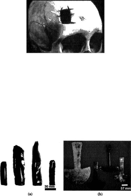
cultures practiced the art of trephination (alternately spelled trepanation), which involves drilling holes in the head, presumably to release “evil spirits” or “bad humors”. Figure 6.2 presents a photograph of a trephined skull that was recovered from an archeological site in South America. Although the resulting health effects of trephination are difficult to assess today, archaeological evidence of bone healing in trephined skulls suggests that ancient patients not only survived the operation, but also went on to live for a long while afterwards [4, 27, 30]. Early papyrus writings from Egypt demonstrate evidence of brain surgery in Africa as early as 3000 BC [8, 9, 14]. Pre-historic evidence of brain surgery existed in the Pre-Incan civilization in Peru, as early as 2000 BC [4]. The early surgical tools were made
MEMS AND NEUROSURGERY |
97 |
FIGURE 6.2. Photograph of a prehistoric human skull from Peru showing evidence of trephination (arrow).
(Courtesy: Museum of Man, San Diego, CA)
from volcanic rock or ancient metals (Figure 6.3) [21]. Archeological evidence indicates brain surgery was used for the treatment of organic diseases, osteomyelitis, and head injuries [4, 8, 27, 32].
The early Greeks used trephination in the treatment of head injuries [21, 23, 31]. Hippocrates (460–370 BC) documented many of their early medical accomplishments. He was also the first to describe numerous neurologic conditions, which most likely developed from battlefield injuries, as well as the use of a trephine for brain contusions. He was aware of the underlying dural sheath covering the brain and recommended such techniques as irrigation during surgery to reduce overheating and injury to intracranial structures.
Aulus Cornelius Celsus was the brain surgeon for ancient Rome during the first century AD. Celsus advanced neurosurgery from the point where Hippocrates left off [31]. Unlike Hippocrates, Celsus performed surgery on depressed skull fractures. He demonstrated that injury to the lower spine could cause paralysis of the leg as well as incontinence (9). Celsus was also responsible for describing the symptoms of brain injury in great detail [21].
FIGURE 6.3. Photographs of instruments for ancient brain surgery showing: (a) obsidian blades; and (b) copper and bronze tumi knives. (Courtesy: Museum of Man, San Diego, CA)
98 |
SHUVO ROY ET AL. |
Paul of Aegina (625–690 AD) was the last great Byzantine physician. His most notable writings, The Seven Books of Paul of Aegina, contained discussions on the treatment of head injuries and the use of a trephine [31]. He also categorized skull fractures into groups: fissure, incision, expression, depression, arched fracture, and in infants, dent. Paul also endorsed surgery for spine fractures [9]. He classified skin incisions and was the first to postulate that hemorrhage may cause hydrocephalus (increased cerebrospinal fluid volume within the brain) [18].
The practice of neurological surgery and Arabic medicine was introduced to medieval Europe around 1050 AD by Constantinus Africanus [15, 31]. Roger of Salerno was the first to write on the topic of surgery in Italy. He studied in Baghdad where he was influenced by Arabic medicine. His work on neurosurgery significantly influenced medieval medical practices. His writings, Practica Chirurgiae, discuss dural tear assessment and cerebrospinal fluid leakage in depressed skull fractures by having the patients hold their breath (Valsalva maneuver) while watching for leaks and air bubbles [21]. Roger was also a pioneer in the techniques for managing nerve injury, and, in particular, he emphasized the rejoining of severed nerves [9].
The seventeenth century exhibited rapid growth in science and medicine. Scientists like Isaac Newton, Francis Bacon, William Harvey, and Robert Boyle made significant advances in physics, experimental design, knowledge of the circulatory system, and physiologic chemistry. Education regarding scientific thought and open public forums were used to transmit information and to discuss the ideas developed in scientific societies. One of the most notable scientists of this era, Thomas Willis (1621–1675), published an accurate anatomic study of the brain [31]. In fact, the circle of Willis bears his name. Willis also introduced the concept of the “neurology”, or the doctrine of neurons, but the term did not enter general medical usage until the eighteenth century.
The nineteenth century was the age of anesthesia, antisepsis and aseptic technique, and cerebral localization [21, 31, 33]. These innovations were great advances for the establishing neurosurgery as an independent field. Anesthesia provided freedom from pain during surgery. Antisepsis and aseptic techniques were used to minimize the risk of infection by creating sterile fields surrounding the surgical site. Cerebral localization techniques were used as diagnostic and decision making tools [33]. It was cerebral localization, the concept that the brain is divided into segments that correspond to particular functions, which aided in the diagnosis of brain lesions or injury. Paul Broca (1824–1880) localized the speech to a particular region of the brain, Broca’s area [35]. He based his discovery on the early work of Ernest Auburtin (1825–1893) who had a patient with a frontal lobe defect [31]. Auburtin abolished speech by applying pressure to the frontal lobe, which returned when the pressure was released.
Finally, it was Harvey Cushing (1869–1939), the father of American Neurosurgery, who ushered in the era of modern neurosurgery [3, 10, 19]. Cushing studied at Johns Hopkins under William Halsted (1852–1922), a premier general surgeon, where he excelled in meticulous surgical technique. Cushing contributed extensively to the field of neurosurgery. Among his many innovations are the introduction of the practice of recording the vitals signs during anesthesia (devised while still a medical student), and the use of silver clips to control bleeding [19]. He was an early proponent of the then-new technique of x-raying [3, 19]. His most memorable work was that on pituitary surgery, which was
MEMS AND NEUROSURGERY |
99 |
published as a monograph in 1912 and formed the basis for further studies on the pituitary gland and classification of brain tumors [31].
6.3. CONVENTIONAL NEUROSURGICAL TREATMENTS
Today, neurosurgery is considered a medical discipline that provides the operative and non-operative care of disorders that affect the central, peripheral and autonomic nervous system (including their supportive structures and vascular supply), and the operative and non-operative management of pain. It encompasses disorders of the brain, skull, pituitary gland, spinal cord, cranial and spinal nerves, and the autonomic nervous system. Some common neurosurgical strategies that are currently used to treat disorders of the brain, nerves, and spine are described below.
6.3.1. Hydrocephalus
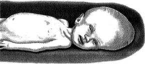
Hydrocephalus is a condition where excess cerebrospinal fluid (CSF) results in enlargement of ventricles of the brain due to conditions such as cerebral atrophy and/or failure of development of the brain [31]. The cerebrospinal fluid (CSF), which is normally clear and colorless, bathes the brain and spinal cord and protects them from injury [35]. This fluid normally flows through one ventricle to the next, and is kept over the surface of the brain and down the spinal cord before being finally absorbed into the blood stream. When the circulation or absorption of CSF is blocked, or if there is excess production, the volume of the brain becomes excessively larger then normal. This increased volume manifests as enlarged heads in babies since their skulls are not completely fused (Figure 6.4).
There are several types of hydrocephalus. An obstructive type of hydrocephalus where the CSF is under increased pressure is termed tension hydrocephalus [35]. The terms communicating and non-communicating hydrocephalus differentiate between a complete or incomplete obstruction of the aqueduct of the brain. Communicating hydrocephalus refers to communication between the ventricles and the spinal subarachnoid space within the brain, whereas, non-communicating is the contrary. Chronic hydrocephalus is a similar condition that has been present for months or years in an individual. Causes of hydrocephalus may
FIGURE 6.4. Graphical illustration showing the effect of hydrocephalus in infants. The increased volume of cerebrospinal fluid (CSF) within the brain causes head enlargement.
100 |
SHUVO ROY ET AL. |
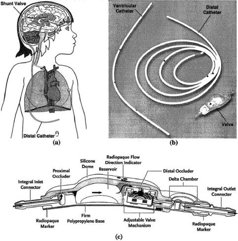
FIGURE 6.5. Treatment of hydrocephalus. Excess CSF is drained from the brain to the peritoneal cavity via a surgically implanted shunt (a). The shunt (b) comprises a short catheter inserted into the ventricle, a one-way valve underneath the skin, and a longer catheter that typically drains into the abdominal cavity. Recent advances in shunt design include the development of valves with features to prevent overdrainage and allow for telemetric adjustment (c). (Courtesy: Medtronic Inc)
include tumors, cysts, trauma, subarachnoid hemorrhages, infection, subdural hematomas, and Paget’s disease [28, 35]. The result of hydrocephalus is the accumulation of cerebrospinal fluid within the brain and increased intracranial pressure (ICP).
The goal of hydrocephalus treatment is to minimize or prevent brain damage by improving CSF flow. Surgical interventions are the primary treatment for hydrocephalus. This includes direct removal of the obstruction, if possible. However, the most common conventional approach involves the surgical placement of a CSF shunt within the brain to bypass the obstructed area (Figure 6.5) [6, 34]. The CSF shunt consists of three primary components: