

2. Upper limb
General plan
The upper limb of humans is built for prehension and manipulation and the range of movements available at the joints of the upper limb enhances the dexterity of the fingers. Four fingers flexing against an opposed thumb enable the hand to function as a grasping mechanism, in which the thumb is equal in functional value to all four fingers. The hand is furthermore the main tactile organ, with a rich nerve supply.
In order to enable the hand to function in any position, the forearm is provided with a range of about 140° of pronation and supination, the elbow has a range of flexion and extension of like amount, and very free mobility is provided at the shoulder joint. This mobility is further increased by the mobility of the pectoral girdle through which the upper limb articulates with the axial skeleton.
Although the upper limb is commonly called the arm, this term strictly refers to the upper part of the limb between the shoulder and elbow, while the part between the elbow and wrist is the forearm. Both arm and forearm have anterior or flexor and posterior or extensor compartments. The hand has an anterior (flexor) surface, or palm, and a posterior (extensor) surface, or dorsum.

Part one. Pectoral girdle
The bones of the pectoral or shoulder girdle, the clavicle and scapula, connect the upper limbs to the axial skeleton. Only one small joint connects the girdle to the rest of the skeleton—the sternoclavicular joint—and the two girdle bones are joined to one another by an even smaller joint, the acromioclavicular. The remaining attachment to the axial skeleton is mainly muscular, and this helps to account for the mobility of the shoulder girdle. The strong coracoclavicular ligament attaches the clavicle and scapula to each other, and the clavicle is anchored to the first costal cartilage by the costoclavicular ligament. Forces from the upper limb are transmitted by the clavicle to the axial skeleton through these ligaments, and neither end of the clavicle normally transmits much force.
Almost all movement between humerus and glenoid cavity at the shoulder joint is accompanied by an appropriate movement of the scapula itself. Furthermore, the scapula cannot move without making its supporting strut, the clavicle, move also. Generally speaking the shoulder joint, the acromioclavicular and sternoclavicular joints all move together in harmony, providing a kind of ‘thoracohumeral articulation’. Defects in any part of the ‘thoracohumeral articulation’ must impair the function of the whole.
The bones of the pectoral girdle are described on pages 97–101, the shoulder joint on page 46 and the clavicular joints on page 43.
Muscles of the pectoral girdle
The muscular attachments between pectoral girdle and trunk are direct and indirect.
Direct attachment of the pectoral girdle to the trunk is provided by muscles that are inserted into the clavicle or scapula from the axial skeleton. These muscles are pectoralis minor, subclavius, trapezius, the rhomboids, levator scapulae and serratus anterior. Indirect attachment to the axial skeleton is secured by the great muscles of the axillary folds (pectoralis major and latissimus dorsi); these muscles, by way of the upper end of the humerus, move the pectoral girdle on the trunk.
The muscular attachments between upper limb and pectoral girdle include the deltoid and short scapular muscles, which are inserted about the upper end of the humerus, and the biceps and long head of triceps which, running over the humerus, are inserted beyond the elbow joint into the bones of the forearm. These muscles are important factors in giving stability to the very mobile shoulder joint across which they lie, and are described with the shoulder region (see p. 44).
Pectoralis major
From clavicular and sternocostal heads this large triangular muscle converges on the upper humerus, folding on itself where it forms the anterior axillary wall to become attached to the humerus by means of a bilaminar tendon.
The clavicular head arises from the medial half of the anterior surface of the clavicle. Running almost horizontally laterally the fibres of this head lie on the manubrial part of the muscle, from which they are separate. They are inserted by the anterior lamina of the tendon into the lateral lip of the intertubercular (bicipital) sulcus of the humerus.
The sternocostal head arises from the lateral half of the anterior surface of the manubrium and body

of sternum, the upper six costal cartilages and the aponeurosis of the external oblique muscle over the upper attachment of rectus abdominis. The manubrial fibres are inserted by the anterior lamina of the tendon into the lateral lip of the intertubercular sulcus behind (deep to) the clavicular fibres. The lower sternocostal and abdominal fibres course upwards and laterally to be inserted progressively higher into the posterior lamina of the tendon, producing the rounded appearance of the anterior axillary fold. The fibres which arise lowest of all are thus inserted highest, and by a crescentic fold blend with the capsule of the shoulder joint (Fig. 2.1). The lower medial part of the muscle is thinner and in danger of being perforated when a subpectoral pocket is created for insertion of a prosthesis during breast reconstruction . Perforating branches of the internal thoracic artery pierce the deep surface of the muscle at the sternal edge and are at risk of being torn during subpectoral dissection. These branches and others from the superior and lateral thoracic arteries supplement the dominant vascular pedicle for pectoralis major from the pectoral branch of the thoracoacromial artery. A musculocutaneous flap based on this dominant pedicle is used in reconstructive procedures after surgical resections for head and neck cancer.
Figure 2.1 Insertion of the right pectoralis major. Part of the clavicular head has been removed and deltoid incised and retracted to show how the lowest fibres of the sternocostal origin twist upwards deep to the manubrial fibres.
Nerve supply. From the brachial plexus via the lateral and medial pectoral nerves, so named because of their origins from the lateral and medial cords of the plexus. The lateral pectoral nerve pierces the clavipectoral fascia medial to the pectoralis minor. Branches of the medial pectoral nerve pierce the pectoralis minor, but may pass round its lateral border, to reach the pectoralis major. The muscle is the only one in the upper limb to be supplied by all five segments of the brachial plexus; C5, 6 supply the clavicular head and C6–8, T1 the sternocostal part. The degree of paralysis of pectoralis major may be helpful in gauging the extent of a brachial plexus injury (see p. 95).
Action. The muscle is a powerful adductor and a medial rotator of the arm. The sternocostal fibres are the chief adductors. The clavicular head assists in flexion at the shoulder joint. With the upper limb fixed in abduction the muscle is a useful accessory muscle of inspiration, drawing the ribs upwards towards the humerus.
Test. For the clavicular head the arm is abducted to 90° or more and the patient pushes the arm

forwards against resistance. For the sternocostal head the arm is abducted to 60° and then adducted against resistance. The contracting heads can be seen and felt.
Pectoralis minor
This small triangular muscle arises from the third, fourth and fifth ribs under cover of pectoralis major (Fig. 2.14). The insertion is by a short thick tendon into the medial border and upper surface of the coracoid process of the scapula (not to the tip of the process, which is fully occupied by biceps and coracobrachialis).
Of no great functional significance, the muscle forms a tight band across the front of the axillary neurovascular and lymphatic contents; division of its tendon facilitates surgical clearance of the axillary lymph nodes (see p. 56).
Nerve supply. By both pectoral nerves (C6–8).
Action. It assists serratus anterior in protraction of the scapula, keeping the anterior (glenoid) angle in apposition with the chest wall as the vertebral border is drawn forwards by serratus anterior. The muscle is elongated when the scapula rotates in full abduction of the arm; its subsequent contraction assists gravity in restoring the scapula to the rest position.
Subclavius
This small muscle arises from the costochondral junction of the first rib and is inserted into the subclavian groove on the inferior surface of the clavicle.
Nerve supply. By its own nerve from the upper trunk of the brachial plexus (C5, 6).
Action. It assists in stabilizing the clavicle in movements of the pectoral girdle. It may prevent the jagged ends of a fractured clavicle from damaging the adjacent subclavian vein.
The pectoral fascia is a thin lamina of deep fascia that covers the anterior surface of pectoralis major. It is attached medially to the sternum, above to the clavicle and is continuous laterally with the axillary fascia. It forms the floor of the retromammary space and gives origin to the platysma muscle from its upper part.
The clavipectoral fascia is a strong fascial sheet deep to pectoralis major. Its upper part, also known as the costocoracoid membrane, is attached laterally to the coracoid process and medially blends with the external intercostal membrane of the upper two spaces. It splits above to enclose subclavius and is attached to the edges of the subclavian groove on the undersurface of the clavicle.
At the lower border of subclavius the two layers fuse and form a well-developed band, the costocoracoid ligament, stretching from the knuckle of the coracoid to the first costochondral junction. From this ligament the fascia stretches as a loosely felted membrane to the upper border of pectoralis minor, where it splits to enclose this muscle. Below pectoralis minor the fascia, also known as the suspensory ligament of the axilla, extends downwards and is attached to the axillary fascia on the floor of the axilla; its tension maintains the concavity of the axilla (Fig. 2.2).
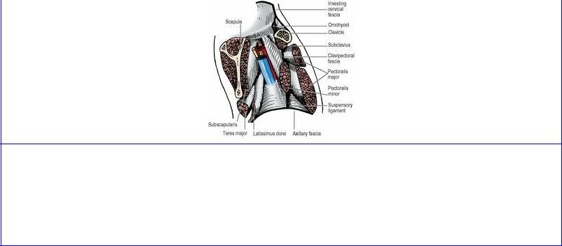
Figure 2.2 Vertical section of the left axilla, looking laterally towards the arm. The clavipectoral fascia encloses subclavius and pectoralis minor, below which it becomes the suspensory ligament of the axilla, joining the axillary fascia which arches upwards between pectoralis major and latissimus dorsi. The neurovascular bundle of the upper limb lies between the anterior and posterior axillary walls.
The clavipectoral fascia is pierced by four structures: two passing inwards, two passing outwards. Passing inwards are lymphatics from the infraclavicular nodes to the apical nodes of the axilla, and the cephalic vein; passing outwards are the lateral pectoral nerve and the thoracoacromial artery, or its branches (pectoral, acromial, deltoid and clavicular); their corresponding veins join the cephalic vein anterior to the fascia.
Trapezius
This large flat muscle, the most superficial of the upper part of the back, arises in the midline from skull to lower thorax and converges on the outer part of the pectoral girdle. Its origin extends from the medial third of the superior nuchal line to the spine of C7 vertebra, finding attachment to the ligamentum nuchae between the external occipital protuberance and the vertebral spine. Below this the origin extends along the spinous processes and supraspinous ligaments of all 12 thoracic vertebrae. Opposite the upper thoracic spines the muscle shows a triangular aponeurotic area, which makes a diamond with that of the opposite side (Fig. 2.5).
The upper fibres are inserted into the posterior border of the lateral third of the clavicle at its posterior border. The middle fibres are inserted along the medial border of the acromion and the superior lip of the crest of the scapular spine. The part of the muscle which arises from the lower six thoracic spines is inserted by a narrow recurved tendon into the medial end of the spine (Fig. 2.5).
Nerve supply. From the spinal part of the accessory nerve (C1–5) and branches from the cervical plexus (C3 and 4); the latter are usually only proprioceptive, although in some cases they contain motor fibres as well (see p. 334). These nerves cross the posterior triangle to enter the deep surface of trapezius. The accessory nerve can be distinguished from the cervical branches by the fact that it emerges from within the substance of sternocleidomastoid; the cervical nerves emerge from behind sternocleidomastoid.
Action. All fibres help to retract the scapula, while the upper and lower fibres are important in scapular rotation, tilting the glenoid cavity upwards, an essential component of abduction of the shoulder. In this action upper fibres elevate the acromion while lower fibres depress the medial end

of the spine, like turning a wing nut (Fig. 2.3), and they are strongly assisted by the lowest four digitations of serratus anterior (see p. 42). The upper fibres can elevate the whole scapula (shrug the shoulder) or prevent its depression (as when carrying something heavy). They can also produce lateral flexion of the neck, but acting with the upper fibres of the opposite side they can extend the neck.
Figure 2.3 Rotation of the scapula. The upper and lower parts of trapezius pull on the scapular spine in different directions, twisting it like a wing-nut, while serratus anterior pulls on the inferior angle.
Test. The shoulder is shrugged against resistance and the upper border of the muscle is seen and felt.
Latissimus dorsi
This muscle, covering such a large area of the back, is characterized by its very wide origin and its very narrow insertion. The muscle arises from the spines of the lower six thoracic vertebrae and the posterior layer of the lumbar fascia, by which it is attached to the lumbar and sacral vertebral spines and to the posterior part of the crest of the ilium (Fig. 2.4). Lateral to this it also arises by muscular fibres from the outer lip of the iliac crest. The upper part of the flat sheet of muscle runs horizontally, covered medially by the lower triangular part of trapezius, and passes over the inferior angle of the scapula, from which a few fibres may arise (Fig. 2.5). The middle part of the muscle runs obliquely upwards and outwards. The lower part of the muscle runs vertically upwards, being reinforced by four slips from the lowest four ribs, whose fibres of origin interdigitate with those of the external oblique. The lateral border of latissimus dorsi forms a boundary of the lumbar triangle (see p. 222). The muscle converges towards the posterior axillary fold, of which it forms the lower border. The muscle sweeps spirally around the lower border of teres major with some intermingling of their fibres. The muscle is then replaced by a flattened, shiny, white tendon about 3 cm broad which is inserted into the floor of the intertubercular sulcus (Fig. 2.9). As a result of the spiral turn around teres major the surfaces of the muscle, anterior and posterior, are reversed at the tendon; and the fibres that originate lowest at the midline insert highest at the humerus, while those that originate highest insert lowest. This glistening white tendon contrasts with adjacent muscle and is a useful landmark at the lateral margin of the posterior wall of the axilla during surgical dissection in the axilla (Fig. 2.18). Occasionally some muscle fibres from the edge of latissimus dorsi cross in front of the axillary vessels and nerves to blend with the tendon of pectoralis major forming a muscular axillary arch.
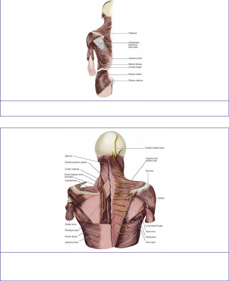
Figure 2.4 Muscles of the left side of the back of the trunk.
Figure 2.5 Muscles of the pectoral girdle from behind. On the left most of trapezius, deltoid and the rhomboids have been removed to show the dorsal scapular nerve accompanied by the dorsal scapular artery, and the axillary nerve with the (unlabelled) posterior circumflex humeral artery.
Nerve supply. By the thoracodorsal nerve (C6–8) from the posterior cord of the brachial plexus. It is vulnerable in operations on the axilla, for in its course down the posterior wall it slopes forwards to
enter the medial surface of the muscle just behind its anterior border in front of the thoracodorsal vessels (Fig. 2.16).
Action. It extends the shoulder joint and medially rotates the humerus (e.g. folding the arms behind the back, or scratching the opposite scapula), but in combination with pectoralis major it is a powerful adductor. When adducting the upper limb from a position of abduction above the shoulder, it plays an integral role in climbing.
Its costal fibres of origin can assist in deep inspiration, elevating the lower four ribs towards the fixed humerus. But the remainder of the muscle, sweeping from the vertebral column around the convexity of the posterolateral chest wall, compresses the lower thorax in violent expiratory efforts such as coughing or sneezing.
In spinal injury the muscle may move the pelvis and trunk; it is the only muscle of the upper limb to have a pelvic attachment (via the lumbar fascia). The muscle is used in reconstructive breast surgery. A part of the muscle, sometimes with an overlying paddle of skin as a musculocutaneous flap, is rotated around to the front and used to create a mound simulating the breast. The thoracodorsal artery is an important source of blood supply to the flap.
Test. The arm is abducted to a right angle and then adducted, extended and medially rotated against resistance; the lateral part of the muscle below the posterior axillary fold can be seen and felt contracting. The muscle can also be felt to contract here when the patient coughs.
Rhomboid major and minor
Rhomboid major arises from four vertebral spines (T2–5), and the intervening supraspinous ligaments. It is inserted into the medial border of the scapula between the root of the spine and the inferior angle (Fig. 2.5).
Rhomboid minor is a narrow ribbon of muscle parallel with the above, arising from two vertebral spines (C7, T1) and inserted into the medial border of the scapula at the root of the spine.
Nerve supplies. By the dorsal scapular nerve (nerve to the rhomboids) from the C5 root of the brachial plexus which passes through scalenus medius, runs down deep (anterior) to levator scapulae (which it supplies) and lies on the serratus posterior superior muscle to the medial side of the descending branch of the transverse cervical artery (Fig. 2.5). It supplies each rhomboid on the deep surface.
Actions. The rhomboids draw the vertebral border of the scapula medially and upwards. With trapezius they contract in squaring the shoulders, i.e. retracting the scapula.
Test. With the hand on the hip or behind the back the patient pushes the elbow backwards against resistance and braces the shoulder back. The muscles are palpated at the vertebral border of the scapula. If the rhomboids of one side are paralysed, the scapula of the affected side remains further from the midline than that of the normal side.
Levator scapulae
This strap-like muscle, which appears in the floor of the posterior triangle, arises from the transverse
processus of the atlas and axis and from the posterior tubercles of the third and fourth cervical vertebrae. It is inserted into the medial border of the scapula from the superior angle to the spine.
Nerve supply. From the cervical plexus (C3, 4, anterior rami), reinforced by the dorsal scapular nerve (C5).
Action. With the upper part of trapezius, it can elevate the scapula and laterally flex the neck.
Serratus anterior
This is a broad sheet of thick muscle (Fig. 2.16) which clothes the side wall of the thorax and forms the medial wall of the axilla. It arises by a series of digitations from the upper eight ribs. The first digitation arises from the first and second ribs (Fig. 2.6). All the other digitations arise from their corresponding ribs and the lower four interdigitate with external oblique. The muscle is inserted on the costal (inner) surface of the scapula: the first and second digitations at the superior angle, the third and fourth as a thin sheet to the length of the vertebral border, and the lowest four at the inferior angle. The muscle is covered by a strong well-developed fascia.
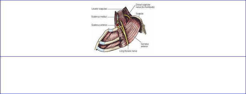
Figure 2.6 Left long thoracic nerve (to serratus anterior). The branches from C5 and 6 fuse within scalenus medius and emerge as a single trunk which is joined in the axilla over the first digitation of serratus anterior by the branch from C7. The second rib gives origin to half of the first digitation and all the second digitation of the muscle.
Nerve supply. By the long thoracic nerve from the C5, 6 and 7 roots of the brachial plexus. The nerve lies behind the midaxillary line (i.e. behind the lateral branches of the intercostal arteries) on the surface of the muscle (Fig. 2.16), deep to the fascia, and is thus usually protected in operations on the axilla.
Action. The whole muscle contracting en masse protracts the scapula (punching and pushing), thus effectively elongating the upper limb. A further highly important action is that of the lower four digitations, which powerfully assist trapezius in rotating the scapula laterally and upwards in raising the arm above the level of the shoulder. In this action it is a more powerful rotator than trapezius. In all positions of the upper limb the muscle keeps the vertebral border of the scapula in firm apposition with the chest wall.
Test. With the arm flexed and the elbow extended the outstretched hand is pushed against a wall. Paralysis results in ‘winged scapula’, where the vertebral border becomes prominently raised off the
posterior chest wall.
Joints of the pectoral girdle
Sternoclavicular joint
This is a synovial joint between the bulbous medial end of the clavicle, the superolateral part of the manubrium of the sternum and the adjoining first costal cartilage (Fig. 2.7). The joint is separated into two cavities by an intervening disc of fibrocartilage, which is attached at its periphery to the capsule of the joint. Although synovial, it is atypical as the bony surfaces are covered by fibrocartilage, not the usual hyaline variety. The sternal end of the clavicle projects above the upper margin of the manubrium so that only about the lower half of the clavicular articular surface lies opposite the sternal articular facet.
Figure 2.7 Left sternoclavicular joint, sectioned and viewed from the front. The clavicle extends well above the bony socket of the manubrium, and is bound down by the disc and costoclavicular ligament.
The capsule invests the articular surfaces like a sleeve. The articular disc is attached to the capsule. The disc is also firmly attached to the medial end of the clavicle above and behind, and to the first costal cartilage below. The capsule is thickened in front and behind as the anterior and posterior sternoclavicular ligaments.
The interclavicular ligament joins the upper borders of the sternal ends of the two clavicles and is attached to the suprasternal (jugular) notch of the manubrium. The costoclavicular ligament binds the clavicle to the first costal cartilage and the adjacent end of the first rib, just lateral to the joint. It is in two laminae. The fibres of the anterior lamina run upwards and laterally, and those of the posterior lamina upwards and medially (these are the same directions as those of the external and internal intercostal muscles). The ligament is very strong and is the major stabilizing factor of the sternoclavicular joint.
Nerve supply. By the medial supraclavicular nerves (C3, 4) from the cervical plexus.
Movements. Elevation (shrugging the shoulder) and depression of the acromial end of the clavicle result in movements downwards and upwards respectively between the sternal end of the clavicle and the disc. Forward and backward (squaring the shoulders) movements of the acromial end likewise cause reciprocal movements at the sternal end; these movements occur between the manubrium and the disc. Similarly, in rotary movements (abduction of the arm above the head) the disc moves with
the clavicle. Rotation of the clavicle is passive; there are no rotator muscles. It is produced by rotation of the scapula and transmitted to the clavicle through the coracoclavicular ligaments (see below).
The stability of the joint is maintained by the ligaments, especially the costoclavicular ligament. It takes all strain off the joint, transmitting stress from clavicle to first costal cartilage. The latter is itself immovably fixed to the manubrium by a primary cartilaginous joint (see p. 181). Dislocation is unusual; the clavicle breaks in preference.
Acromioclavicular joint
This is a synovial joint between the flat overhanging lateral end of the clavicle and the underlying medial border of the acromion. The articulating surfaces are covered (like those of the sternoclavicular joint) by fibrocartilage (so it is an atypical synovial joint).
A sleeve-like capsule surrounds the articular surfaces; it is not strong, but on top there is a thickening of fibres which constitutes the acromioclavicular ligament. An incomplete disc of fibrocartilage hangs down into the upper part of the joint cavity.
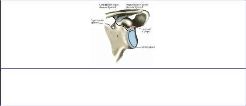
The coracoclavicular ligament, extremely strong, is the principal factor in providing stability to the joint. It consists of two parts, conoid and trapezoid (Fig. 2.8). The conoid ligament, an inverted cone, extends upwards from the knuckle of the coracoid process to a wider attachment around the conoid tubercle, on the undersurface of the clavicle (Fig. 2.51). The trapezoid ligament is attached to the ridge of the same name on the upper surface of the coracoid process and extends laterally, in an almost horizontal plane, to the trapezoid ridge on the undersurface of the clavicle. The two ligaments are connected to each other posteriorly, forming an angle that is open anteriorly.
Figure 2.8 Glenoid cavity of the left scapula and the coracoclavicular ligament. The black line marks the site of the epiphyseal plate between the scapula proper and the coracoid component. The glenoid labrum continues above into the long head of biceps. The trapezoid part of the coracoclavicular ligament lies in front of and lateral to the conoid part.
Nerve supply. By the suprascapular nerve (C5, 6) from the brachial plexus.
Movements. These are passive; muscles which move the scapula cause it to move on the clavicle. Scapular movements on the chest wall fall into three groups: (1) protraction and retraction around the
chest wall, (2) rotation, and (3) elevation or depression. These basic movements can be combined in varying proportions, and each of these transmits, through ligaments, corresponding movements to the clavicle. All movements of the scapula involve movements in the joint at either end of the clavicle.
Horizontally, in protraction and retraction of the tip of the shoulder, the scapula hugs the thoracic wall, held to it by serratus anterior and pectoralis minor. The acromion glides to and fro with the end of the clavicle.
In abduction of the arm the total range of scapular rotation on the chest wall is about 60°, but only 20° of this occurs between the scapula and the clavicle. The two parts of the coracoclavicular ligament are then taut, and transmit the rotating force to the clavicle, whose rotation then accounts for the remainder of scapular rotation on the chest wall.
Elevation (shrugging the shoulders) is produced by the upper fibres of trapezius together with levator scapulae. Depression of the scapula is produced by gravity, assisted when necessary by serratus anterior and pectoralis minor. Elevation and depression move the medial end of the clavicle (see above), but they scarcely move the acromioclavicular joint.
The stability of the joint is provided by the coracoclavicular ligament. The scapula and upper limb hang suspended from the clavicle by the conoid ligament (assisted by the deltoid, biceps and triceps muscles). Forces transmitted medially from the upper limb to the glenoid cavity are transmitted from scapula to clavicle by the trapezoid ligament and from clavicle to first rib by the costoclavicular ligament. Thus a fall on outstretched hand or elbow puts no strain on either end of the clavicle at the joints. If the clavicle fractures as a result, it always does so between these ligaments. Falls on the shoulder may dislocate the acromioclavicular joint, forcing the acromion under the clavicle and tearing the coracoclavicular ligament.