

Chapter 6. Head and neck and spine

Part one. General topography of the neck
The first thoracic vertebra lies at the highest part of the sloping thoracic inlet. From its upper border rises the cervical spinal column, gently convex forwards, and supporting the skull. A mass of extensor musculature lies behind the vertebrae, supplied segmentally by posterior rami of cervical nerves that emerge from intervertebral foramina. A much smaller amount of prevertebral flexor musculature, covered by prevertebral fascia, lies in front of the vertebrae and behind the pharynx, supplied segmentally by anterior rami.
Projecting forwards and downwards from the base of the skull in front of the upper part of the pharynx is the face. The hard palate lies on a level with the anterior arch of the atlas (C1 vertebra); the lower border of the mandible lies between C2 and 3 vertebrae. The pharynx extends from the base of the skull to the level of the cricoid cartilage (C6) and then continues on as the oesophagus.
In front of the lower pharynx and upper oesophagus lie the larynx and trachea. Lying above the larynx is the hyoid bone at C3 vertebra level. It is connected to the mandible by the mylohyoid muscles, which are at the upper limit of the anterior part of the neck and form the floor of the mouth. The hyoid bone is suspended by muscles from the skull and the larynx is suspended from the hyoid by a membrane and muscles. Inferiorly they are connected by muscles to the sternum and the scapula. Deep to these muscles the thyroid gland, enclosed in the pretracheal fascia, lies alongside the larynx and trachea.
On each side of the pharynx is the carotid sheath, containing the common and internal carotid arteries and the internal jugular vein, with the cervical sympathetic trunk behind it. The external carotid artery lies outside the sheath and gives several branches in the neck. Descending into the neck are the ninth, tenth, eleventh and twelfth cranial nerves; the ninth and twelfth pass forwards to the oropharynx and tongue, the eleventh runs backwards to the sternocleidomastoid and trapezius muscles, the vagus continues down in the carotid sheath.
Surrounding the whole neck is a collar of fascia, the investing layer of deep cervical fascia, which encloses the trapezius and sternocleidomastoid muscles; the fascia and the muscles are attached above to the base of the skull and below to the clavicle at the root of the neck.
Superficial structures
The platysma is a broad flat sheet of muscle that lies superficial to the investing layer of deep cervical fascia. It extends from the fascia over the upper parts of pectoralis major and deltoid to the lower border of the mandible; some fibres continue on to the face, blending with the muscles of facial expression. The muscle covers the external and anterior jugular veins. The two muscles are separated below, but converge above towards the midline just beneath the chin.
Nerve supply. By the cervical branch of the facial nerve; afferent (proprioceptive) fibres run with the transverse cervical nerve.
Action. It plays a part in facial expression and may assist in opening the mouth.
The anterior jugular veins commence beneath the chin and pass downwards, side by side beneath the platysma, to the suprasternal region (Fig. 6.5). Here they pierce the deep fascia and come to lie in the

suprasternal space, where they are often connected by a short anastomotic vein. Each now angles laterally and passes deep to sterno-cleidomastoid, but superficial to the strap muscles, to open into the external jugular vein near its termination.
Deep cervical fascia
The deep cervical fascia consists of four parts: the investing layer, pretracheal fascia, prevertebral fascia, and the carotid sheath.
Investing layer
This fascia, comparable in every way to the deep fascia that underlies the subcutaneous fat in the limbs and elsewhere, surrounds the neck like a collar (Fig. 6.1). It splits around sternocleidomastoid and trapezius and posteriorly it blends with the ligamentum nuchae, which is attached to the spines of the cervical vertebrae. Anteriorly it is attached to the hyoid bone; and above to the lower border of the mandible and to the mastoid process, superior nuchal line and external occipital protuberance at the base of the skull.
Figure 6.1 Deep cervical fascia of one half of the neck, showing its four components: the investing, pretracheal and prevertebral layers and the carotid sheath.
Between the angle of the mandible and the tip of the mastoid process the investing layer is strong and splits to enclose the parotid gland. The superficial part extends superiorly as the parotidomasseteric fascia and reaches up to the zygomatic arch. The deep part extends to the base of the skull; between the styloid process and the angle of the mandible it is thickened as the stylomandibular ligament.
Below, the investing layer is attached to the spine and acromion of the scapula and the clavicle with the trapezius, and to the clavicle and the manubrium of the sternum with the sternocleidomastoid. In the intervals between these muscles, it is attached to both clavicles and to the jugular (suprasternal) notch by two layers into which it splits a short distance above them. The layers are attached to the anterior and posterior borders of the jugular notch, enclosing between them the suprasternal space

which contains the lower parts of the anterior jugular veins, an anastomotic arch between them, the sternal heads of the sternocleidomastoids and sometimes a lymph node. Of the two layers that adhere to the middle third of the clavicle, the deeper splits around the inferior belly of the omohyoid, forming a fascial sling which keeps this muscle belly low down in the neck (see Fig. 2.2, p. 39). The two layers are pierced by the external jugular vein.
Prevertebral fascia
This is a firm, tough membrane that lies in front of the prevertebral muscles (Fig. 6.1). It extends from the base of the skull, in front of the longus capitis, rectus capitis lateralis and longus colli muscles, downwards to blend with the anterior longitudinal ligament on the body of T4 vertebra. It extends sideways across the scalenus anterior, scalenus medius and levator scapulae muscles (Fig. 6.8), getting thinner further out and fading under cover of the anterior border of trapezius. It covers the muscles that form the floor of the posterior triangle of the neck and all the cervical nerve roots (thus the cervical plexus and trunks of the brachial plexus lie deep to it). The lymph nodes of the posterior triangle and the accessory nerve lie superficial to it. The third part of the subclavian artery lies deep to the fascia, which becomes prolonged over the artery and the brachial plexus below the clavicle as the axillary sheath to a varying extent in the axilla. It does not invest the subclavian or axillary vein; these lie in loose areolar tissue anterior to it, free to dilate during times of increased venous return from the upper limb. The fasica is pierced by the four cutaneous branches of the cervical plexus (great auricular, lesser occipital, transverse cervical and supraclavicular nerves).
Pretracheal fascia
This thin fascia lies deep to the infrahyoid strap muscles (sternothyroid, sternohyoid and omohyoid) so that its upward attachment is limited by the respective attachments of those muscles, namely, the body of the hyoid bone and the oblique line of the thyroid cartilage. It splits to enclose the thyroid gland, to which it is not adherent except to the back of the isthmus where it is also attached to the second, third and fourth rings of the trachea. Laterally, it fuses with the front of the carotid sheath on the deep surface of the sternocleidomastoid and inferiorly it passes behind the brachiocephalic veins to blend with the adventitia of the arch of the aorta and the fibrous pericardium. The pretracheal fascia is also described in some accounts as being part of a cervical visceral fascia that surrounds the pharynx, oesophagus, larynx and trachea.
Carotid sheath
This is not a fascia in the sense of a demonstrable membranous layer, but consists of a feltwork of areolar tissue that surrounds the common and internal carotid arteries, internal jugular vein, vagus nerve and some deep cervical lymph nodes (Fig. 6.1). It is thin where it overlies the internal jugular vein, allowing the vein to dilate during increased blood flow. The sheath is attached to the base of the skull at the margins of the carotid canal and jugular fossa, and is continued downwards along the vessels to blend with the adventitia of the aortic arch. In front the lower part of the sheath fuses with the fascia on the deep surface of the sternocleidomastoid. Where they lie alongside, the sheath blends with the pretracheal fascia. Behind the carotid sheath there is a minimum of loose areolar tissue between it and the prevertebral fascia; the cervical sympathetic trunk lies here in front of the prevertebral fascia (Fig. 6.8). The carotid sheath is described further on page 366.
Tissue spaces of the neck

Behind the prevertebral fascia is the closed prevertebral space from which an anterior escape can only be made by a perforation in the fascia. Hence pus from an abscess in a cervical vertebra can lift the prevertebral fascia as far down as the superior mediastinum.
Immediately in front of the prevertebral fascia is a space that extends from the base of the skull to the diaphragm passing through the superior into the posterior mediastinum as it does so (see Fig. 4.9, p. 189). Its upper part is the retropharyngeal space, which is continuous laterally with a parapharyngeal space at the side of the pharynx; the upper part of this space is in the infratemporal fossa (see p. 361), bounded laterally by the pterygoid muscles and the parotid sheath.
In the upper part of the neck is the submandibular space below the mylohyoid muscle and deep to the investing layer of fascia between the hyoid bone and the mandible. This space communicates around the posterior border of mylohyoid with a sublingual space under the mucous membrane of the floor of the mouth. Ludwig's angina is a rare but severe form of cellulitis that involves these spaces and spreads backwards into the parapharyngeal space.
Three or four small submental lymph nodes lie beneath the chin, some superficial and others deep to the investing layer of deep cervical fascia (Fig. 6.6). They drain, across the midline, a wedge of tissue in the floor of the mouth opposite the four lower incisor teeth, including those teeth, gums and lip, and the tip of the tongue (Fig. 6.34). In their turn they drain to submandibular nodes or directly to the upper deep cervical group.
Figure 6.34 Lymph drainage of the tongue. Of the deep cervical nodes, only the jugulodigastric and jugulo-omohyoid nodes are shown; other channels drain to other nodes of the group.
About half a dozen submandibular lymph nodes lie on the surface of the submandibular gland, some embedded within the gland (Fig. 6.6). They drain the submental nodes, the lateral parts of the lower lips, all the upper lip and external nose, and the anterior part of the tongue, mainly but not exclusively from their own side. They also receive lymph from the anterior half of the nasal walls and the paranasal sinuses that drain there (frontal, anterior and middle ethmoidal, and maxillary), and from all the teeth (except lower incisors).
Part two. Triangles of the neck
To assist the description of the topographical anatomy of the neck and the location of pathological lesions, each side is divided into anterior and posterior triangles by the obliquely placed sternocleidomastoid muscle (Fig. 6.2). The posterior triangle lies between the posterior border of sternocleidomastoid, the anterior border of trapezius and the clavicle, and the anterior triangle between the anterior border of sternocleidomastoid, the lower border of the mandible and the midline. The anterior triangle can be subdivided into submental, digastric, carotid and muscular triangles (see p. 344).
Figure 6.2 The position of underlying structures in the right side of the neck have been indicated. The subject is turning her face to the left by contracting her right sternocleidomastoid.
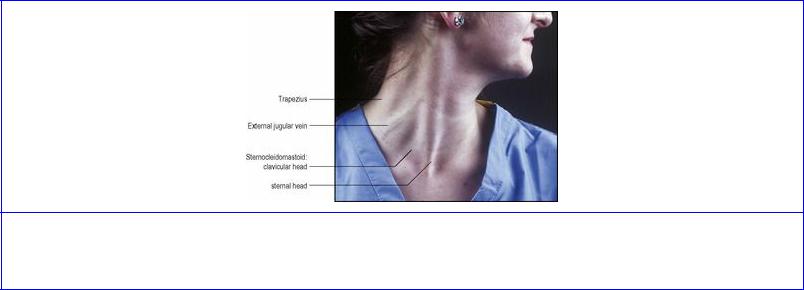
Sternocleidomastoid
This prominent neck landmark has two heads of origin below: that from the sternal manubrium is a rounded tendon, that from the clavicle a flat, fleshy mass (Fig. 6.2). A triangular interval exists between the two above the sternoclavicular joint, and the lower end of the internal jugular vein lies here, where it can be entered by needle or catheter. The manubrial tendon is attached to the front of the bone below the jugular notch; the clavicular head arises from the superior surface of the medial third of the clavicle. The muscle is attached by a tendon to the lateral surface of the mastoid process and by a thin aponeurosis to the lateral half of the superior nuchal line of the occipital bone. The clavicular fibres spiral behind the sternal fibres with the deep aspect of which they blend. The clavicular fibres are directed mainly to the mastoid process, while the sternal fibres run more obliquely chiefly to the occipital bone. The spinal accessory nerve enters the muscle under cover of the lobule of the ear, about 3 cm below the tip of the mastoid process, accompanied by a branch to the muscle from the occipital artery. It runs through the deep part of the muscle to emerge from between a third and a half of the way down the posterior border of the muscle.
Sternocleidomastoid is enclosed within a sheath of the investing layer of deep cervical fascia, which splits to surround it (Fig. 6.1). The muscle is crossed superficially by the great auricular nerve, the external jugular vein and the transverse cervical nerve, in that order from above downwards. Deep to the upper half of the muscle lies the cervical plexus; deep to its lower part lies the carotid sheath and its contents, overlying scalenus anterior.
The blood supply of the muscle is from branches of the occipital and superior thyroid arteries.

Nerve supply. By the spinal part of the accessory nerve, from a branch which leaves the nerve proximal to its point of entry into the muscle. The pathway for innervation by the cerebral cortex of the anterior horn cells of the segments concerned (mostly C2 and 3) is disputed; projection to the muscle from either or both hemispheres has been described. Branches from the cervical plexus (C2, 3) carrying proprioceptive fibres enter the muscle directly or by joining the accessory nerve.
Action. Contraction of one muscle tilts the head towards the ipsilateral shoulder, and rotates the head and face to the opposite side. Both muscles acting together from below draw the head forwards. With the head fixed, the muscles can assist in raising the roof of the thorax in forced inspiration.
Test. The face is turned to the opposite side against resistance and the muscle palpated.
Posterior triangle
This is an area enclosed between the sternocleidomastoid and trapezius muscles. Its apex lies high up at the back of the skull on the superior nuchal line, where there is a small gap between the attachments of the two muscles. Its base is the middle third of the clavicle at the side of the root of the neck. Its roof is formed by the investing layer of deep cervical fascia. Its floor consists of the prevertebral fascia lying on, from above downwards, splenius, levator scapulae and scalenus medius. Depending on the size of the sternocleidomastoid and the degree of depression of the shoulder, scalenus anterior and the first digitation of serratus anterior may contribute to the floor, and at the apex of the triangle, splenius may be low enough to expose a little of semispinalis capitis.
Although the subclavian artery, the three trunks of the brachial plexus and branches of the cervical plexus are deep to the prevertebral fascia, they are listed as contents of the posterior triangle; in operations on the triangle all these structures are safe provided the prevertebral fascia is left intact. The pulsation of the subclavian artery can be felt by pressing downwards behind the clavicle at the posterior border of sternocleidomastoid.
The cutaneous branches of the cervical plexus pierce the investing fascia at the posterior border of sternocleidomastoid. The cervical branches to trapezius pass across the floor of the triangle deep to the prevertebral fascia.
Lying between the roof and floor are the lymph nodes of the posterior triangle. Two or three occipital nodes lie in the subcutaneous tissue at the apex and several supraclavicular nodes lie above the clavicle; the latter are really outlying members of the lower group of deep cervical nodes (see p. 410).
The accessory nerve emerges from sternocleidomastoid, about a third of the way or a little lower down its posterior border. It passes downwards and backwards, with a characteristic wavy course adherent to the inner surface of the fascia of the roof of the triangle, to disappear beneath the anterior border of trapezius, about a third of the way from its lower end and 3–5 cm above the clavicle. These points of reference to the borders of sternocleidomastoid and trapezius enable the surface marking of the accessory nerve in the posterior triangle, where it is particularly liable to injury in operations involving the removal of lymph nodes, one or two of which may lie in contact with the nerve. More proximally, the nerve lies in front of the transverse process of the atlas (palpable between the mastoid process and mandibular ramus) and it enters the substance of sternocleidomastoid between the upper
two quarters of the muscle.
The inferior belly of omohyoid crosses the lower medial part of the triangle and is kept in place by its sling of investing fascia. Deep to the omohyoid are the transverse cervical and suprascapular vessels, just above the clavicle. The external jugular vein pierces both split layers of the lower part of investing fascia to enter the posterior triangle on its way to the subclavian vein, which itself is too low to be a content of the triangle; the wall of the vein is adherent to the fascia as it passes through.
Cervical plexus
The cervical plexus (Fig. 6.3) is formed by loops between the anterior rami of the upper four cervical nerves, after each has received a grey ramus communicans from the superior cervical ganglion. It lies in series with the brachial plexus, on the scalenus medius, behind the prevertebral fascia. It is covered by the upper part of sternocleidomastoid, and does not lie in the posterior triangle. The upper three cervical nerves have meningeal branches for the posterior cranial fossa. C1 fibres ascend with the hypoglossal nerve, C1 and 2 fibres ascend with the vagus nerve and C2 and 3 fibres ascend through the foramen magnum.
Figure 6.3 Right cervical plexus.
Muscular branches. Muscular branches are given off segmentally to the prevertebral muscles (longus capitis, longus colli and the scalenes). Other muscular branches are:
•A branch from C1 to the hypoglossal nerve, by which the fibres are carried to the superior root of the ansa cervicalis and the nerves to thyrohyoid and geniohyoid.
•Branches from C2 and 3 to sternocleidomastoid, and from C3 and 4 to trapezius. These fibres are mainly proprioceptive, but occasionally the whole of trapezius is not paralysed when the accessory nerve is damaged, as some of the cervical fibres may be motor.
•The inferior root of the ansa cervicalis is formed by union of a branch each from C2 and C3. The nerve spirals around the lateral side of the internal jugular vein and descends to join the superior root (C1) at the ansa (see p. 344).
•The phrenic nerve is formed mainly from C4 with contributions from C3 and C5 and runs down vertically over the obliquity of the scalenus anterior muscle, passing from lateral to medial borders, beneath the prevertebral fascia, lateral to the ascending cervical branch of the inferior thyroid artery. It passes behind the subclavian vein into the mediastinum (see p. 195). It may be

joined below the vein by a branch (the accessory phrenic nerve) from the nerve to subclavius; this branch may descend in front of the subclavian vein. The phrenic nerve is one of the most important in the body, being the sole motor supply to its own half of the diaphragm (see p. 187), and it also has an extensive afferent distribution, not only to the diaphragm but to the pericardium, pleura and peritoneum (see pp. 197, 212 and 238).
Cutaneous branches. Cutaneous branches of the plexus (Fig. 6.4) supply the front and sides of the neck and contribute to the supply of the scalp, face and chest.
Figure 6.4 Right lower face and posterior triangle of neck. The fat in the lower part of the triangle overlies deeper structures. The accessory nerve has been depicted taking a lower than usual course through the posterior triangle. The transverse cervical and suprascapular arteries are usually deep to the inferior belly of omohyoid.
The lesser occipital nerve (C2) is a slender branch that hooks around the accessory nerve and runs up along the posterior border of sternocleidomastoid to supply the posterior part of the upper neck and adjacent scalp behind the auricle. It may contribute to the supply of the auricle.
The great auricular nerve (C2, 3, mostly 2) is a large trunk passing almost vertically upwards over sternocleidomastoid; it is distributed to an area of skin on the face over the angle of the mandible and the parotid gland and to the parotid fascia. It also supplies the skin of the auricle over the whole of its cranial surface and on the lower part of its lateral surface below the external acoustic meatus, and skin over the mastoid region. Branches passing deep to the parotid gland supply the deep layer of the parotid fascia.
The transverse cervical nerve (C2, 3) curves round the posterior border of sternocleidomastoid, perforates the investing fascia and divides into ascending and descending branches that innervate the skin of the front of the neck from chin to sternum. The ascending branch communicates with the cervical branch of the facial nerve.
The supraclavicular nerve (C3, 4, but mostly 4) emerges with the other three nerves at the posterior border of sternomastoid and soon divides into several branches. They are distributed in three main

groups (see Fig. 1.8, p. 12). The medial group supply the skin as far down as the sternal angle. The intermediate group proper pass anterior to the clavicle and supply skin as far down as the second rib. The lateral group cross the acromion to supply skin halfway down the deltoid muscle, and pass posteriorly to supply skin as far down as the spine of the scapula.
Dermatomes of the neck
In addition to the cutaneous branches of the cervical plexus described above, which supply the anterior and lateral skin of the neck, the greater occipital and third occipital nerves from posterior rami of C2 and C3 respectively provide sensory fibres for the back of the neck (Fig. 6.15). The first cervical nerve does not supply any skin. C2 supplies most of the superior part of the neck, extending into the occipital region of the scalp and forwards to the auricle and the face over the parotid gland. C3 supplies the cylindrical part of the neck, C4 extends over the clavicle to the sternal angle, across the top of the shoulder and down to the scapular spine at the back. There is much overlap across dermatome boundary lines.
Anterior triangle
Beneath the investing layer of deep cervical fascia, between the mandible and the manubrium of the sternum, are longitudinal muscles supplied by the anterior rami of the upper three cervical nerves. They lie above or below the hyoid bone and there are four muscles in each group. The suprahyoid muscles comprise digastric, stylohyoid, mylohyoid and geniohyoid; the mylohyoids of each side unite to form the floor of the mouth, with the digastrics and stylohyoids superficial (below) and geniohyoids deep (above) to them. The infrahyoid muscles are sternohyoid and omohyoid, lying side by side in the same plane, and more deeply a wider sheet of muscle attached to the thyroid cartilage, namely thyrohyoid and sternothyroid. The last four are called the ‘strap muscles’ from their flat shape.
Suprahyoid muscles
Digastric
This arises as the posterior belly, from the digastric groove on the medial surface of the base of the mastoid process (Fig. 6.35). The triangular fleshy belly tapers down to the intermediate tendon, which is held beneath a fibrous sling attached to the junction of the body and the greater horn of the hyoid bone. The tendon is lubricated by a synovial sheath within the fibrous sling. The bifurcated tendon of insertion of stylohyoid which embraces the tendon plays no part in holding it down. The anterior belly lies on the inferior surface of mylohyoid, and connects the intermediate tendon to the digastric fossa on the inner surface of the mandible near the midline.

Figure 6.35 External surface of the central and left part of the base of the skull.
Nerve supply. The posterior belly is supplied by the facial nerve, by a branch arising between the stylomastoid foramen and the parotid gland, and the anterior belly by the nerve to mylohyoid.
Action. To depress and retract the chin, and to assist the lateral pterygoid in opening the mouth.
Stylohyoid
This arises from the back of the styloid process, high up near the base of the skull, and slopes down along the upper border of digastric. Its lower end divides to embrace the digastric tendon and is inserted by two slips into the junction of the greater horn and body of the hyoid bone.
Nerve supply. By the facial nerve, by a branch from that to the posterior belly of digastric.
Action. To retract and elevate the hyoid bone when swallowing.
Mylohyoid
The muscles of each side unite to make a thin sheet forming the ‘diaphragm’ of the floor of the mouth (Fig. 6.6). Each arises from the whole length of the mylohyoid line of its own side on the inner aspect of the mandible from as far back as medial to the third molar tooth to below the mental spines (see Fig. 8.5B, p. 510). The two muscles slope downwards towards each other, and the posterior quarter of each is inserted into the anterior surface of the body of the hyoid bone. In front of this the anterior three-quarters of each interdigitate in a midline raphe which extends from the chin to the hyoid bone.
Nerve supply. By its own nerve, a branch of the inferior alveolar (from the mandibular division of the trigeminal nerve), which arises just before the parent nerve enters the mandibular foramen, pierces the sphenomandibular ligament and runs forward on the inferior surface of the mylohyoid, supplying it and the anterior belly of the digastric.
Action. It forms a mobile but stable floor of the mouth. The two muscles together form a gutter; contraction makes the gutter more shallow, thus elevating the tongue and the hyoid bone as when swallowing or protruding the tongue.
Geniohyoid

This slender muscle extends from the inferior mental spine (genial tubercle) of the mandible (see Fig. 8.5B, p. 510) to the upper border of the body of the hyoid bone (see Fig. 8.6, p. 512). The two muscles lie side by side between the mylohyoids and the base of the tongue (genioglossus), on the floor of the mouth.
Nerve supply. By a branch from the hypoglossal nerve, consisting of fibres from the C1 nerve and not from the hypoglossal nucleus.
Action. To protract and elevate the hyoid bone in swallowing, or if the hyoid is fixed, to depress the mandible.
Infrahyoid muscles
Sternohyoid
This flat strap of muscle is attached to the back of the upper part of the manubrium and the adjoining sternoclavicular joint and clavicle. Its upper attachment is to the lower border of the body of the hyoid bone. The two muscles lie edge to edge at the hyoid bone, but diverge from each other below.
Nerve supply. By a branch from the ansa cervicalis which enters the lower part of the muscle.
Omohyoid
This flat strap of muscle lies edge to edge with sternohyoid at its attachment to the lateral part of the inferior border of the hyoid bone (Fig. 6.5). As it descends it diverges somewhat from the sternohyoid and, passing deep to sternocleidomastoid, it comes to lie over the carotid sheath. Where it lies over the internal jugular vein, the muscle fibres are replaced by a flat tendon, a useful guide at operation to the underlying vein. A change of direction now occurs, and the inferior belly runs almost horizontally just above the level of the clavicle to pass back to its attachment to the upper border of the scapula and the transverse scapular ligament. The intermediate tendon and supraclavicular portion of the muscle are bound down close to the clavicle in a fascial sling derived from the deep layer of the investing layer of deep cervical fascia (see Fig. 2.2, p. 39), which results in the angulated course of the muscle.

Figure 6.5 Thyroid gland and the front of the neck.
Nerve supply. The superior root of the ansa cervicalis supplies the superior belly and the ansa supplies the inferior belly.
Thyrohyoid
This is a broader and shorter muscle that lies under cover of the upper ends of sternohyoid and omohyoid. It arises from the greater horn of the hyoid bone, and is inserted into the oblique line of the thyroid cartilage alongside sternothyroid.
Nerve supply. By a branch of the hypoglossal nerve, but the fibres are all ‘hitch-hiking’ from C1.
Sternothyroid
Broader than sternohyoid and lying deep to it, this muscle is attached lower down than sternohyoid to the posterior surface of the manubrium and the adjacent first costal cartilage. Its upper attachment is to the oblique line of the thyroid cartilage.
Nerve supply. By the ansa cervicalis, which gives a branch to the lower part of the muscle.
Actions of the infrahyoid muscles
They are all depressors of the larynx. Sternothyroid acts directly on the thyroid cartilage, the others act indirectly via the hyoid bone. Depression of the larynx increases the volume of the resonating chambers during phonation and thus affects the quality of the voice. The infrahyoid muscles also oppose the elevators of the larynx (mylohyoid, palatopharyngeus, stylopharyngeus, salpingopharyngeus), enabling them to act progressively and gradually. The infrahyoid muscles prevent ascent of the hyoid bone when the digastric and geniohyoid lower the mandible.
Submandibular gland
The submandibular gland, mixed mucous and serous in type, consists of a large superficial part and a

small deep part which are continuous with one another round the free posterior margin of mylohyoid (Fig 6.24).
The superficial part (Fig. 6.6) has three surfaces: lateral, inferior and medial. The lateral surface lies against the submandibular fossa of the mandible (see Fig. 8.5B, p. 510), overlapping the front of the medial pterygoid insertion and being deeply grooved posteriorly by the facial artery which hooks under the mandible to reach the face at the front of the masseter muscle. The inferior or superficial surface is covered by skin, platysma and the investing fascia and is crossed by the facial vein and the cervical branch of the facial nerve, and sometimes by the marginal mandibular branch of the facial nerve (see p. 353), the nerves lying outside the investing fascia. Submandibular lymph nodes lie in contact with the surface of the gland and within its substance, hence the need to remove the gland as well as nodes in the operation of radical neck dissection. The medial surface lies against the mylohyoid, and behind it on the hyoglossus, lingual nerve, hypoglossal nerve and its accompanying veins. The facial artery is at first deep to the gland, and then grooves the posterosuperior part as it hooks over the top of the gland on to its lateral surface.
Figure 6.6 Left submandibular region.
T he deep part of the gland extends forwards for a variable distance, between mylohyoid and hyoglossus, below the lingual nerve and above the hypoglossal nerve.
The submandibular duct (of Wharton) is 5 cm long (the same length as the parotid duct) and emerges from the medial surface of the superficial part of the gland near the posterior border of mylohyoid. It runs with the deep part, forwards and slightly upwards, first between mylohyoid and hyoglossus, and then between the sublingual gland and genioglossus, to open into the floor of the mouth on the sublingual papilla beside the frenulum of the tongue. As it lies on hyoglossus, the duct is crossed laterally by the lingual nerve which then turns under the duct to pass medially to the tongue (Fig. 6.7).

Figure 6.7 Coronal section of the left side of the mandible and adjacent structures, just behind the first molar tooth, viewed from behind.
Blood supply
From the facial artery, with veins draining into the facial vein.
Lymph drainage
To the submandibular lymph nodes.
Nerve supply
Secretomotor fibres to the gland have their cell bodies in the submandibular ganglion (see p. 22), which hangs suspended from the lingual nerve on the surface of hyoglossus. The preganglionic fibres pass from cell bodies in the superior salivary nucleus in the pons by way of the nervus intermedius, facial nerve, chorda tympani and the lingual nerve (see pp. 365–366). Postganglionic fibres pass to the submandibular gland and also to the lingual nerve for transmission to the sublingual gland. Sympathetic (vasoconstrictor) fibres come from the plexus around the facial artery.
Development
An ectodermal groove in the floor of the mouth becomes converted into a tunnel whose blind end proliferates to form the secreting acini.
Surgical approach
The gland is exposed by a skin crease incision about 4 cm below the mandible, which continues through platysma and investing fascia on to the gland, to avoid the marginal mandibular branch of the facial nerve which may lie over the gland (see p. 353). Removal of the gland requires ligation of the facial vein which lies on the gland surface. The facial artery needs to be separated from its groove on the posterosuperior part of the gland, or a segment of the artery removed with the gland. The hypoglossal nerve and lingual nerve need to be safeguarded, the latter particularly when the duct is ligated and divided. The removal of a stone from the duct is carried out from within the mouth by incising the mucous membrane and duct over the stone; the mucosal and duct incisions are not sutured.
Thyroid gland
The thyroid gland is situated low down at the front of the neck. It consists of two symmetrical lobes
united by an isthmus that lies in front of the second, third and fourth tracheal rings (Fig. 6.5). The lobes lie on either side of the larynx and trachea, extending from the oblique line of the thyroid cartilage to the sixth tracheal ring. It weighs about 25 g. In addition to its own capsule, the gland is enclosed by an envelope of pretracheal fascia.
Each lateral lobe is pear-shaped with a narrow upper pole and a broader lower pole, and appears approximately triangular on cross-section with lateral, medial and posterior surfaces. The lateral (superficial) surface is under cover of sternothyroid and sternohyoid. The medial surface lies against the lateral side of the larynx and upper trachea, with the lower pharynx and upper oesophagus immediately behind. This surface is related to the cricothyroid muscle of the larynx and the inferior constrictor of the pharynx, as well as to the external and recurrent laryngeal nerves. The posterior surface overlaps the medial part of the carotid sheath, i.e. the part containing the common carotid artery; if enlarged, the lobe may extend across the more laterally placed internal jugular vein. The parathyroid glands usually lie in contact with this surface, between it and the fascial sheath.
The relationship of the recurrent laryngeal nerves (see p. 368) to the thyroid lobes has importance in thyroid surgery. As they approach the medial surface of the gland from below, the nerves lie in or in front of the groove between the trachea and oesophagus. The left nerve, which recurves around the arch of the aorta in the superior mediastinum, is more likely to have entered the groove and lies posterior (though occasionally anterior) to the inferior thyroid artery. The right nerve recurves around the right subclavian artery at the root of the neck and may be more lateral to the trachea, passing anterior or posterior to the inferior thyroid artery or in between its branches. Each nerve is behind the pretracheal fascia, and runs medial or lateral or through a thickening of the fascia attached to the cricoid cartilage and upper tracheal rings (the suspensory ligament of Berry). The nerve runs behind the cricothyroid joint and passes upwards under cover of the inferior constrictor. At the level of the upper border of the isthmus the nerve often divides into two. If so, the anterior (larger) branch is the motor branch to laryngeal muscles, and the posterior branch is sensory only. The rare non-recurrent right laryngeal nerve (see p. 27) may be a hazard during thyroid surgery.
The smaller external laryngeal nerve lies on the inferior constrictor, close behind the superior thyroid artery, as it runs down medial to the upper pole to supply cricothyroid.
The isthmus joins the anterior parts of the lobes, towards their lower poles. The posterior surface of the isthmus is firmly adherent to the second, third and fourth rings of the trachea, and the pretracheal fascia is here fixed between them. This fixation and the investment of the whole gland by pretracheal fascia are responsible for the gland moving up and down with the larynx during swallowing. An anastomosis between the two superior thyroid arteries runs across the upper border of the isthmus, and tributaries of the inferior thyroid veins emerge from its lower border.
A small portion of gland substance often projects upwards from the isthmus, generally to the left of the midline, as the pyramidal lobe and represents a devel-opment of glandular tissue from the caudal end of the thyroglossal duct (see p. 26). It may be attached to the inferior border of the hyoid bone by fibrous tissue; muscle fibres sometimes present in it are named levator glandulae thyroideae and are innervated by a branch of the external laryngeal nerve. Separate small masses of thyroid tissue (accessory thyroid glands) are not uncommonly found near the hyoid bone, in the tongue, in the superior mediastinum, or anywhere along the path of descent of the thyroglossal duct, though their
presence may only be revealed by histological study.
Blood supply
The superior thyroid artery, the first branch from the anterior aspect of the external carotid (see p. 342), after giving off its sternocleidomastoid and superior laryngeal branches, pierces the pretracheal fascia as a single vessel to reach the summit of the upper pole. The external laryngeal nerve is immediately behind the artery as the vessel approaches the pole, but they part company as the artery reaches the gland and the nerve descends to supply the cricothyroid muscle. The artery divides on the gland into an anterior branch that runs down to the isthmus and a posterior branch that runs down the back of the lobe and anastomoses with an ascending branch of the inferior thyroid artery from the lower pole. In thyroidectomies the artery is ligated close to the upper pole, or its anterior and posterior branches are ligated instead, to avoid damage to the external laryngeal nerve.
The inferior thyroid artery, from the thyrocervical trunk (see p. 349), arches upwards and medially behind the carotid sheath and then loops downwards to the lower pole. It divides outside the pretracheal fascia into branches that pierce the fascia separately to reach the lower part of the gland. As described above, the recurrent laryngeal nerve has a variable relationship to the artery but always lies behind the pretracheal fascia. Ligating the inferior thyroid artery well lateral to the gland, or carefully ligating its small branches on the surface of the gland, helps to safeguard the nerve. The inferior thyroid artery gives off the ascending cervical artery and small pharyngeal, oesophageal, laryngeal and tracheal branches before its terminal distribution to the thyroid gland; the small inferior laryngeal artery ascends with the recurrent nerve.
A thyroidea ima artery enters the lower part of the isthmus in 3% of individuals. It arises from the brachiocephalic trunk, arch of the aorta or right common carotid artery.
From a venous plexus on the surface of the gland the superior thyroid vein follows the superior thyroid artery and enters either the internal jugular or facial vein in about equal proportions. The middle thyroid vein crosses anterior to the common carotid artery to drain into the internal jugular vein; early ligature and division of this vein during thyroid surgery facilitates mobilisation of the gland. The inferior thyroid veins are multiple and drain downwards mainly into the left brachiocephalic vein; one may enter the right brachiocephalic vein.
Lymph drainage
The lymphatics from the thyroid gland drain mainly to deep cervical nodes. A few pass into prelaryngeal, preand paratracheal nodes, and a few drain directly into the thoracic duct.
Nerve supply
Sympathetic (vasoconstrictor) nerves from the superior, middle and inferior cervical ganglia accompany the thyroid arteries.
Structure
The thyroid consists essentially of a mass of more or less rounded follicles containing varying amounts of colloid produced by the single layer of epithelial (follicular) cells that form the walls of the follicles. The thyroid is unique in being the only endocrine gland to store its secretion outside the
cells. The colloid is iodinated when in the follicle and reabsorbed |
by the cells before being |
discharged into blood capillaries. The main hormonal products |
are thyroxine (T4) and |
triiodothyronine (T3). Less than 2% of the epithelial cells are the C or parafollicular cells which secrete calcitonin. The C cells are scattered on the outer aspects of the follicles and do not reach the lumina of the follicles. The thyroid gland is highly vascular.
Development
The gland develops as a proliferation of cells from the caudal end of the thyroglossal duct (see p. 26). The parafollicular calcitonin-producing cells develop from the ultimobranchial body (fifth pharyngeal pouch), under the influence of neural crest cells.
Surgical approach
The thyroid gland is approached surgically through a transverse incision in a low skin crease on the front of the neck. The investing fascia is divided vertically and the sternohyoid and the sternothyroid muscles retraced, or divided at a high level, to safeguard their nerve supply and to prevent adherence of their subsequent suture line to the closure of the skin incision. The pretracheal fascia needs to be divided to expose the gland proper.
Parathyroid glands
The small parathyroid glands normally lie behind the lobes of the thyroid gland. There are usually four glands (in 90% of subjects), two on each side. Each weighs about 50 mg.
The superior gland is the more constant in position; it is usually within the thyroid's pretracheal fascial capsule, at the middle of the back of the thyroid lobe, level with the first tracheal ring and above the inferior thyroid artery. The inferior gland is less constant in position. It is usually within the pretracheal fascial sheath behind the lower pole; but it may be in the gland itself, or outside the fascial sheath in a variable position in the neck, or in the superior or posterior mediastinum. The glands are not necessarily on the same level on each side. They are brownish-yellow, which helps to distinguish them from the deep red of the thyroid gland. They are easily subject to subcapsular haematoma formation on handling.
Blood supply
Both upper and lower parathyroids are usually supplied by the inferior thyroid artery, otherwise by an anastomosis between the superior and inferior arteries. Their minute veins join thyroid veins.
Lymph drainage
As for lymph drainage of the thyroid gland (above) and thymus see page 196.
Nerve supply
Sympathetic vasoconstrictor fibres enter with the arteries.
Structure
The gland is a mass of small closely packed chief or principal cells which secrete the parathyroid hormone (PTH). The mass of cells bears a superficial resemblance to lymphoid tissue, but the number

of blood capillaries in the gland provides a clear distinction. Scattered among the chief cells are small groups of slightly larger oxyphil cells.
Development
The superior gland is termed parathyroid IV because it develops from the fourth pharyngeal pouch. The inferior gland is parathyroid III, developed from the third pouch, but displaced caudally by the descent of the thymus from the same pouch (see p. 26); hence its liability to end up in unusual positions.
Surgical approach
For parathyroidectomy the lobes of the thyroid gland are exposed as for thyroidectomy, and then retracted forwards and medially so that the posterior surfaces can be inspected for the parathyroids. If not obvious, branches of the inferior thyroid artery are followed and should lead to the glands. Exposure of the thymus through a median sternotomy may be necessary.
Trachea
The trachea (Fig. 6.5) begins at the level of C6 vertebra in continuity with the larynx, being attached to the lower margin of the cricoid cartilage by the cricotracheal ligament. Of the total length of 10 cm, 5 cm are in the neck from the cricoid cartilage to the jugular notch. From the neck the trachea passes into the thorax.
The cervical part lies in the midline of the neck, in contact with the front of the oesophagus. In the groove between trachea and oesophagus runs the recurrent laryngeal nerve. To the side of the trachea is the carotid sheath. The isthmus of the thyroid gland is adherent to the second, third and fourth tracheal rings and the lobes of the gland lie against the lateral sides of the trachea as far down as the sixth ring. The inferior thyroid veins and anterior jugular venous arch lie in front, and also (if present) the thyroidea ima artery and the upper end of a persistently large thymus. On account of the shape of the lower cervical and upper thoracic parts of the vertebral column, the trachea passes downwards and backwards as it enters the thorax. Although close to the skin at the upper end, it is 2 cm or more deep to the front of the jugular notch.
The thoracic part of the trachea and other features are described on page 194.
Tracheotomy and tracheostomy
Tracheotomy implies making an incision in the trachea; tracheostomy involves removing a small part of the wall (making a stoma), but the strict distinction between these terms is often ignored. In an emergency, laryngotomy (incising the cricothyroid ligament, see p. 393) provides a subglottic airway; the procedure also gives access to the trachea for sucking out excess secretions. Depending on the speed with which tracheostomy needs to be performed, the skin incision is vertical from the lower border of the thyroid cartilage to the jugular notch, or (preferably) transverse 2 cm below the cricoid cartilage. The sternohyoid and sternothyroid muscles are retracted laterally. The isthmus of the thyroid gland is usually divided and an opening made by removing part of the second and third tracheal rings so that a tracheostomy tube can be inserted. In the narrow trachea of children only a vertical incision is made in the trachea (tracheotomy), as removal of any segment of tracheal wall tends to lead to tracheal stenosis.

Oesophagus
The oesophagus commences in continuity with the pharynx at the level of the lower border of the cricoid cartilage (C6 vertebra). It lies in front of the prevertebral fascia behind the trachea. It is overlapped on either side by the lower poles of the thyroid gland, beyond which, on the left side, its edge is visible posterolateral to the trachea. The thoracic duct runs upwards behind the lower part of its left border. The recurrent laryngeal nerves are on each side in the groove between trachea and oesophagus.
The oesophagus continues into the thorax and is further described on page 208.
Great vessels of the neck
Common carotid artery
The common carotid artery arises on the left side from the arch of the aorta, where it lies in front of the subclavian artery up to the sternoclavicular joint. Here the two arteries diverge. On the right the brachiocephalic trunk bifurcates behind the sternoclavicular joint into common carotid and subclavian arteries. The common carotid gives off no branches proximal to its bifurcation. It lies within the medial part of the carotid sheath, with the internal jugular vein lateral to it and the vagus nerve deeply placed between the two vessels. The sympathetic trunk is behind the artery and outside the sheath, which is overlapped superficially by the infrahyoid muscles and sternocleidomastoid. Medial to the sheath is the trachea and oesophagus and, at a higher level, the larynx and pharynx. The thyroid gland overlaps the sheath anteromedially and the inferior thyroid artery crosses from the thyrocervical trunk to the gland behind the sheath.
The common carotid artery usually bifurcates at the level of the upper border of the lamina of the thyroid cartilage (upper border of C4 vertebra) into the external and internal carotids; it may do so higher near the tip of the greater horn of the hyoid bone (C3 vertebra). The terminal portion of the artery often shows a slight dilatation, the carotid sinus, which includes the commencement of the internal carotid artery.
The carotid pulse can be felt by pressing backwards between the trachea and lower larynx medially and sternocleidomastoid laterally, pressing the artery against the anterior tubercle of the transverse process of C6 vertebra (carotid tubercle of Chassaignac). The surface marking of the common carotid artery is along a vertical line from the sternoclavicular joint to the level of the upper border of the thyroid cartilage. The vessel can be surgically exposed by retracting the lower part of sternocleidomastoid backwards and incising the carotid sheath. The middle thyroid vein is divided between ligatures.
External carotid artery
The external carotid artery at its commencement lies against the side wall of the pharynx somewhat anteromedial to the internal carotid artery. It then ascends in front of the internal carotid deep to the posterior belly of digastric and stylohyoid, above which it pierces the deep lamina of the parotid fascia and enters the gland. It divides within the gland behind the neck of the mandible into the maxillary and superficial temporal arteries. As the external carotid artery lies in the parotid gland it is separated from the internal carotid by the deep part of the gland and its fascia, styloid process and
its continuation the stylohyoid ligament, styloglossus and the ‘pharyngeal’ structures: stylopharyngeus muscle, glossopharyngeal nerve and pharyngeal branch of the vagus (and, if present, the track of a branchial fistula, see p. 26). At the commencement of the artery the internal jugular vein lies lateral, but higher up it is posterior and deep to the artery. The facial vein crosses the artery, with the hypoglossal nerve lying between. Except at its commencement the vessel lies in front of the anterior border of sternocleidomastoid.
The surface marking of the external carotid is along a line from the bifurcation of the common carotid passing up behind the angle of the mandible to a point immediately in front of the tragus of the ear.
Surgical approach. The vessel can be exposed in front of the upper part of sternocleidomastoid before it enters the parotid gland by ligating the facial vein. The hypoglossal nerve which crosses the external and internal carotids superficially must not be damaged.
Branches. Before it divides into its two terminal branches the external carotid artery gives off six branches, three from in front, two from behind and one from its deep (medial) aspect. The three from in front are the superior thyroid, lingual and facial, and they diverge widely. The two from behind are the occipital and posterior auricular, which pass up deep to and above the posterior belly of digastric respectively. The branch from the medial side is the ascending pharyngeal, which ascends to the base of the skull on the side wall of the pharynx, alongside the internal carotid artery.
The superior thyroid artery arises at the commencement of the external carotid. It runs almost vertically downwards, with the vein, to the upper pole of the thyroid gland (see p. 339), with the external laryngeal nerve close behind it, alongside the larynx (Fig. 6.24). Before reaching the thyroid gland it gives off infrahyoid, sternocleidomastoid, superior laryngeal and cricothyroid branches. The superior laryngeal artery pierces the thyrohyoid membrane with the internal laryngeal nerve. The cricothyroid artery crosses the upper part of the crico-thyroid membrane to anastomose with the contralateral artery.
The lingual artery arises from the front of the external carotid above the superior thyroid, near the tip of the greater horn of the hyoid bone. It forms a short loop (Fig. 6.22), then passes forwards along the upper border of the greater horn, deep to hyoglossus (p. 381). It is accompanied by the lingual vein. The loop of the artery is crossed laterally by the hypoglossal nerve and its companion vein, the latter opening into the facial vein.
The facial artery arises from the front of the external carotid above the lingual artery (sometimes by a common linguofacial trunk with the lingual) and runs upwards on the superior constrictor, deep to the digastric and stylohyoid muscles, then deep to the submandibular salivary gland. It grooves the posterosuperior part of the gland. As the artery lies on the superior constrictor muscle it gives off a tonsillar and an ascending palatine branch to the tonsil and soft palate. The facial artery then makes an S bend, curling over the submandibular gland (Fig. 6.7) and crossing the inferior border of the mandible, where its pulsation can be felt, at the anterior border of masseter. Before passing to the face it gives off the submental artery, which accompanies the mylohyoid nerve into the submandibular fossa and sends perforating branches through the mylohyoid to anastomose with a sublingual branch of the lingual artery.
The occipital artery arises from the back of the external carotid on a level with the facial artery. It
courses backwards deep to the lower border of the posterior belly of digastric. It grooves the base of the skull at the occipitomastoid suture, deep to the digastric notch on the mastoid process (Fig. 6.35), and passes through the apex of the posterior triangle to supply the back of the scalp (see p. 356). The artery gives off two branches to sternocleidomastoid. The upper branch is a guide to the accessory nerve in front of the upper border of the muscle. At its origin the occipital artery crosses lateral to the hypoglossal nerve, which hooks around it from behind (Fig. 6.24), the nerve being held down here by the lower sternocleidomastoid branch of the artery (Fig. 6.23).
The posterior auricular artery arises above the level of the digastric muscle, often within the substance of the parotid gland. It runs up superficial to the styloid process above the digastric posterior belly and crosses the surface of the mastoid process to supply the scalp. Auricular branches supply the pinna of the ear. Its stylomastoid branch enters the stylomastoid foramen and supplies the facial nerve; this branch may arise instead from the occipital artery.
The ascending pharyngeal artery arises just above the commencement of the external carotid, from its deep aspect. It runs up along the side wall of the pharynx in front of the prevertebral fascia, deep to the internal carotid artery. It supplies the pharyngeal wall and the soft palate and sends meningeal branches through the nearby foramina in the base of the skull (foramen lacerum, jugular foramen, hypoglossal canal).
Internal carotid artery
The internal carotid artery arises at the bifurcation of the common carotid (see p. 342) and continues upwards within the carotid sheath (see p. 366). At its commencement it shows a slight bulge, the carotid sinus. Here the arterial wall is thin and its contained baroreceptors are supplied by the glossopharyngeal and vagus nerves, which mediate blood pressure impulses to medullary centres. The carotid body is a small structure lying behind the bifurcation of the common carotid artery, or between its branches, from which it receives two or three very small glomic arteries. Its cells are chemoreceptors concerned (like the aortic bodies, see p. 192) with respira-tory reflexes, and are innervated by the glossopharyngeal and vagus nerves. Carotid body tumours form a swelling at the anterior border of sternocleidomastoid at the level of the carotid bifurcation, and exhibit transmitted pulsation from the arteries.
The internal carotid artery is lateral to the external carotid at its origin, but soon passes up posteriorly to a medial and deeper level. It has no branches and passes straight up in the carotid sheath, beside the pharynx, to the carotid canal in the base of the skull; its intracranial course is considered on pages 445 and 449.
Behind the internal carotid artery in the neck is the sympathetic trunk (outside the carotid sheath), pharyngeal veins and the superior laryngeal branch of the vagus. The ascending pharyngeal artery is medial to it. The internal jugular vein is lateral, with the vagus nerve deeply placed between artery and vein. Superficially near its origin it is crossed by the lingual and facial veins, the occipital artery and hypoglossal nerve. The superior root of the ansa cervicalis runs downwards along it, embedded in the carotid sheath. At a higher level it is crossed by the posterior belly of digastric and stylohyoid and the posterior auricular artery, and by the structures that separate it from the external carotid (see above).
The surface marking of the internal carotid artery in the neck is along a line from the bifurcation of the common carotid artery to the head of the mandible.
Surgical approach. The internal carotid is exposed in the neck by an incision along the anterior border of sternocleidomastoid. The muscle is retracted backwards, the facial and lingual veins divided between ligatures, and the carotid sheath is incised. The hypoglossal nerve must be safeguarded and this may require division of the lower sternocleidomastoid branch of the occipital artery. The emergence of branches from the external carotid artery ensures its differentiation from the internal carotid.
Internal jugular vein
The internal jugular vein emerges from the jugular bulb at the posterior compartment of the jugular foramen (Fig. 6.35). At first behind the internal carotid artery, it lies on the transverse process of the atlas, crossed by the accessory nerve. It receives the inferior petrosal sinus as its first tributary, just below the base of the skull; the sinus passing back lateral or medial to the glossopharyngeal, vagus and accessory nerves. The vein passes down to gain the lateral side of first the internal carotid artery and then the common carotid artery, within the loose lateral part of the carotid sheath, with the vagus nerve deeply placed between the vein and the arteries. In the lower part of its course the vein is overlaid by the sloping sternocleidomastoid. Deep cervical lymph nodes within the sheath are closely adjacent to the vein throughout its course. Its posterior relations include the cervical plexus lying on levator scapulae and scalenus medius, and the phrenic nerve on scalenus anterior. The thoracic duct crosses behind the left vein at the level of C7 vertebra. The inferior root of the ansa cervicalis curls round its lateral border, to unite with the superior root (from the hypoglossal nerve) at a variable level in front of the vein. Low down the tendon of omohyoid crosses the vein, providing a useful guide to its position. The terminal part of the vein lies deep to the triangular interval between the sternal and clavicular heads of sternocleidomastoid (Fig. 6.2). It joins the subclavian to form the brachiocephalic vein behind the sternal end of the clavicle.
The tributaries of the internal jugular vein below the inferior petrosal sinus are the pharyngeal, lingual, facial and superior and middle thyroid veins. The lingual and superior thyroid veins may join the facial vein and other variations are possible.
At its commencement and termination the vein is slightly dilated to form superior and inferior bulbs. There is a pair of valves above the inferior bulb.
The surface marking of the internal jugular vein is along a line from the lobule of the ear to the sternal end of the clavicle, between the two heads of sternocleidomastoid. The jugular venous pulse is a guide to jugular venous pressure, which is the same as right atrial or central venous pressure and thus an indicator of cardiovascular function. With a patient reclining at 45° the jugular venous pulse should be visible just above the clavicle.
Catheterization. The right internal jugular vein (on a direct path to the right atrium) can be cannulated for the insertion of a central venous line, for measurements of central venous pressure, or the rapid administration of drugs when a peripheral approach would be too slow. The vein is usually approached through the centre of the triangle formed by the two heads of sternocleidomastoid and the clavicle. The needle is directed caudally, parallel to the sagittal plane at a 30° posterior angle with

the coronal plane, entering the vein at about 4–5 cm depth. (The subclavian vein is an alternative, see p. 348). The most common complication is haematoma formation; others include common carotid artery puncture, vagus nerve injury and pneumothorax due to pleural perforation.
The ansa cervicalis lies on the front of the internal jugular vein and gives branches to the infrahyoid muscles. It is usually embedded within the anterior wall of the carotid sheath and classified as one of its contents. It is formed by union of superior and inferior roots. The superior root is a branch of the hypoglossal nerve containing only C1 fibres, which have hitch-hiked along the hypoglossal nerve. It runs down on the front of the internal and common carotid arteries, giving a branch to the superior belly of omohyoid.
The inferior root is formed by union of a branch each from C2 and C3 anterior rami in the cervical plexus. The single nerve so formed spirals from behind around the internal jugular vein and runs down to join the superior root in a wide loop over the lower part of the vein, from which branches arise for the infrahyoid muscles (sternohyoid, sternothyroid and inferior belly of omohyoid). Sometimes the inferior root passes forwards between internal jugular vein and internal carotid artery.
The uppermost part of the hypoglossal nerve in the neck is described on page 368. It emerges between the internal carotid artery and internal jugular vein deep to the posterior belly of digastric. Hooking round the occipital artery, which runs backwards lateral to the nerve, it curves forwards lateral to the internal and external carotids and the loop of the lingual artery (Fig. 6.23). As it crosses these arteries it lies just below the posterior belly of the digastric, behind its tendon and just above the tip of the greater horn of the hyoid bone. It gives off the superior root of the ansa cervicalis as it crosses the internal carotid and the branch to thyrohyoid (C1 fibres) as it crosses the lingual artery. It passes forwards on the lateral surface of hyoglossus deep to mylohyoid to enter the mouth, giving off the branch to geniohyoid (C1 fibres) as it does so (Fig. 6.24).
Summary of triangles of the neck
Posterior triangle
Boundaries. Sternocleidomastoid, trapezius, clavicle.
Contents. Occipital, transverse cervical, suprascapular and subclavian arteries; transverse cervical, suprascapular and external jugular veins; accessory nerve; cervical plexus branches and brachial plexus trunks (deep to prevertebral fascia); inferior belly of omohyoid; lymph nodes.
Anterior triangle
Boundaries. Sternocleidomastoid, mandible, midline. Subdivided into:
Carotid triangle
Boundaries. Sternocleidomastoid, posterior belly of digastric, superior belly of omohyoid.
Contents. Bifurcation of common carotid artery and branches of external carotid (except posterior auricular); lingual, facial and superior thyroid veins; hypoglossal, internal and external laryngeal nerves, and superior root of ansa cervicalis; lymph nodes.
Digastric triangle
Boundaries. Mandible, anterior and posterior bellies of digastric.
Contents. Submandibular gland and lymph nodes; facial, submental and mylohyoid vessels; hypoglossal and mylohyoid nerves.
Submental triangle
Boundaries. Anterior bellies of digastric, body of hyoid bone. (Triangle crosses midline.)
Contents. Anterior jugular veins; lymph nodes.
Muscular triangle
Boundaries. Sternocleidomastoid, superior belly of omohyoid, midline from hyoid bone to jugular notch.
Contents. Parts of larynx, trachea, pharynx, oesophagus, thyroid and parathyroid glands; their vessels and nerves; lymph nodes.