


Part eight. Posterior compartment of the forearm
A dozen muscles occupy the extensor compartment. At the upper part are anconeus (superficial) and supinator (deep). From the lateral part of the humerus arise three muscles that pass along the radial side (brachioradialis, and extensors carpi radialis longus and brevis), and three that pass along the posterior surface of the forearm (extensors digitorum, digiti minimi and carpi ulnaris). At the lower end of the forearm these two groups are separated by three muscles that emerge from deeply in between them and go to the thumb (abductor pollicis longus and extensors pollicis longus and brevis). Finally, one muscle for the forefinger runs deeply to reach the back of the hand (extensor indicis).
The nerve of the extensor compartment is the posterior interosseous nerve, which reaches it by passing around the radius (compare the peroneal nerve in the leg); the artery is the posterior interosseous, which gains the extensor compartment by passing between the two bones (compare the anterior tibial artery in the leg). The artery is small and the blood supply of the posterior compartment is reinforced by the anterior interosseous artery.
The six long muscles that come from the lateral side of the humerus have not enough area available at the lateral epicondyle. Two of them arise above this, from the lateral supracondylar ridge and the lateral intermuscular septum.
Brachioradialis
Arising from the upper two-thirds of the lateral supracondylar ridge, the muscle forms the lateral border of the cubital fossa (Fig. 2.27). At the mid-forearm level the muscle fibres end in a flat tendon, which is inserted into the base of the radial styloid (Fig. 2.30). The muscle and its tendon overlie the radial nerve and the radial artery as they pass down the forearm and the nerve deviates posteriorly. The lower part of the tendon is covered by abductor pollicis longus and extensor pollicis brevis as they spiral down to the thumb (Fig. 2.33).
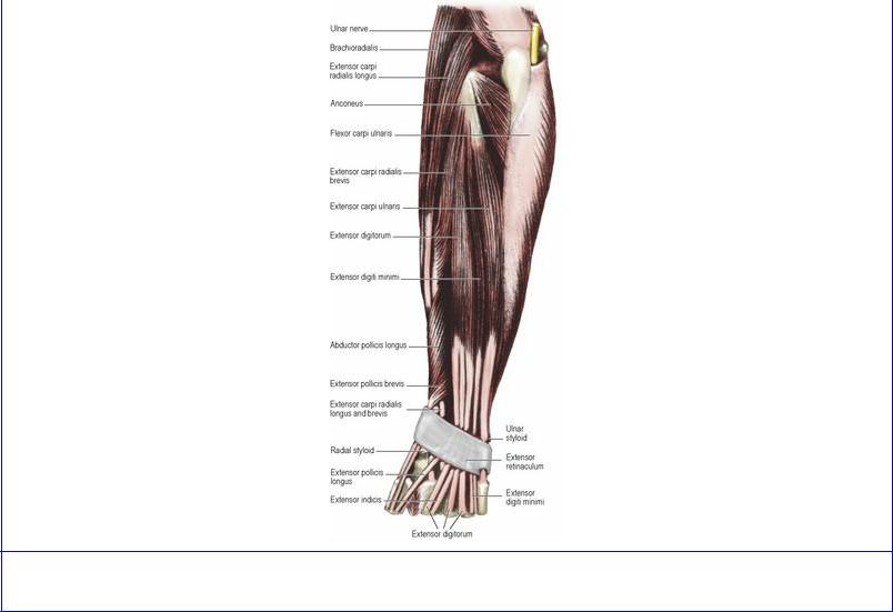
Figure 2.33 Superficial extensor muscles of the left forearm.
Nerve supply. By the radial nerve (C5, 6) by a branch arising above the elbow joint.
Action. Its action is to flex the elbow joint. It acts most powerfully when the forearm is semipronated.
Test. With the forearm in the midprone position the elbow is flexed against resistance; the muscle can be seen and felt.
Extensor carpi radialis longus
Arising from the lower third of the lateral supracondylar ridge of the humerus the muscle passes down the forearm, behind brachioradialis (Fig. 2.33) and then deep to the thumb muscles, to be inserted as a flattened tendon into the base of the second metacarpal.
Nerve supply. By the radial nerve (C6, 7) by a branch arising above the elbow.
Action. It is an extensor and abductor of the wrist. It is indispensable to the action of ‘making a fist’, acting as a synergist during finger flexion (see p. 90). It assists in flexion of the elbow. In paralysis of forearm flexor muscles it can be transferred into flexor digitorum profundus.
Test. With the forearm pronated the wrist is ex-tended and abducted against resistance and the muscle is palpated below and behind the lateral side of the elbow.
Common extensor origin
The common extensor origin is attached to the smooth area on the front of the lateral epicondyle (Fig. 2.24). From it arise the fused tendons of extensor carpi radialis brevis, extensor digitorum, extensor digiti minimi and extensor carpi ulnaris. All four muscles pass to the posterior surface of the forearm. When the forearm is extended and supinated they spiral around the upper part of the shaft of the radius; behind this rounded mass of muscle is an elongated pit in which lies the head of the radius. In the usual working position of the forearm (flexed and half pronated) these muscles pass straight from the front of the lateral epicondyle into the forearm. Repetitive use of the extensor muscles of the forearm, however, may strain or tear fibres of the common extensor origin from the lateral epicondyle causing pain and tenderness over the lateral epicondyle (tennis elbow).
Extensor carpi radialis brevis
This muscle arises from the common extensor origin on the front of the lateral epicondyle of the humerus, passes down behind and deep to its fellow longus (Fig. 2.33), and is inserted by a flattened tendon into the base of the third metacarpal. It and the longus are inserted into the same metacarpals as flexor carpi radialis. The lower part of both tendons are crossed by abductor pollicis longus and the two extensor muscles of the thumb (Fig. 2.34).
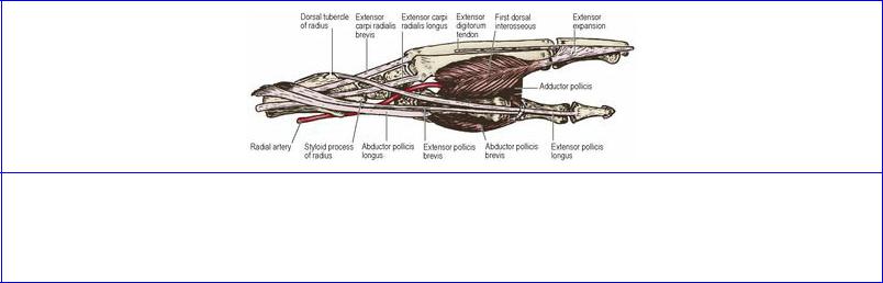
Figure 2.34 Deep extensor muscles of the left forearm. Compare with Figure 2.57.
Nerve supply. By a branch in the cubital fossa from the posterior interosseous nerve (C7, 8), before
the nerve pierces the supinator muscle.
Action. As a wrist extensor like its longus companion it contracts in making a fist.
Extensor digitorum
Arising from the common extensor origin the muscle expands into a rounded belly in the middle of the forearm, diverging from the three muscles on the radial side and separated from them by the emergence of thumb muscles (Fig. 2.33). Its four tendons pass under the extensor retinaculum crowded together, overlying the tendon of extensor indicis. On the back of the hand the tendons spread out towards the fingers. Commonly the fourth tendon is fused with that to the ring finger, and reaches the little finger only by a tendinous band that passes across near the metacarpophalangeal joint. Other bands join adjacent tendons in a variable manner. The extensor expansions and their insertions into the phalanges are considered with the hand (see p. 90).
Nerve supply. By the posterior interosseous nerve on the back of the forearm (C7, 8).
Action. It is an extensor of the wrist, metacarpo-phalangeal and interphalangeal joints. Its action on the fingers is discussed in detail on page 90.
Test. With the forearm in pronation and the fingers extended, the patient tries to keep the fingers extended at the metacarpophalangeal joints while pressure from the examiner on the proximal phalanges tries to flex these joints.
Extensor digiti minimi
Arising in common with the extensor digitorum the belly of the muscle separates after some distance (Fig. 2.33) and then becomes tendinous. Passing beneath the extensor retinaculum on the dorsal aspect of the radioulnar joint the tendon usually splits into two, which lie side by side on the fifth metacarpal bone as they pass to the little finger (Fig. 2.37). The tendon of extensor digitorum to the little finger commonly joins them as a band near the metacarpophalangeal joint and they all form an expansion on the dorsum of the little finger, which behaves as the other extensor expansions (see p. 90).
Nerve supply. By the posterior interosseous nerve (C7, 8).
Action It assists extensor digitorum in extension of the little finger and wrist joint.
Extensor carpi ulnaris
This muscle arises from the common extensor origin and by an aponeurotic sheet from the subcutaneous border of the ulna (Fig. 2.33). This aponeurosis arises in common with that of flexor carpi ulnaris, the two passing in opposite directions into the extensor and flexor compartments. The tendon of the muscle lies in the groove beside the ulnar styloid as it passes deep to the extensor retinaculum to be inserted into the base of the fifth metacarpal.
Nerve supply. By the posterior interosseous nerve (C7, 8) at the back of the forearm.
Action. It is an extensor and adductor of the wrist. It acts as a synergist during finger flexion and is indispensable in ‘making a fist’ (see p. 90).
Test. With the forearm pronated and the fingers extended, the wrist is extended and adducted against resistance. The muscle can be seen and felt in the upper forearm and the tendon palpated proximal to the head of the ulna.
Anconeus
This small muscle arises from the posterior surface of the lateral epicondyle. It fans out to its insertion on the lateral side of the olecranon and adjacent shaft of the ulna (Fig. 2.34).
Figure 2.35 Left anatomical snuffbox. It lies between the extensor tendons of the thumb. In its bony floor are the radial styloid, scaphoid, trapezium and base of the first metacarpal. The floor is crossed by the radial artery.
Nerve supply By the radial nerve (C7, 8) by a branch that leaves the trunk in the radial groove and passes through triceps, supplying it as well.
Action. The muscle produces the small amount of posterolateral movement of the ulna that occurs during pronation (see p. 73).
Supinator
This muscle arises from the distal border of the lateral epicondyle, the lateral ligament of the elbow joint, the annular ligament of the radius, the supinator crest of the ulna and the fossa in front of it. The muscle fibres run behind the radius (Fig. 2.34) and are inserted on its lateral surface, between the anterior and posterior oblique lines. It has superficial and deep layers and the posterior interosseous nerve passes between these two parts as it leaves the cubital fossa to enter the back of the forearm. The deep fibres are wrapped around the proximal third of the radial shaft.
Nerve supply. By the posterior interosseous nerve in the cubital fossa before the nerve enters the muscle (C6, 7).
Action. While the biceps is the powerful supinator of the forearm, supinator fixes the forearm in supination. Only when the elbow is completely extended is the supinator the prime mover for the action of supination, which is much weaker in this position.
Abductor pollicis longus
This arises obliquely from the back of both bones of the forearm and the intervening interosseous membrane, the ulnar origin being more proximal than the radial (Figs 2.34 and 2.57). The tendon of the muscle usually divides into two slips, one being attached to the base of the first metacarpal, and
the other to the trapezium.
Nerve supply. By the posterior interosseous nerve (C7, 8).
Action. Despite its name this muscle extends the thumb at the carpometacarpal joint, displacing it laterally in the plane of the palm (see p. 85). It can assist in abducting and flexing the wrist, producing a ‘trick’ flexion when other flexors are paralysed.
Test. The thumb is extended at the carpometacarpal joint against resistance. The tendon is seen and felt at the radial side of the snuffbox and on the radial side of the adjacent extensor pollicis brevis tendon.
Extensor pollicis brevis
This arises below abductor pollicis longus from the radius and the adjacent interosseous membrane (Figs 2.34 and 2.57). In contact with abductor pollicis longus it spirals from the depths of the forearm around the radial extensors and brachioradialis to reach the radial border of the snuffbox. Its slender tendon is inserted into the base of the proximal phalanx.
Nerve supply. By the posterior interosseous nerve (C7, 8).
Action It extends the carpometacarpal and metacarpophalangeal joints of the thumb (Fig. 2.35). It prevents flexion of the metacarpophalangeal joint when flexor pollicis longus is flexing the terminal phalanx, as in pinching index and thumb pads together (e.g. threading a needle).
Test. The thumb is extended at the metacarpophalangeal joint against resistance. The tendon is seen and felt at the radial side of the snuffbox on the ulnar side of the adjacent abductor pollicis longus tendon.
Extensor pollicis longus
This arises from the ulna just distal to abductor pollicis longus (Figs 2.34 and 2.57). Thus it extends higher into the forearm than extensor pollicis brevis. It extends more distally also into the thumb, being inserted into the base of the distal phalanx. Its long tendon changes direction as it hooks around the dorsal tubercle of the radius (Lister's tubercle), and forms the ulnar boundary of the snuffbox (Fig. 2.35). In this situation the tendon is supplied with blood by local branches of the anterior interosseous artery. Their occlusion after Colles' fracture may lead to necrosis and spontaneous rupture of the tendon; unopposed action of flexor pollicis longus then produces a flexion deformity of the distal phalanx of the thumb, known as hammer thumb. Such a rupture is not due to wearing through of the tendon as it grates over the bone fragments.
There is no extensor expansion on the thumb; the tendon of extensor pollicis longus is stabilized on the dorsum of the thumb by receiving expansions from abductor pollicis brevis and adductor pollicis.
Nerve supply. By the posterior interosseous nerve (C7, 8).
Action. It extends the terminal phalanx of the thumb, and draws the thumb back from the opposed position. It assists in extension and abduction of the wrist.
Test. The thumb is extended at the interphalangeal joint against resistance. The tendon is seen and felt on the ulnar side of the snuffbox.
Extensor indicis
This small muscle arises from the ulna distal to the former muscle (Fig. 2.34). Its tendon remains deep and passes across the lower end of the radius covered by the tendons of extensor digitorum, with which it shares a common synovial sheath. From here it passes over the dorsal surface of the metacarpal bone of the index finger lying to the ulnar side of the digitorum tendon (Fig. 2.37). It joins the dorsal expansion of the index finger.
Nerve supply By the posterior interosseous nerve (C7, 8).
Action. It extends the index finger, as in pointing.
Anatomical snuffbox
If the thumb is fully extended the extensor tendons are drawn up, and a concavity appears between them on the radial side of the wrist. This ‘snuffbox’ lies between the extensor pollicis longus tendon on the ulnar side and the tendons of extensor pollicis brevis and abductor pollicis longus on the radial side (Fig. 2.35). The cutaneous branches of the radial nerve cross these tendons, and they can be rolled on the tight tendon of extensor pollicis longus. The cephalic vein begins in the roof of the snuffbox, from the radial side of the dorsal venous network. The radial artery, deep to all three tendons, lies on the floor. Bony points palpable in the snuffbox from proximal to distal are the radial styloid, scaphoid, trapezium and the base of the thumb metacarpal.
Posterior interosseous nerve
After passing through the supinator muscle between its two layers the nerve appears in the extensor compartment of the forearm (Fig. 2.17) and passes downwards over the abductor pollicis longus origin. It now dips deeply to reach the interosseous membrane on which it passes between the muscles as far as the wrist joint. Here it ends in a small nodule from which branches supply the wrist joint. The nerve supplies the muscles which arise from the common extensor origin and the deep muscles of the extensor compartment.
Posterior interosseous artery
This vessel gains the extensor compartment by passing between the bones of the forearm above the interosseous membrane and below the oblique cord. This small vessel accompanies the posterior interosseous nerve and supplies the deep muscles of the extensor compartment. The arterial supply of the extensor compartment is supplemented by the anterior interosseous artery, which pierces the interosseous membrane just above the upper border of pronator quadratus. The anterior interosseous artery then passes distally to end on the back of the wrist in the dorsal carpal arch.
Extensor retinaculum
The extensor retinaculum is a band-like thickening in the deep fascia of the forearm, about 2.5 cm wide, which lies obliquely across the extensor surface of the wrist (Fig. 2.37). Its proximal attachment is to the anterolateral border of the radius above the styloid process. It is not attached to the ulna; its distal attachment is to the pisiform and triquetral bones.
From the deep surface of the extensor retinaculum fibrous septa pass to the bones of the forearm, dividing the extensor tunnel into six compartments. The most lateral compartment lies over the lateral surface of the radius at its distal extremity, and through it pass the tendons of abductor pollicis longus and extensor pollicis brevis, each usually lying in a separate synovial sheath. The next compartment extends as far as the dorsal tubercle, and conveys the tendons of the radial extensors of the wrist (longus and brevis), each lying in a separate synovial sheath (Fig. 2.57). The groove on the ulnar side of the radial tubercle lodges the tendon of extensor pollicis longus, which lies within its own compartment invested with a synovial sheath. Between this groove and the ulnar border of the radius is a shallow depression in which all four tendons of extensor digitorum lie, crowded together over the tendon of extensor indicis. All five tendons in this compartment are invested with a common synovial sheath. The next compartment lies over the radioulnar joint and transmits the tendon of extensor digiti minimi in a synovial sheath. Lastly, the groove near the base of the ulnar styloid transmits the tendon of extensor carpi ulnaris in its synovial sheath.