


Part nine. Posterior compartment of the leg
This is commonly called the calf. The skin of the upper half of the calf is supplied by the termination of the posterior femoral cutaneous nerve. Below this level the sural and sural communicating nerves, from tibial and common peroneal nerves, supply the back and lateral side of the calf, and the saphenous nerve supplies the medial side (Fig. 3.47).
The small (short) saphenous vein, draining the lateral side of the dorsal venous arch and the lateral margin of the foot, lies with the sural nerve behind the lateral malleolus. It passes upwards in the subcutaneous fat to the midline of the calf and pierces the deep fascia anywhere from midcalf to the roof of the popliteal fossa. It usually runs within and then beneath the deep fascia for some distance before it enters the popliteal vein. It communicates by several channels with the great saphenous vein.
The deep fascia is thickened above the heel, where it is attached to the tibia and fibula, across the back of the tendo calcaneus (Achilles' tendon), forming a ‘pulley’ for the tendon and separated from it by a bursa. A further thickening of fascia, the flexor retinaculum, bridges the deep flexor tendons and neurovascular bundle; it extends posteriorly from the tip of the medial malleolus to the medial process of the calcaneus.
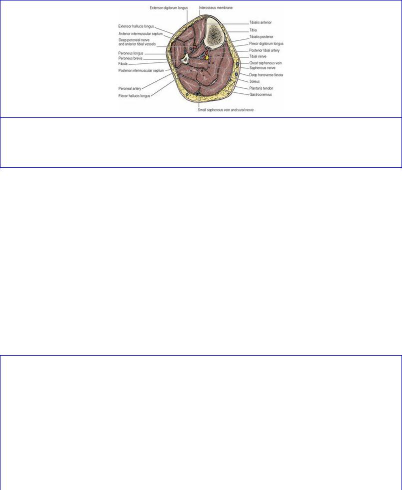
The muscles of the calf, the posterior compartment of the leg, fall into superficial and deep groups, with the deep transverse fascia of the leg between them. This fibrous septum extends transversely from the soleal line and posterior border of the tibia to the posterior border of the fibula; it is continuous above with the fascia covering the popliteus. Both the superficial and deep compartments require decompression by incising the deep fascia and the deep transverse fascia when local conditions predispose to the development of the compartment syndrome in the calf (p. 142). The superficial muscles consist of gastrocnemius, plantaris and soleus which all converge on a thick tendon at the back of the heel, the tendo calcaneus or Achilles' tendon. They are the main plantarflexors of the ankle joint. The deep group includes popliteus (see p. 134), and three muscles— flexor digitorum longus, flexor hallucis longus and tibialis posterior—whose tendons pass under the flexor retinaculum into the sole of the foot. The nerve of the posterior compartment is the tibial part of the sciatic, and the arteries are the posterior tibial (from the popliteal) and its peroneal branch.
Superficial muscles of the calf
Gastrocnemius and plantaris
The lateral head of gastrocnemius arises on the lateral surface of the lateral femoral condyle, from a smooth pit above that of popliteus; the pits are separated by the epicondyle (Fig. 3.21). The medial head of gastrocnemius arises from the back of the medial condyle and the popliteal surface of the shaft of the femur (Fig. 3.19). There is a bursa between each head and the capsule of the knee joint. The medial bursa communicates with the joint cavity and the lateral bursa may do so. The medial bursa may also communicate with the semimembranosus bursa.
The two heads converge to lie side by side (Fig. 3.33), and the larger medial head extends to a lower level than the lateral head. The broad bellies of the muscle insert into a dense aponeurosis on their anterior surfaces, bearing on the soleus muscle (Fig. 3.30). The aponeurosis blends with that of soleus to form the tendo calcaneus, which is inserted into a smooth transverse area on the middle third of the posterior surface of the calcaneus. A bursa lies between it and the upper part of the calcaneus. A
second bursa lies between it and the thickened deep fascia 5 cm above its insertion.
Figure 3.33 Cross-section of the middle of the right leg, looking towards the knee. Tibialis posterior is the deepest calf muscle, immediately behind the interosseous membrane. Note the many veins associated with soleus—a potential site of dangerous deep venous thrombosis.
Plantaris arises from the lower part of the lateral supracondylar line of the femur. Its slender tendon runs distally and medially, deep to the medial head of gastrocnemius (Fig. 3.18), and continues along the medial border of the tendo calcaneus with which it fuses. It can be harvested for use as a tendon graft but is absent unilaterally or bilaterally in about 10–20% of subjects.
Soleus
The muscle arises from the upper quarter of the back of the fibula, including the head of the bone, whence a fibrous arch (which bridges over the popliteal vessels and tibial nerve) carries it in continuity to the soleal line of the tibia and the middle third of the posterior border of the tibia (Fig. 3.34). The muscle has a dense aponeurosis upon either surface and muscle fibres that slope downwards from the anterior to the posterior lamella; these fleshy fibres are visible at the medial and lateral borders of the muscle. The posterior (superficial) lamella is continued at its lower end into the tendo calcaneus, and the muscle fibres of soleus are received into its deep surface down to within a short distance of the calcaneus.
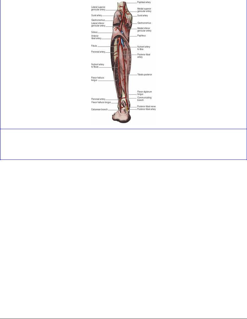
Figure 3.34 Left popliteal, posterior tibial and peroneal arteries: posterior aspect. A high origin of the peroneal artery from the posterior tibial artery makes the popliteal artery appear in some arteriograms as though it ends by trifurcating.
Perforating veins from the great saphenous vein enter the substance of soleus. The muscle contains a rich plexus of veins (Fig. 3.33), and these are pumped empty by contraction of the muscle, thus aiding venous return. Stagnation in these veins predisposes to deep venous thrombosis and the danger of pulmonary embolism. The ‘soleal pump’ is aided by the ‘sole pump’ (see p. 155).
Nerve supply. All three muscles are supplied by the tibial nerve (S1, 2). Each head of gastrocnemius receives a branch from the nerve in the popliteal fossa, and the lateral branch usually supplies plantaris as well. Soleus receives two branches, one from above the muscle in the popliteal fossa and one on its deep surface in the calf. In cases of intractable intermittent claudication both branches must be cut if soleus is to be completely denervated.
Actions. Soleus and gastrocnemius are the chief plantar flexors of the foot, and gastrocnemius is also a flexor of the knee. The powerful multipennate soleus is an antigravity muscle. In standing it contracts alternately with the extensor muscles of the leg to maintain balance. It is a very strong but relatively slow plantar flexor of the ankle joint, a necessary mechanical result of the obliquity of its multipennate fibres. The gastrocnemius bellies provide the necessary rapid contraction required for propulsion in fast walking, running and leaping. One strolls along quietly mainly with soleus; one wins the long jump mainly with gastrocnemius.
Test. The foot is plantarflexed against resistance; the tendo calcaneus and muscles above it can be seen contracting and can be palpated.
Deep muscles
The deep muscles of the calf consist of flexor digitorum longus, flexor hallucis longus and tibialis posterior. Their three tendons pass under the flexor retinaculum into the sole of the foot. Those of tibialis posterior and flexor hallucis longus are parallel throughout their course, the tibialis medial to the hallucis. The tendon of flexor digitorum longus takes an oblique course superficial to both. In the calf it lies medially, crosses tibialis posterior to lie under the flexor retinaculum between the two, and in the sole it crosses flexor hallucis longus to pass to the lateral four toes.
Flexor digitorum longus
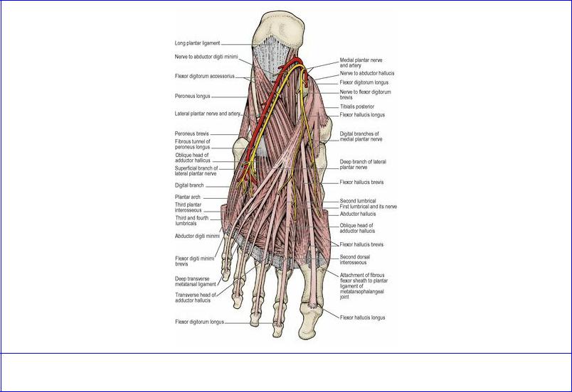
This muscle arises from the posterior surface of the tibia below the soleal line. The tendon of the muscle slopes downwards across the posterior surface of the tendon of tibialis posterior in the lower part of the leg. Enclosed in a synovial sheath it passes deep to the flexor retinaculum. It passes along the medial side of the sustentaculum tali of the calcaneum and enters the sole of the foot, where it crosses the inferior surface of the tendon of flexor hallucis longus (Fig. 3.37). At this point it divides into four tendons, the medial two of which receive a strong slip from the tendon of flexor hallucis longus. The four tendons at their commencement receive the insertion of the flexor accessorius muscle. More distally each gives origin to a lumbrical muscle. The tendons pass into the fibrous flexor sheaths of the lateral four toes, perforate the tendons of flexor digitorum brevis, and are inserted into the bases of the distal phalanges.
Figure 3.37 Plantar muscles of left foot: second and third layers.
Nerve supply. By the tibial nerve (S1, 2).
Action. Its principal action is to plantarflex the lateral four toes and, secondarily, to plantarflex the ankle joint. Its tonus, with that of the other deep calf muscles, assists in maintaining the longitudinal arch of the foot (see p. 160).
Flexor hallucis longus
This is the bulkiest and most powerful of the three deep muscles of the calf. It arises from the flexor surface of the fibula and from the interosseous membrane (Fig. 3.51).
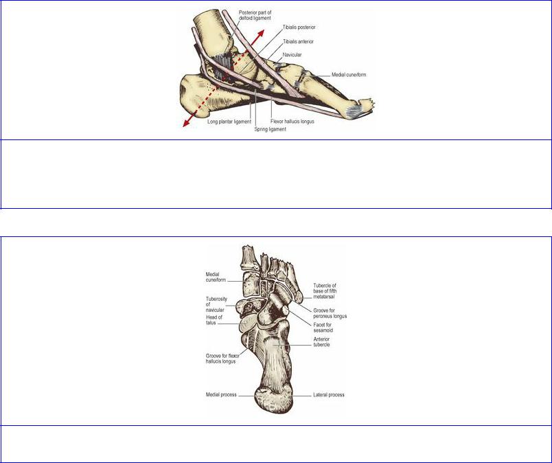
The fibres spiral down to be inserted into a central tendon which escapes from the muscle just at the lower end of the tibia, hence ‘beef to the heel’ describes the flexor hallucis longus ( Fig. 3.34). The tendon passes deep to the flexor retinaculum, enclosed in a synovial sheath. It grooves the posterior surface of the talus and the undersurface of the sustentaculum tali (Figs 3.44 and 3.57), from where it passes directly forwards like a bowstring beneath the arched medial border of the foot to be inserted into the base of the distal phalanx of the great toe.
Figure 3.44 Left foot from the medial side, with the axis of inversion and eversion indicated by the interrupted arrow. The inverting tendons pull at right angles around the axis. The everting peroneal tendons act in a similar way on the lateral side.
Figure 3.57 Left tarsal bones, from below.
It is crossed in the sole by the tendons of flexor digitorum longus and gives a strong slip to the medial two of these (those for the second and third toes). In the calf the peroneal artery runs down deep to the muscle on the tibialis posterior.
Nerve supply. By the tibial nerve (S1, 2).
Action. Its principal action is to flex the great toe, and it is significant that this is the ‘take-off’ point, the last part to leave the ground in propulsion (see p. 162). It plantarflexes the ankle joint simultaneously. The pull of this powerful muscle is an important factor in maintaining the medial longitudinal arch of the foot.
Tests. The terminal phalanges of the great toe (for flexor hallucis longus) and of other toes (for flexor digitorum longus) are flexed against resistance.
Tibialis posterior
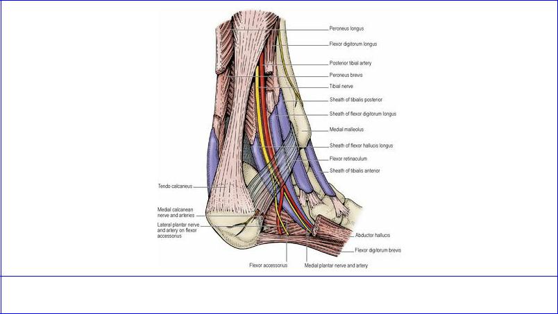
Tibialis posterior is the most deeply placed muscle in the calf. It arises from the interosseous membrane and the adjoining surface of both bones of the leg below the origin of soleus. Enclosed in a synovial sheath, the tendon passes deep to the flexor retinaculum and grooves the back of the medial malleolus (Fig. 3.35). It passes forward above the medial side of the sustentaculum tali, superficial to the deltoid ligament (see p. 156) and is inserted mainly into the tuberosity of the navicular. Tendinous slips also pass to the sustentaculum tali, all three cuneiforms, the cuboid and the second, third and fourth metatarsals. The proximal attachment of the muscle consists of two pointed processes, between which the anterior tibial vessels pass.
Figure 3.35 Left ankle region: medial aspect.
Nerve supply. By the tibial nerve (L4).
Action. It inverts and adducts the forefoot and, since it passes behind the medial malleolus, it plantarflexes the ankle joint. It contributes to maintaining the medial longitudinal arch of the foot.
Test. With the foot in slight plantarflexion, it is inverted against resistance; the tendon can be felt behind the medial malleolus.
The posterior tibial artery arises at the lower border of the popliteus, where the popliteal artery divides into anterior and posterior tibial branches. It passes under the fibrous arch in the origin of soleus and runs down on tibialis posterior, between flexor digitorum longus and flexor hallucis longus. It ends under the flexor retinaculum by dividing into medial and lateral plantar arteries. It is accompanied throughout its course by a pair of venae comitantes which frequently communicate with each other around the artery.
The pulsation of the artery can be palpated behind the medial malleolus, 2.5 cm in front of the medial border of the tendo calcaneus. It can be exposed here, e.g. for making an arteriovenous shunt with the great saphenous vein for haemodialysis.
Branches. The peroneal (fibular) artery arises 2.5 cm distal to popliteus. Its proximal course is in line with the popliteal artery (Fig. 3.34), and in some arteriograms the latter may appear to have trifurcated into anterior and posterior tibial and peroneal arteries. It runs distally in a fibrous canal between flexor hallucis longus and tibialis posterior, giving branches to the calf muscles and others that wind around the fibula to supply peroneus longus and brevis. It gives a nutrient artery to the fibula. It ends by dividing into a perforating branch which pierces the interosseous membrane to enter the extensor compartment and a lateral calcanean branch to the lateral side of the heel. The perforating branch may replace or supplement the dorsalis pedis artery.
The circumflex fibular artery passes laterally around the fibular neck to join the arterial anastomosis around the knee. The large nutrient artery to the tibia pierces tibialis posterior and runs downwards to enter the bone just distal to the soleal line. Muscular branches supply soleus and the deep flexors. Medial calcanean branches pierce the flexor retinaculum and supply the medial side of the heel.
The tibial nerve runs straight down the midline of the calf, deep to soleus. The posterior tibial artery is at first lateral to it, but passes anterior to it and continues downwards on its medial side. The nerve ends under the middle of the flexor retinaculum by dividing into the medial and lateral plantar nerves (Fig. 3.35); its surface marking is from the middle of the popliteal fossa to midway between the medial malleolus and the tendo calcaneus.
It is the nerve of the flexor compartment, giving branches to soleus, flexors digitorum longus, hallucis longus and tibialis posterior. It gives medial calcanean nerves which pierce the flexor retinaculum to supply the skin of the heel, including the weight-bearing surface (Fig. 3.35).