

Part two. Medial compartment of the thigh
The contents of this (adductor) compartment of the thigh are separated from the anterior (extensor) compartment by the medial intermuscular septum, but there is no septum dividing them from the posterior (flexor or hamstring) compartment. The muscles consist of gracilis and the three adductors, longus, brevis and magnus, while deeply lies obturator externus. The nerve of the compartment is the obturator, and the artery is the profunda femoris, assisted proximally by the obturator artery.
The medial intermuscular septum is a thin fascia that lies between vastus medialis and the adductors and pectineus. It is attached to the fascia lata and the linea aspera of the femur, and is continuous above with the fascia on pectineus (see p. 115).
Gracilis
This, the most superficial muscle of the medial side of the thigh (Fig. 3.2), arises as a flat sheet from the edge of the inferior ramus of the pubis and the adjoining ischial ramus (Fig. 3.48). The muscle narrows in triangular fashion and is replaced by a cylindrical tendon which is inserted into the upper part of the medial surface of the shaft of the tibia just behind sartorius. The gracilis receives a segmental blood supply from the medial circumflex, profunda and femoral arteries in succession distally. A gracilis muscular flap based on the profunda pedicle is used in perineal reconstructive surgery.
Adductor longus
This, the most superficial of the three adductors, arises from a circular area on the body of the pubis, in the angle between the pubic crest and symphysis (Fig. 3.48), by a strong round tendon, sometimes ossified (‘rider's bone’). The muscle rapidly becomes fleshy and flattens out to be inserted by an aponeurotic flat tendon into the middle third of the linea aspera of the femur (Fig. 3.11).
Adductor brevis
This muscle arises from the body and inferior ramus of the pubic bone, deep to pectineus and adductor longus (Fig. 3.48). It widens in triangular fashion to be inserted into the upper part of the linea aspera immediately behind the insertion of pectineus and adductor longus. The anterior division of the obturator nerve passes vertically downwards on its anterior surface (Fig. 3.11); the posterior division passes down behind it. The upper border of adductor brevis thus lies between the two divisions of the obturator nerve in the same way as the upper border of adductor longus lies between the femoral and the profunda femoris vessels.
Adductor magnus
This is a composite muscle formed by the fusion of adductor and hamstring muscle masses, each with their own nerve supply.
The hamstring part arises from the ischial tuberosity (Fig. 3.48), and the fibres pass vertically downwards to a tendinous attachment to the adductor tubercle of the femur, with an expansion to the medial supracondylar line. In continuity with the ischial origin, the adductor part arises from the ischiopubic ramus. These fibres are inserted progressively higher along the medial supracondylar line, the linea aspera and up to the gluteal tuberosity (Fig. 3.50B). The upper border of the muscle is horizontal, lying edge to edge with the lower border of quadratus femoris, and the medial circumflex

femoral artery passes between the two muscles from front to back to reach the cruciate anastomosis (Fig. 3.13). Near the top of the medial supracondylar line there is a gap in the muscle attachment through which the femoral vessels pass, changing their name to popliteal as they do so. Along the linea aspera attachment there are four small openings, the lowest for the end of the profunda femoris vessels, and the others for their perforating branches.
Figure 3.50 Right femur: A anterior aspect; B posterior aspect.
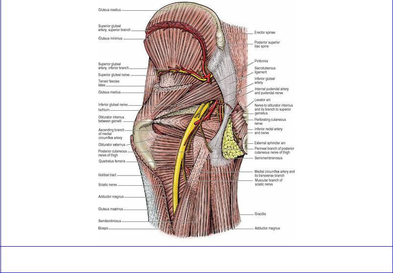
Figure 3.13 Left gluteal region, with much of gluteus maximus removed.
Nerve supplies of adductors. Gracilis and adductor longus and brevis are supplied by the anterior division of the obturator nerve, the hamstring part of magnus by the tibial part of the sciatic nerve and the rest of magnus by the posterior division of the obturator (L2, 3 for all muscles).
Actions of adductors. The adductor mass of muscles, though large, is less important in the prime movement of adduction than in synergic activities associated with posture and gait.
Tests for adductors. While lying on the back with the knee straight, the patient adducts the thigh against resistance, and the upper ends of gracilis and adductor longus are palpated.
Obturator externus
This muscle arises from the whole of the obturator membrane and from the anterior bony margin of the obturator foramen. Both membrane and muscle fall short of the obturator notch above, thereby forming a canal for the passage of the obturator nerve and vessels (Fig. 3.11). The muscle passes laterally and posteriorly beneath the neck of the femur where it narrows into a tendon that spirals in contact with the back of the femoral neck to be inserted on the medial surface of the greater trochanter into a deep pit, the trochanteric fossa. The capsule of the hip joint encloses the back of the neck of the femur only as far as the place where obturator externus tendon is in contact with periosteum, namely half the neck of the femur (Fig. 3.12), whereas in front the capsule of the hip joint includes the whole of the neck of the femur (Fig. 3.8).
Nerve supply. By the posterior division of the obturator nerve (L3, 4).
Action. With the other short muscles around the hip joint, it stabilizes and supports the proximal part of the limb. As its line of pull passes behind the hip joint, it is a lateral rotator of the femur.
Obturator artery and nerve
The obturator artery, on emerging from the obturator foramen with the nerve, divides into anterior and posterior branches that encircle the foramen between the obturator externus and the membrane. They anastomose with each other and with the medial circumflex artery. From the posterior branch the articular twig to the hip joint arises; it enters the acetabular notch and runs in the ligament of the head of the femur to supply a small scale of bone in the region of the pit for the attachment of the ligament (see p. 129).
The obturator nerve divides in the obturator notch into anterior and posterior divisions; the anterior passes above obturator externus, the posterior passes through the muscle, giving off a branch to supply it before doing so.
The anterior division, giving an articular branch to the hip joint, descends in the thigh behind the adductor longus, which it supplies. Passing over the anterior surface of adductor brevis (Fig. 3.11), which it usually supplies, it goes on to supply gracilis and end in the subsartorial plexus, whence branches supply the skin over the medial side of the thigh. Direct branches to the skin are often given off at a level above the subsartorial plexus.
The posterior division emerges through obturator externus (having already supplied that muscle), and passes vertically downwards on adductor magnus deep to the other adductor muscles. It supplies adductor magnus and gives a terminal branch which runs with the femoral artery through the hiatus in the muscle to the popliteal fossa and supplies the capsule of the knee joint by passing in with the middle genicular artery. The posterior division may supply adductor brevis when this is not supplied by the anterior division.