


Part two. Brainstem
The brainstem is the part of the brain connecting the cerebrum and diencephalon with the spinal cord, and consists of the midbrain, pons and medulla oblongata (Figs 7.3B, 7.6 and Figure 7.18, Figure 7.19 and Figure 7.20). It extends from just above the aperture in the tentorium cerebelli to C1 vertebra below the foramen magnum. The cerebellum projects from its dorsal surface.
The brainstem consists of fibres and cells. Most of the fibres in the brainstem ascend or descend longitudinally, as in the spinal cord, and most of the cells are aggregated into nuclei. These nuclei consist of three groups:
•The nuclei of the third to the twelfth cranial nerves.
•Other named nuclei which are demonstrable, such as the colliculi, the red nucleus, the substantia nigra, the pontine nuclei and the olivary nucleus.
•The reticular formation, a diffuse system of cells and fibres which is intermingled with the named nuclei and tracts, continues into the spinal cord, and is described further on page 481. Some of its cells form the so-called ‘vital centres’—cardiac, respiratory, vasomotor, etc.—which are not anatomically demonstrable as distinct ‘nuclei’, but are of great physiological importance.
The levels of the cranial nerve nuclei are as follows.
Those of the third and fourth are in the midbrain.
The motor nucleus of the fifth, and the sixth and seventh, are in the pons. The three sensory nuclei of the fifth are distributed between the midbrain, pons, medulla and upper spinal cord. The salivary (parasympathetic) and sensory (taste) nuclei of the seventh are in the lower pons and upper medulla, respectively.
The eighth nerve nuclei overlap the junction of pons and medulla and lie partly in each.
The nuclei of the ninth to the twelfth are in the medulla (with the eleventh having a spinal part derived from the cervical region of the cord).
Midbrain
The midbrain connects the diencephalon and cerebrum to the pons (Figs 7.3B and 7.18). It is the shortest segment of the brainstem, being not more than 2 cm in length. Most of it lies in the posterior cranial fossa, with its upper part passing through the tentorial notch.
The midbrain consists of right and left halves, each half forming a cerebral peduncle made up of a ventral part, the base (basis pedunculi) and a dorsal part, the tegmentum. Running through the tegmentum is the aqueduct of the midbrain (aqueduct of Sylvius), joining the third and fourth ventricles. The part of the tegmentum dorsal to the aqueduct is the tectum.
On the ventral surface are seen the bases of the peduncles (often called the crura), which lie in V- shaped manner cranial to the pons, enclosing the posterior perforated substance of the diencephalon between them (Fig. 7.18). They converge down towards the upper border of the pons from their point of emergence below the thalamus.

Dorsally the midbrain shows two pairs of low rounded eminences, the superior and inferior colliculi (formerly called corpora quadrigemina, Fig. 7.20). The superior colliculi lie below the pineal body and the splenium of the corpus callosum, overlapped by the pulvinar of the thalamus. Lateral to each superior colliculus is the medial geniculate body which, although appearing to belong to the brainstem, is part of the thalamus (see p. 470). Below the inferior colliculi the superior cerebellar peduncles converge into the dorsal surface of the midbrain from the cerebellum.
The third and fourth cranial nerves leave the brainstem at the midbrain (Figs 7.19, 7.21 and 7.22). The oculomotor nerve leaves through the medial surface of the crus, on the ventral surface of the midbrain, and passes forwards between the posterior cerebral and superior cerebellar arteries in the interpeduncular cistern to reach the roof of the cavernous sinus. The trochlear nerve leaves the dorsal surface of the midbrain just below the inferior colliculus. This nerve is unique in three respects: it is the smallest cranial nerve, the only one to emerge from the dorsal surface of the brainstem, and the only one to decussate within the brainstem. The nerve curls round the lateral side of the peduncle and passes forwards between the same two arteries as the third nerve (poster-ior cerebral and superior cerebellar) but farther laterally, to reach the roof of the cavernous sinus. The optic tract and the basal vein also curl round the peduncle and the posterior communicating artery lies on the medial surface of the peduncle.
Figure 7.21 Cross-section of the midbrain at the level of the superior colliculi.
Figure 7.22 Cross-section of the midbrain at the level of the inferior colliculi.
Internal structure
Sections of the midbrain are recognized by the colliculi of the tectum on the dorsal surface, the aqueduct, and the rectangular crura on the ventral surface, delimited by a dark line of pigmented cells, the substantia nigra. Other naked-eye features at superior colliculus level are the red nucleus with fibres of the third nerve sweeping though it, while at inferior colliculus level is the centrally placed decussation of the superior cerebellar peduncles (Figs 7.21 and 7.22).
Each crus contains corticospinal, corticonuclear and corticopontine fibres; the first two occupy the middle two-thirds of the crus. The medial sixth of the crus is occupied by frontopontine fibres and the lateral sixth by temporopontine and other corticopontine fibres (see p. 461).
The superior colliculus contains cells involved in general light reflexes, while the inferior colliculus is concerned with sound reflexes. They receive inputs from the retina and cochlea respectively and project to the motor nuclei of cranial and spinal nerves (via tectobulbar and tectospinal tracts) for reflex movements of the eyes, head, body and limbs away from or towards light and sound stimuli. The pupillary light reflexes (see p. 407) involve the pretectal nuclei, which lie just cranial to the superior colliculi at the junction of the midbrain and diencephalon.
The oculomotor nucleus lies close against the midline ventral to the aqueduct at superior colliculus level (Fig. 7.21), in line vertically with the other cranial somatic motor nuclei (fourth, sixth and twelfth). The parasympathetic part (Edinger–Westphal or accessory oculomotor nucleus) lies near the midline in the cranial part of the nucleus; its axons run out with the third nerve and relay in the ciliary ganglion, from which postganglionic fibres innervate the sphincter pupillae and ciliary muscles (see p. 407). The third nerve passes ventrally through the red nucleus to emerge from the brainstem on the medial side of the base of the peduncle.
The trochlear nucleus lies caudal to the oculomotor nucleus, ventral to the aqueduct at inferior colliculus level (Fig. 7.22). The nerve proceeds dorsally and crosses the midline, where it decussates with its fellow dorsal to the aqueduct. It emerges through the superior medullary velum (see p. 482) on the dorsal aspect of the midbrain, below the inferior colliculus (Fig. 7.20).
The red nucleus lies in the tegmentum just ventral to the third nerve nucleus (Fig. 7.21). It is slightly larger than a full-sized pea. It receives fibres coming from the dentate nucleus in the opposite cerebellar hemisphere via the superior cerebellar peduncle. Its efferent fibres decussate and descend to motor nuclei of cranial and spinal nerves, as part of the extrapyramidal system (see p. 489).
Although in the midbrain at the junction of the tegmentum and crus, the substantia nigra belongs functionally to the basal nuclei (see p. 458). Many of its cells contain melatonin, responsible for its naked-eye dark appearance. Some of its cells give rise to nigrostriatal fibres which are dopaminergic and project to the caudate nucleus and putamen. The loss of about 80% of its dopaminergic cells is the fundamental defect in parkinsonism.
The mesencephalic nucleus of the trigeminal nerve lies in the central grey matter, lateral to the aqueduct, throughout the whole length of the midbrain. This long slender nucleus receives proprioceptive fibres from the muscles supplied by the mandibular branch of the trigeminal (muscles of mastication) and from the muscles of the orbit and face and, perhaps, the muscles of the tongue (see p. 382). It is unique in being a collection of first neuron cells buried in the central nervous system (see p. 489). It is accompanied by the mesencephalic tract which descends to the upper pons (Fig. 7.23).
The spinal lemniscus and the medial lemniscus, which are ascending sensory tracts (see p. 482), lie in the lateral part of the tegmentum. Between the medial lemniscus and the central grey matter the tegmentum contains fragments of grey matter broken up by criss-cross bundles of white fibres. The ‘network’ appearance so produced gives it the name reticular formation. It is traceable through the

pons (see p. 479) and medulla into the upper spinal cord, and is described further on page 480.
Blood supply
The midbrain is supplied by the posterior cerebral and superior cerebellar arteries as they curl around the cerebral peduncle. The veins drain for the most part into the basal vein as it passes around the peduncle. From the colliculi some blood enters the great cerebral vein.
Pons
The pons is a broad transverse mass between the midbrain and medulla (Fig. 7.18), curving at the sides into the middle cerebellar peduncle (Fig. 7.6). The only cranial nerve to emerge from the pons itself, the fifth, does so by a large sensory and small motor root. They emerge laterally from the middle of the ventral aspect of the pons, with the motor root slightly cranial and medial to the sensory root. The two nerve roots pass forwards together in the posterior cranial fossa (i.e. below the tentorium) to run over the groove on the apex of the petrous bone into the trigeminal cave in the middle cranial fossa (see Fig. 6.105, p. 446).
The ventral surface of the pons shows a shallow midline groove with a bulge on either side. The bulge is due to the underlying mass of pontine nuclei, intermingled with corticospinal and corticonuclear fibres (see below). This ventral surface lies along the clivus (see p. 510), separated from the bone by the subarachnoid pontine cistern, in which the basilar artery runs upwards. The artery may or may not lie in the midline groove; usually it has a gentle curve to one side. The superior cerebellar artery curls round the upper margin of the pons. The labyrinthine artery passes laterally to reach the internal acoustic meatus. Emerging from the junction of pons and medulla, the sixth nerve runs upwards across the ventral surface to enter the dura on the clivus. The seventh and eighth nerves emerge more laterally at the junction of pons and medulla, in the region often referred to as the cerebellopontine angle; the nervus intermedius part of the seventh nerve lies separately between the main part of the seventh and eighth nerves. Most laterally in this region lies the flocculus of the cerebellum (see p. 485) and the choroid plexus that has emerged from the lateral recess of the fourth ventricle (Fig. 7.18).
The dorsal surface of the pons is concealed by the attached cerebellum. The aqueduct of the midbrain opens out at the upper border of the pons into the cavity of the fourth ventricle, which is mostly pontine but medullary at its lower end (see p. 482). The pontine part of the roof of the ventricle consists only of a thin sheet of white matter, the superior medullary velum (Figs 7.20 and 7.29), upon which lies the lingula of the cerebellum. The velum is attached at each side to the superior cerebellar peduncles.

Figure 7.29 Sagittal section of the cerebellum.
Internal structure
In the ventral part of the pons are the pontine nuclei, from which fibres emerge to cross to the opposite side and form the middle cerebellar peduncle. With the various corticopontine fibres that have travelled down in the cerebral peduncles to synapse with the pontine nuclei, they complete an extensive corticopontocerebellar pathway.
In the dorsal part of the pons are the nuclei of the fifth to eighth nerves and the salivary nuclei.
The motor nucleus of the trigeminal nerve is in the upper pons below the lateral part of the floor of the fourth ventricle. The fibres pass ventrally and laterally to emerge as already noted as a small motor root at the junction of the pons and the middle cerebellar peduncle.
Lateral to the motor nucleus is the main sensory nucleus of the trigeminal nerve. It receives those incoming fibres of the sensory root subserving touch. Its caudal continuation into the lower pons, medulla and upper cervical spinal cord is the spinal nucleus, which receives pain and temperature fibres; its upward continuation is largely a small bundle of fibres but with some cell bodies, the mesencephalic tract of the trigeminal, leading to the mesencephalic nucleus for proprioception (Figs 7.21 and 7.23).
Figure 7.23 Cross-section of the upper pons.
In the lower part of the pons the abducens nucleus lies near the midline just below the floor of the fourth ventricle, but with fibres of the facial nerve overlying it. The abducens nucleus plus the overlying facial nerve fibres form a small swelling, the facial colliculus, in the ventricular floor (Figs 7.20 and 7.24). The facial nucleus itself lies deeper and farther from the midline.
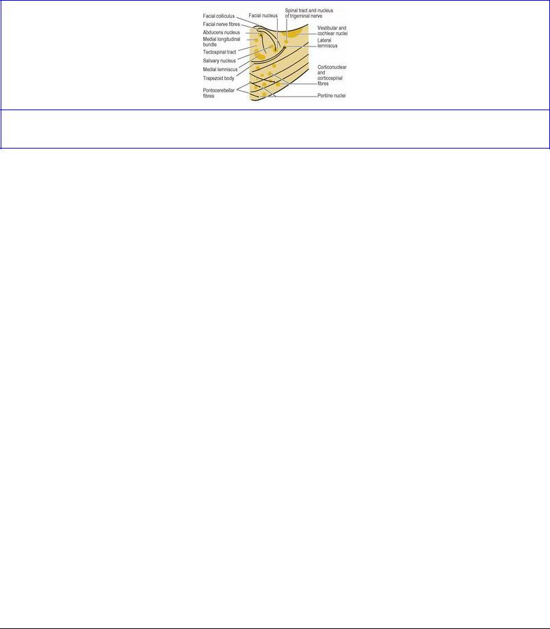
Figure 7.24 Cross-section of the lower pons.
The upper part of a collection of cells alongside the facial nucleus forms the superior salivary nucleus (parasympathetic). It provides axons which pass out in the nervus intermedius part of the facial nerve and reach the pterygopalatine and submandibular ganglia. The lower part forms the inferior salivary nucleus, just above the pontomedullary junction; its fibres join the glossopharyngeal nerve to reach the otic ganglion.
The nuclei of the vestibulocochlear nerve lie beneath the floor of the lateral angle of the fourth ventricle (the vestibular area, see Fig. 7.20) in both the pons and medulla. The vestibular fibres that emerge from the internal acoustic meatus pass anterior to the inferior cerebellar peduncle of the medulla and synapse in the vestibular nuclei, many of whose fibres pass back into that peduncle. Other fibres join the medial longitudinal bundle and connect with extraocular muscle nuclei and cervical anterior horn cells. Some of these connections provide the basis for vestibulo-ocular reflexes.
The spiral ganglia of the cochlea send their axons to dorsal and ventral cochlear nuclei on the corresponding aspects of the inferior cerebellar peduncle, which are continuous medially with the vestibular area. Axons from these nuclei form the decussating trapezoid body (see p. 482), part of the auditory pathway.
The very uppermost ends of the dorsal nucleus of the vagus and the nucleus of the tractus solitarius also extend into the pons but essentially belong to the medulla, where they are described.
The pontine part of the reticular formation (see p. 480) lies dorsal to the pontine nuclei and their intermingled fibres.
Blood supply
The pons is supplied mainly by pontine branches from the basilar artery, with contributions from the superior cerebellar and anterior inferior cerebellar vessels. Venous return is into the inferior petrosal sinuses and the basilar plexus.
Medulla oblongata
The medulla oblongata is the part of the brainstem between the pons and spinal cord and it extends through the foramen magnum to the level of the atlas. Above the foramen magnum it is embraced dorsally by the cerebellar hemispheres. The lower end which contains the upward continuation of the central canal of the spinal cord is the ‘closed part of the medulla’, while the upper end, where the canal comes to the surface as the lower part of the floor of the fourth ventricle, is the ‘open part’.

Ventrally ( Fig. 7.18) the upper part of the medulla is deeply grooved in the midline, with a bold convexity on either side, the pyramid, due to the contained corticospinal fibres. Lateral to the pyramid is another convexity, the olive, due to the underlying inferior olivary nucleus. Lateral to the olive the lateral surface of the medulla is formed by the inferior cerebellar peduncle, which enters the cerebellum medial to and below the middle peduncle.
The sixth, seventh and eighth cranial nerves emerge between the pons and the medulla, the sixth nerve between the pons and the pyramid, the main part of the seventh nerve between the pons and the olive, and the nervus intermedius part of the seventh and the eighth nerve between the pons and the inferior cerebellar peduncle (Figs 7.6 and 7.8). The rootlets of the ninth, tenth and cranial part of the eleventh nerves emerge lateral to the olive, and those of the twelfth by two small groups of rootlets between the pyramid and the olive.
Dorsally the lower part of the floor of the fourth ventricle forms the upper part of the medulla (Fig. 7.20). Here the roof of the ventricle is ependyma and pia mater. At the lower corner of the diamond- shaped floor the hypoglossal trigone is adjacent to the midline, with the vagal trigone lateral to it. Higher up and at the lateral corners of the diamond is the vestibular area and the medullary striae (see p. 483).
In the lower or closed part of the medulla, the fourth ventricle has become narrowed to the tiny central canal, and the external dorsal surface shows small elevations, the gracile and cuneate tubercles, the former being medial to the latter (Fig. 7.20).
Internal structure
Decussation of the pyramids characterizes the lowest part of the medulla, and at a slightly higher level is seen the central decussation of the fibres forming the medial lemnisci (Figs 7.25 and 7.26; see also p. 482).
Figure 7.25 Cross-section of the lower part of the closed medulla.

Figure 7.26 Cross-section of the upper part of the closed medulla.
Most of the nuclei of the medulla are below the floor of the fourth ventricle. The hypoglossal nucleus underlies the hypoglossal trigone and the dorsal nucleus of the vagus is under the vagal trigone; more laterally are the nucleus of the tractus solitarius and the spinal nucleus and tract of the trigeminal nerve. At a deeper level is the nucleus ambiguus, with the inferior olivary nucleus ventrally (Fig. 7.27).
Figure 7.27 Cross-section of the open part of the medulla.
The hypoglossal nucleus adjacent to the midline gives rise to the hypoglossal nerve fibres which pass ventrally to emerge between the pyramid and olive (Fig. 7.27).
Lateral to the hypoglossal nucleus is the dorsal nucleus of the vagus, which contains motor cell bodies for cardiac and visceral muscle and the cells of secretomotor fibres for glands. The corresponding afferent cell bodies lie in the nucleus of the tractus solitarius lateral to the dorsal nucleus; the afferent fibres form the tract which is almost surrounded by cells of the nucleus. The upper part of the nucleus receives taste fibres from the chorda tympani (via the nervus intermedius part of the facial nerve), glossopharyngeal nerve and internal laryngeal branch of the vagus. The rest of the nucleus receives many afferent fibres of the glossopharyngeal and vagus nerves from thoracic and abdominal viscera, as well as those from the baroreceptors and chemoreceptors of the carotid sinus and carotid body and the aortic arch and aortic bodies (see pp. 343 and 193). It has extensive connections with the dorsal nucleus of the vagus and the reticular formation.
The nucleus ambiguus contains motor cell bodies for the skeletal muscle of the larynx, soft palate, pharynx and upper oesophagus, all distributed by branches of the vagus except for the supply to stylopharyngeus which is by the glossopharyngeal nerve. The cells of the upper end of the nucleus send their fibres to the glossopharyngeal nerve, while those from the lower part send theirs to the cranial root of the accessory nerve, which joins the vagus below the skull.
The spinal nucleus (and tract) of the trigeminal nerve continues down into the medulla from the pons (see p. 478) and lies lateral to the nucleus of the tractus solitarius (Fig. 7.27). It receives somatic sensory fibres from the glossopharyngeal and vagus nerves.
The inferior olivary nucleus is a crenated C-shaped lamina of grey matter, in section like a wrinkled sac with an open end facing towards the opposite inferior cerebellar peduncle. Its fibres
(olivocerebellar) decussate across the midline to enter this peduncle (see p. 486).
The cochlear and vestibular nuclei extend into the medullary part of the floor of the fourth ventricle, and have been considered with the pons.
The gracile and cuneate nuclei underlie the corresponding tubercles of the dorsal surface of the lower medulla. They contain the cell bodies on which the incoming fibres of the gracile and cuneate tracts of the spinal cord terminate, and the nuclei give origin to the medial lemniscus (see p. 482).
The medullary reticular formation, continuous upwards with that of the pons and extending downwards into the spinal cord at the lateral margin of the central grey matter, is the irregular mass of cells and fibres occupying much of the area between the inferior olivary nucleus and the floor of the fourth ventricle, intermingled with other cell groups and tracts. Although they are not anatomically demonstrable as distinct nuclei, pools of neurons of the reticular formation (‘centres’) are functionally associated with vasoconstrictor, cardioaccelerator, cardiopressor, inspiratory and expiratory effects.
Although anatomically such a diffuse entity, the brainstem reticular formation is responsible for the ‘alert’ or ‘wakeful’ component of consciousness. It plays a part in the control of many other functions, including motor activity (as part of the extrapyramidal system, see p. 489), sensory function (by modifying sensory input to the thalamus), autonomic activity (via the medullary centres), circadian rhythms and endocrine secretion (via the hypothalamus). Among its connections are those from the cerebral cortex of the same side via the corpus striatum (see p. 458), from the opposite cerebellar hemisphere via the dentate nucleus (see p. 486), and from the hypothalamus and other components of the limbic system (see p. 466). Some reticular formation cells give origin to the reticulospinal tracts (see p. 491) which are part of the extrapyramidal system, and others receive spinoreticular fibres which are part of the pain pathway from the spinal cord (see p. 490). Other cells provide communications between cranial nerve nuclei, e.g. for eye movements, the corneal reflex, swallowing, etc. However, the conduction paths of the reticular formation are difficult to define, complex, and partly crossed and uncrossed; unilateral stimulation often results in bilateral responses.
Blood supply
The medulla is supplied ventrally by branches of the vertebral and basilar arteries, and laterally and dorsally by the posterior inferior cerebellar artery (Fig. 7.19). The anterior spinal branch of the vertebral gives penetrating branches which supply the region next to the midline, i.e. the part containing the pyramid, medial lemniscus and hypoglossal nucleus. Damage to these vessels produces the medial medullary syndrome—paralysis of the tongue on the same side and hemiplegia with loss of touch and kinaesthetic sense on the opposite side. Damage to the vessels of the lateral and dorsal part gives rise to the lateral medullary syndrome or ‘syndrome of the posterior inferior cerebellar artery’. The loss of nucleus ambiguus function paralyses laryngeal, palatal and pharyngeal muscles on that side, causing dysphonia and dysphagia. Loss of the uncrossed spinal tract of the trigeminal and of the crossed spinal lemniscus results in loss of pain and temperature sensation on the same side of the face and opposite side of the body. There will also be a Horner's syndrome (see p. 408) on the ipsilateral side due to interruption of descending hypothalamospinal fibres of the sympathetic pathway. Involvement of the vestibular nuclei causes vertigo and nystagmus with nausea and vomiting.
The veins drain dorsally to the occipital sinus and ventrally into the basilar plexus of veins and the inferior petrosal sinus. The medullary veins communicate with the spinal veins.
Brainstem tracts
Some tracts in the brainstem begin from cell groups therein, but most are passing through from the rest of the brain to the spinal cord or vice versa. The most important of the long descending tracts are the corticonuclear and corticospinal fibres concerned with voluntary movement, supplemented for posture and coordination of movement by reticulospinal and vestibulospinal tracts which begin in the brainstem. The main ascending tracts are the medial lemniscus for fine touch and kinaesthetic sensations, beginning in the medulla, and the spinal lemniscus and spinoreticulothalamic fibres for pain, temperature and crude touch, beginning in the cord and brainstem.
Descending tracts
Cells in layer V of the sensorimotor area MsI (see pp. 462 and 464) give rise to corticospinal (pyramidal) and corticonuclear fibres. However, only 40% of such fibres come from this motor area (though they appear to be the ones that matter most); the remainder come from widely scattered areas of the cortex and not just other parts of the frontal lobes. They pass through the corona radiata, and the corticonuclear fibres then run down through the genu of the internal capsule into the brainstem. Some will go straight to the oculomotor and trochlear nuclei, and the others collect in the most medial part of the central two-thirds of the crus of the midbrain. From there they run down to reach the motor nuclei of the rest of the brainstem. Cranial nerve nuclei which send their fibres to skeletal muscle are mostly bilaterally innervated (i.e. from the cortex of both hemispheres, although there are individual variations). The most important exception is the lower part of the facial nucleus, which is only supplied by the opposite cortex.
The corticospinal fibres lie in the anterior two-thirds of the posterior limb of the internal capsule. Continuing down to the brainstem, they occupy the central and lateral parts of the central two-thirds of the crus of the midbrain peduncle, with the ‘arm’ fibres medial to the ‘leg’ fibres. In the pons the fibres become broken up into small bundles among the pontine nuclei. Passing on to the medulla the bundles collect into a single large mass forming the bulging pyramid adjacent to the midline. Each pyramid contains about 1 million nerve fibres of which 700 000 are small and myelinated (1–4 μm diameter). In the lowest part of the medulla 75–90% of the fibres cross to the opposite side in the pyramidal decussation to form the lateral corticospinal tract of the cord (see p. 491). Some of the decussating fibres may be seen on the surface. The few uncrossed fibres continue downwards as the anterior corticospinal tract, but most of them also eventually cross in the spinal cord, where all fibres end in the anterior horn (see p. 491).
After arising from many areas of all four lobes of the cortex, corticopontine fibres pass through the internal capsule to the crus of the midbrain peduncle. Frontopontine fibres occupy the anterior limb of the capsule and the medial one-sixth of the crus; parietoand occipitopontine fibres occupy the retrolentiform part, and temporopontine fibres the sublentiform part, of the capsule and they descend in the lateral one-sixth of the crus. All end by synapsing with cells of the pontine nuclei (see p. 478) whose axons decussate to form the pontocerebellar fibres of the middle cerebellar peduncle. This is one of the pathways by which the cerebral cortex communicates with the cerebellar cortex.
The superior cerebellar peduncles (see p. 486) enter the midbrain tegmentum and decussate at the level of the inferior colliculi on their way to the red nuclei (at superior colliculus level), whose efferent fibres form the rubrospinal tracts which immediately decussate. Just dorsal to this is the similar decussation of the tectospinal tracts.
The medial longitudinal bundle lies ventral to the grey matter round the aqueduct, and remains adjacent to the midline at lower levels. It extends from the upper border of the midbrain to the upper cervical part of the spinal cord. It links the vestibular nuclei with the oculomotor, trochlear, abducent, spinal accessory and reticular nuclei, to help coordinate head and eye movements as required for fixation of the gaze and maintaining equilibrium.
The lateral and medial reticulospinal tracts arise from the medullary and pontine parts of the reticular formation. They are rather ill-defined in the brainstem, and their fibres become largely mixed with corticospinal fibres in the spinal cord. The lateral and medial vestibulospinal tracts originate from the vestibular nuclei. All these extra-pyramidal tracts end in the anterior horn (see p. 491).
Hypothalamospinal fibres from the hypothalamus run in the region of the spinal lemniscus to sympathetic and parasympathetic neurons in the thoracolumbar and sacral lateral horns (see p. 491).
Ascending tracts
The medial lemniscus, the main ascending pathway of the brainstem for touch and its associated sensations (see p. 489), begins in the lower medulla, formed by the axons of the gracile and cuneate nuclei of the opposite side which have crossed (as internal arcuate fibres ) in the sensory decussation. At first the medial lemniscus lies longitudinally adjacent to the midline, but as it passes up through the pons and midbrain it deviates laterally before reaching the thalamus. On its upward path it is joined by the trigeminal lemniscus, fibres from the main sensory and spinal nuclei of the trigeminal nerve of the opposite side.
The spinal lemniscus is the upward continuation of the lateral spinothalamic (anterolateral) tract of the cord conveying pain, temperature and crude touch sensations (see p. 489). It lies near the middle of the lateral part of the medulla, and runs up the brainstem at first lateral and then dorsal to the medial lemniscus. It quickly becomes very much smaller, since most of its fibres end in the reticular formation rather than continuing all the way to the thalamus.
The lateral lemniscus is formed by the upgoing fibres of the trapezoid body, the name given to transversely decussating fibres from the cochlear nuclei at the pontomedullary junction. Some of the fibres of this lemniscus terminate on cells of the inferior colliculus for auditory reflexes, while the majority relay in the medial geniculate body for passage via the internal capsule to the auditory area of the cerebral cortex. Both the trapezoid body and lateral lemniscus contain cell stations which make connections with the extraocular and spinal accessory nuclei via the medial longitudinal bundle to help coordinate audiovisual reflexes involving the head and neck.
The anterior and posterior spinocerebellar tracts lie at the lateral margin of the lower medulla. The posterior tract enters the inferior cerebellar peduncle but the anterior tract continues up the lateral part of the brainstem to enter the superior peduncle.

Fourth ventricle
The aqueduct within the midbrain is continuous below with the fourth ventricle, which lies behind the pons and the upper medulla. The dorsal aspects of these parts of the brainstem form the diamondshaped floor. The roof, or posterior wall, is projected backwards, like that of a tent lying on its side, and is covered by the cerebellum (Fig. 7.6). Hence the cavity of the ventricle is triangular in sagittal section. The caudal part of each lateral boundary is formed by the gracile and cuneate tubercles and the inferior cerebellar peduncle (Fig. 7.20); the cranial part is formed by the superior cerebellar peduncle. A thin sheet of white matter, the superior medullary vellum, stretched between the superior cerebellar peduncles, forms the cranial part of the roof. The caudal part of the roof is mostly devoid of neural tissue, and is formed only by ependyma and pia mater. This part of the roof is perforated by a midline slit, the median aperture (foramen of Magendie), by which CSF escapes into the cerebellomedullary cistern. The cavity is prolonged laterally as a narrow lateral recess behind and around the inferior cerebellar peduncle. The narrow, tubular lateral recess has a patent extremity, the lateral aperture (foramen of Luschka), which opens anteriorly, just behind the eighth nerve, into the pontine cistern (Figs 7.11B and 7.18). Through these three apertures (one median and two lateral) the CSF escapes from the ventricular system into the subarachnoid space for absorption by the arachnoid villi. These are the only exits from the system and if blocked, e.g. following meningitis, hydrocephalus results.
The choroid plexus of the fourth ventricle is a small T-shaped structure which indents the medullary part of the roof. It receives its blood supply from a branch of the posterior inferior cerebellar artery, which enters through the lateral aperture on each side and passes medially to meet its fellow, and the two turn down towards the median aperture making the vertical part of the T double. The veins from the plexus drain back into the occipital sinus.
The floor of the fourth ventricle is known as the rhomboid fossa (Fig. 7.20). The upper boundaries are the superior cerebellar peduncles, the lower are formed by the gracile and cuneate tubercles and their underlying nuclei and, above them, by the inferior cerebellar peduncles. A midline groove, the median sulcus, runs from the aperture of the aqueduct of the midbrain above to the commencement of the central canal below. On each side of the groove the floor is symmetrical.
The pontine part of the floor is characterized by an elevation adjacent to the median sulcus, the facial colliculus, formed by recurving fibres of the facial nerve over the underlying abducent nucleus. At its widest part, the floor is crossed transversely by glistening white fibres, the medullary striae. They are aberrant fibres from pontine nuclei, which emerge from the median sulcus and run transversely into the inferior peduncle. At the lateral angle of the floor, spanning the lower pons and upper medulla, is the vestibular area, overlying the vestibular nuclei.
The medullary part of the floor is smaller than the pontine part. On each side, from the inferior angle, a faint groove passes up towards the vestibular area, dividing the floor on each side into two small triangular regions. The medial one, with its apex down, is the hypoglossal trigone, overlying the twelfth nerve nucleus; the lateral triangle, apex upwards, is the vagal trigone, overlying the dorsal nucleus of the vagus.
Cerebrospinal fluid

Cerebrospinal fluid (CSF) is largely produced by the choroid plexuses of the lateral third and fourth ventricles, but about 30% comes from other brain capillaries and seeps into the system via the extracellular fluid. The total volume of CSF is about 130 mL (at a pressure of approximately 130 mm of water), of which about 30 mL are within the ventricular system and 100 mL in the subarachnoid space (75 mL in the spinal part and 25 mL in the cranial part). These round numbers are approximations for ease of recall. The total production is over 500 mL per day, but there is constant circulation and resorption which takes place mainly through the arachnoid granulations (see p. 440). There is also some drainage through the cribriform plate of the ethmoid bone in the anterior cranial fossa (see p. 447) into the tissues of the nose and so into the cervical lymphatics. Changes in arterial pressure have little effect on CSF pressure, but increases in venous pressure, with the accompanying distension of veins and venous sinuses within the skull, are quickly reflected in CSF pressure rises.
The CSF provides a protective buffer for neural tissue and a waterbath in which the brain can float, thus effectively reducing the 1500 g weight of the brain to 50 g. It is also an important pathway for the removal of brain metabolites; there is no ‘brain–CSF barrier’, but the ependymal cells of the ventricles, which cover the choroid plexuses, have selective transport mechanisms and tight junctions between adjacent cells that provide a ‘blood–CSF barrier’ (similar to the blood–brain barrier; see p. 472).
Summary of cranial nerve nuclei
Oculomotor nerve nuclei
Two motor.
Somatic efferent: oculomotor nucleus in midbrain level with superior colliculi, for superior, medial and inferior rectus, inferior oblique and levator palpebrae superioris.
General visceral efferent: Edinger–Westphal, or accessory oculomotor nucleus, cranial to somatic part, for sphincter pupillae and ciliary body, via ciliary ganglion.
Trochlear nerve nucleus
Somatic efferent: trochlear nucleus in midbrain level with inferior colliculi, for superior oblique.
Trigeminal nerve nuclei
One motor and three sensory.
Branchial efferent: motor nucleus of trigeminal in upper pons, for mastication muscles, mylohyoid and tensor palati and tensor tympani.
Somatic afferent: three sensory nuclei of trigeminal, continuous throughout the brainstem and extending into upper spinal cord. Mesencephalic nucleus in midbrain, for proprioception from muscles of mastication, face, tongue and orbit. Main sensory nucleus in upper pons, lateral to motor nucleus, for touch from trigeminal area. Spinal nucleus in lower pons, medulla and upper cervical spinal cord, for pain and temperature from trigeminal area; also receives afferent fibres from glossopharyngeal and vagus nerves.
Abducens nerve nucleus
Somatic efferent: abducent nucleus in pons deep to facial colliculus in floor of fourth ventricle, for lateral rectus.
Facial nerve nuclei
Two motor and one sensory.
Branchial efferent: facial nerve nucleus in pons. General visceral efferent: superior salivary nucleus adjacent to facial nucleus, secretomotor to pterygopalatine and submandibular ganglia, mainly for lacrimal and salivary secretion. Special visceral afferent: nucleus of tractus solitarius, lateral to dorsal nucleus of vagus in upper medulla, for taste fibres of chorda tympani from tongue and of greater petrosal nerve from soft palate.
Vestibulocochlear nerve nuclei
Six sensory.
Special somatic afferent: two cochlear nuclei in inferior cerebellar peduncle, for hearing.
Special somatic afferent: four vestibular nuclei in pons and medulla, in lateral angle of floor of fourth ventricle, for equilibrium.
Glossopharyngeal nerve nuclei
Two motor and two sensory.
Branchial efferent: nucleus ambiguus in upper medulla, for stylopharyngeus. General visceral efferent: inferior salivary nucleus in lower pons, secretomotor to otic ganglion for parotid secretion.
Special visceral afferent: nucleus of tractus solitarius, lateral to dorsal nucleus of vagus in upper medulla, for taste fibres from posterior third of tongue and for baroreceptors of carotid sinus and chemoreceptors of carotid body; somatic afferent: spinal nucleus of trigeminal nerve for ordinary sensation from mucous membrane of tongue, palate, pharynx and tonsil.
Vagus nerve nuclei
Two motor and two sensory.
Branchial efferent: nucleus ambiguus in upper medulla, for skeletal muscle of pharynx and upper oesophagus, and for cricothyroid. Special visceral efferent: dorsal motor nucleus of vagus in upper medulla, for cardiac muscle and visceral muscle of thoracic and abdominal viscera.
Special visceral afferent: nucleus of tractus solitarius, lateral to dorsal nucleus of vagus in upper medulla, for afferent fibres from heart, lungs and abdominal viscera, for baroreceptors of aortic arch and chemoreceptors of aortic bodies, and taste fibres from epiglottis; somatic afferent: spinal nucleus of trigeminal nerve, for skin of external acoustic meatus and auricle, and mucous membrane of pharynx and larynx.
Accessory nerve nuclei
Two motor.
Branchial efferent: nucleus ambiguus in upper medulla, for cranial part, fibres joining vagus for skeletal muscle of palate and larynx.
Branchial efferent: anterior horn cells of upper five or six cervical segments of spinal cord, for spinal part, for sternocleidomastoid and trapezius.
Hypoglossal nucleus
Somatic efferent: hypoglossal nucleus in upper medulla, for muscles of tongue.