

Part fifteen. Lymph drainage of head and neck
All the lymph drainage from the head and neck goes to the deep cervical nodes. They receive afferents from other lymph node groups in the head and neck as well as directly from organs in these regions. Efferents from the deep cervical nodes form the jugular trunk which on the left drains into the thoracic duct and on the right into the right lymphatic duct. The thoracic duct and the right lymphatic duct usually empty into the junction of the subclavian and internal jugular veins on their respective sides; otherwise they open into either of these veins.
There is a horizontal, encircling band of lymph node groups at the craniocervical junction. Nodes in all these groups are clinically palpable when enlarged. Submental nodes lie across the midline, below the chin in the submental triangle (see p. 344). The other lymph node groups in the horizontal band are bilaterally represented. Submandibular nodes lie in the digastric triangle in relation to the submandibular salivary gland (see p. 338). Preauricular nodes are found both superficial and deep to the fascial capsule of the parotid, as well as within the gland (see p. 359). A small mandibular node is frequently present where the facial vessels cross the lower border of the mandible, and a s ma l l buccal node may lie on the lateral surface of the buccinator. One or two mastoid (postauricular) nodes lie on the mastoid process and two or three occipital nodes are present at the apex of the posterior triangle of the neck (see p. 333). The organs and areas that drain to all these nodes are mentioned in connection with the descriptions of the relevant regions.
A few superficial cervical nodes lie along the external jugular vein, on the superficial surface of the sternocleidomastoid, and drain the lobule of the auricle, floor of the external acoustic meatus and skin over the lower parotid region, as well as the lateral cervical skin. Anterior cervical skin drains to a few superficially located anterior cervical nodes along the anterior jugular veins; one such node frequently lies in the suprasternal space.
Deep to the investing fascia at the front of the neck are infrahyoid nodes lying on the thyrohyoid membrane, prelaryngeal nodes on the cricothyroid membrane and pretracheal nodes on the tracheal rings. They drain the anterior cervical nodes and receive lymph from the larynx, trachea and thyroid gland. Paratracheal nodes on either side of the trachea and oesophagus receive lymph from pretracheal nodes and directly from the trachea and oesophagus. Retropharyngeal nodes lie posterior to the pharynx and anterior to the prevertebral fascia. They drain the pharynx, soft palate, posterior parts of hard palate and nose, and the cervical vertebrae. When enlarged, these nodes can cause difficulty in swallowing (dysphagia) due to pressure on the pharynx.
Many of the deep cervical nodes are closely related to the internal jugular vein, some within the carotid sheath, some on the surface of the sheath. They are descriptively divided into upper and lower groups (superior and inferior deep cervical nodes) and are mainly under cover of the sternocleidomastoid. Some nodes of the lower group extend into the lower part of the posterior triangle and are related to the brachial plexus and subclavian vessels; these are also termed supraclavicular nodes. One or two nodes lie in contact with the accessory nerve at a higher level in the posterior triangle. One or two nodes of the upper group of deep cervical nodes, the jugulodigastric nodes, lie behind the posterior belly of the digastric in front of the internal jugular vein (Fig. 6.63). When enlarged, as a result for instance of pathology in the palatine tonsil, these are easily palpable behind and below the angle of the mandible. The jugulo-omohyoid node is a lower group node that lies above the intermediate tendon of omohyoid posterior to the internal jugular vein.

All the lymph drainage from the tongue is believed to reach this node on the two sides of the neck before entering the jugular trunks. The node lies deep to sternocleidomastoid and needs to be considerably enlarged to be clinically palpable.
Figure 6.63 Jugulodigastric and jugulo-omohyoid nodes of the deep cervical chain. Sternocleidomastoid is indicated in dotted outline.
Surgical approach
Surgeons treating malignant lymph nodes in the neck tend to classify them by levels. Level I nodes are in the submental and submandibular triangles. Level II–IV nodes are deep cervical nodes, Level II being from the base of the skull to the carotid bifurcation (hyoid bone), Level III from there to the intermediate tendon of omohyoid (cricoid cartilage), and Level IV from there down to the clavicle and including the supraclavicular nodes. Level V nodes are in the posterior triangle of the neck, related to the accessory nerve. Level VI nodes are nodes surrounding the midline visceral structures and include the pretracheal and paratracheal nodes. Level VII nodes are in the superior mediastinum. Classical radical neck dissection removed Level I–V nodes with the sternocleidomastoid muscle, internal jugular vein and accessory nerve. Modified radical neck dissection (also called functional neck dissection) preserves some or all of these latter three structures. Selective neck dissection removes some but not all Level I–V nodes.

Part sixteen. Temporomandibular joint
The temporomandibular joint is a synovial joint between the head (condyle) of the mandible and the mandibular fossa on the undersurface of the squamous part of the temporal bone. The mandible is a single bone with a horizontal horseshoe-shaped body, which is continuous at its posterior ends with a pair of vertical rami, each ramus being surmounted by a head or condyle. The cranium, with which the mandible articulates, is also mechanically a single component, with a mandibular fossa on each side. This complex is in effect one functioning joint, as movement cannot take place at one temporomandibular joint without a concomitant movement occurring at the joint on the opposite side. The temporomandibular joints are thus the bilateral components of a craniomandibular articulation.
The joint is separated into upper and lower cavities by a fibrocartilaginous disc within it. Both bone surfaces are covered with a layer of fibrocartilage identical with that of the disc. Though termed fibrocartilage, the articular cartilage and disc consist mainly of collagen fibres with few cartilage cells. There is no hyaline cartilage in this joint, so it is an atypical synovial joint.
The capsule is attached high up on the neck of the mandible anteriorly, near the articular margin of the head, but lower down the neck posteriorly. Above, it is attached anteriorly just in front of the articular eminence of the temporal bone (Fig. 6.35), posteriorly to the squamotympanic fissure, and medially and laterally to the margins of the mandibular fossa. It is lax above the disc, but taut below. The synovial membrane lines the inside of the capsule and the intracapsular posterior aspect of the neck of the mandible.
The articular disc is attached around its periphery to the inside of the capsule and to the medial and lateral poles of the head of the mandible. Its upper surface is anteroposteriorly concavoconvex in the sagittal plane to fit the articular eminence and fossa; the inferior surface is concave in adaptation to the condyle (Fig. 6.64). Anteriorly the disc is continuous through its capsular attachment with the tendon of lateral pterygoid. Posteriorly the disc divides into two laminae. The upper fibroelastic lamina is attached to the margin of the mandibular fossa; the lower non-elastic fibrous lamina is attached to the neck of the mandible. Between the two laminae is a pad of loosely textured tissue containing many blood vessels and sensory nerve endings. The disc has two transverse thickened bands, the posterior being thickest; between these bands it is thinnest and relatively avascular.
Figure 6.64 Sagittal section of the temporomandibular joint.
The lateral temporomandibular ligament is a stout band of fibrous tissue passing obliquely down

and back from the articular tubercle of the zygomatic arch (see p. 505) to the lateral surface and posterior border of the neck of the mandible (Fig. 6.65). On its deep aspect a narrow band runs transversely from the articular tubercle to the lateral pole of the mandibular head.
Figure 6.65 Capsule and ligaments of the left temporomandibular joint: lateral aspect.
The sphenomandibular ligament, running between the spine of the sphenoid and the lingula of the mandible (Fig. 6.66), is an accessory ligament of the joint.
Figure 6.66 Capsule and ligaments of the left temporomandibular joint: medial aspect.
The nerve supply of the joint is from the auriculotemporal nerve and the nerve to masseter.
Stability
The joint is much more stable with the teeth in occlusion than when the jaw is open.
In occlusion the teeth themselves stabilize the mandible on the maxilla and no strain is thrown on the joint when an upward blow is received on the mandible. In the occluded position apart from the stabilizing effect of the teeth, forward movement of the condyle is discouraged by the prominence of the articular eminence and by contraction of the posterior fibres of temporalis, while backward movement is prevented by the fibres of the lateral ligament and by contraction of the lateral pterygoid.
In the open position the joint is less stable as the condyle lies forward on the slope of the articular eminence. Forward dislocation is the most common form of displacement. Forward dislocation is normally opposed by the articular eminence, by the tension of the lateral ligament and by contraction of the masseter, temporalis and medial pterygoid muscles. But when the condyle is dislocated forwards, reduction is prevented by spasm of these same muscles, which hold the dislocated jaw open with the condyle in front of the eminence. The spasm must be overcome (with or without an anaesthetic) by the operator's thumbs pressing downwards on the molar teeth or alveoli, before the condyle can be guided back into the fossa. Anterior dislocation readily occurs in the edentulous. In addition to the loss of stability resulting from the lack of proper occlusion in the elderly, upward tilting of the edentulous mandibular body lowers the mandibular head and neck and elongates the lateral ligament.
Movements
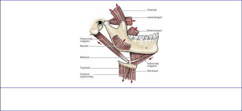
There are three sets of mandibular movements at the temporomandibular joint. These are depression and elevation (opening and closing the jaws), side-to-side (grinding) movements, protraction and retraction (protrusion and retrusion). The group of muscles commonly classified as the muscles of mastication—temporalis, masseter and medial and lateral pterygoids—play major roles in these movements; others taking part can be called accessory muscles of mastication (Fig. 6.67).
Figure 6.67 Muscles producing movements of the temporomandibular joint. The direction of their actions is indicated by the arrows.
When the mouth is opened, the mandibular head rotates around a horizontal axis in a hinge-like movement that occurs in the lower compartment of the temporo-mandibular joint, between the head and the inferior aspect of the disc, while a gliding movement occurs in the upper compartment between the disc and the mandibular fossa of the temporal bone. In this sequence of events, the mandible is depressed by the digastric, mylohyoid and geniohyoid muscles, while the infrahyoid muscles act to stabilize the hyoid bone. Forward movement of the mandibular head on to the articular eminence of the temporal bone is effected by the lateral pterygoid muscle, principally its inferior head, while the superior head draws the disc forwards.
Elevation of the mandible (closing the jaw) is produced by the masseters, medial pterygoids and temporalis muscles.
Side-to-side movements are the result of medial and lateral pterygoid activity on one side, alternating with similar activity on the other side. Simultaneous contraction of lateral and medial pterygoid muscles of one side rotates the mandible in the horizontal plane around a vertical axis passing a little behind the mandibular head on the opposite side, which moves slightly laterally, while the head on the side of the contracting muscles is drawn forwards on to the articular eminence.
During protraction (as when jutting the chin forwards), all four pterygoid muscles contract, such that the head and disc are drawn forwards, without depression or elevation of the mandibular body. The normal position is restored by passive recoil of stretched joint structures, aided by contraction of the posterior horizontal fibres of temporalis and the posterior deep fibres of masseter.

Part seventeen. Ear
The ear, which houses the peripheral parts of the auditory and vestibular apparatus, is descriptively divided into the external, middle and internal ear. The external ear consists of the auricle or pinna and the external acoustic meatus, at the medial end of which lies the tympanic membrane, separating the external ear from the middle ear. The middle ear or tympanic cavity (tympanum) is a small space in the temporal bone containing the auditory ossicles (malleus, incus and stapes) and air that communicates with the nasopharynx by the auditory tube. By its medial wall the middle ear adjoins the inner ear, which is composed of the osseous labyrinth, another space within the temporal bone, inside which is the membranous labyrinth containing the auditory and vestibular nerve receptors.
External ear
The auricle or pinna has a skeleton of resilient yellow elastic cartilage which is thrown into folds. The folds give the auricle its characteristic shape. The cartilage is covered on both surfaces with adherent hairy skin; it does not extend into the lobule of the ear. The lobule is a tag of skin containing soft fibrofatty tissue; it is easily pierced for earrings. The cartilage of the auricle is prolonged inwards in tubular fashion as the cartilaginous part of the external acoustic meatus, whose attachment to bone stabilizes the auricle in position. Small anterior, superior and posterior auricular muscles attach the auricle to the scalp and skull, and all are supplied by the facial nerve.
T he external acoustic meatus is a sinuous tube nearly 3 cm in length; it is straightened for introduction of an otoscope by pulling the auricle upwards and backwards. Due to the obliquity of the tympanic membrane at the deep end of the meatus, separating it from the tympanic cavity, its anteroinferior wall is longest and its posterosuperior wall shortest (Fig. 6.68). Its outer third is cartilage, its inner two-thirds bone; in both zones the skin is firmly adherent.
Figure 6.68 Oblique section through the right external ear, middle ear and pharyngotympanic tube: anterior aspect.
The bony part is formed by the tympanic part of the temporal bone, C-shaped in cross-section, the gap in the C being applied to the under surface of the squamous and petrous parts. The cartilaginous portion is likewise C-shaped; the gap is filled with fibrous tissue. Hairs and sebaceous glands abound in the skin of the cartilaginous part. Here also are the ceruminous glands, long coiled tubules like modified sweat glands, which secrete a yellowish-brown wax. The meatus is narrowest at the

isthmus, a few millimetres from the membrane.
The auricle and external meatus are mainly supplied by the posterior auricular and superficial temporal arteries, with the deeper part of the meatus receiving the deep auricular artery (from the maxillary) which enters the meatus through the squamotympanic fissure. There are corresponding veins.
Lymphatic drainage is to occipital, preauricular and superficial cervical nodes.
The main cutaneous nerves are the great auricular and auriculotemporal nerves, with a small contribution from the vagus. The great auricular supplies the whole of the cranial surface of the auricle (C2, with a little overlap from the lesser occipital at the top) and the lower part of the lateral surface. The auriculotemporal supplies the upper part of the lateral surface and most of the meatal skin. The auricular branch of the vagus (Arnold's nerve) supplies small areas of skin on the cranial auricular surface, posterior wall and floor of the meatus and adjoining part of the tympanic membrane. The facial nerve may also contribute via a communication with the vagus.
Middle ear
The middle ear is an air space in the temporal bone (Fig. 6.68). It contains the three auditory ossicles whose purpose is to transmit sound vibrations from the tympanic membrane in its lateral wall to the inner ear via its medial wall. The cavity of the middle ear, the tympanic cavity or tympanum, is really the intermediate portion of a blind diverticulum from the respiratory mucous membrane of the nasopharynx. From front to back the diverticulum consists of the auditory tube, the tympanic cavity, and the mastoid antrum and air cells.
Tympanic cavity
The tympanic cavity, about 15 mm in anteroposterior and vertical diameters, is the shape of a biconcave lens. Its lateral wall is largely occupied by the tympanic membrane, which extends upwards for 10 mm from the floor and bulges inwards to within a couple of millimetres of the medial wall. Above the membrane the temporal bone is hollowed out into the epitympanic recess.
The tympanic membrane is a thin fibrous structure covered externally with a thin layer of stratified squamous epithelium and internally with low columnar epithelium. The framework consists of collagen fibres. The membrane is circular, 1 cm in diameter, and lies obliquely at 55° with the external acoustic meatus, facing downwards, forwards and laterally (Fig. 6.68). It is concave towards the meatus. At the depth of the concavity is a small depression, the umbo. When the drum is illuminated for inspection, the concavity of the membrane produces a ‘cone of light’ radiating from the umbo over the anteroinferior quadrant. The handle of the malleus is firmly attached to the inner surface of the membrane. From the lateral process of the malleus two thickened fibrous folds (mallear folds) diverge up to the margins of the tympanic bone; between them the small upper segment of the membrane is lax (pars flaccida, Shrapnell's membrane). This part and the neck of the malleus are crossed internally by the chorda tympani. The rest of the membrane, the main part, is the pars tensa. It is held tense by the inward pull of the tensor tympani muscle. Its tension is affected by difference of pressure in the tympanic cavity and external meatus in cases of auditory tube obstruction. The tympanic membrane is thickened at its circumference and slotted into a groove in the tympanic plate.

The tympanic membrane is supplied by the deep auricular artery (maxillary) on the meatal side, and on the mucosal side the stylomastoid artery (posterior auricular) forms a circular anastomosis with the anterior tympanic branch of the maxillary round the margin of the membrane.
On the meatal surface the tympanic membrane is supplied by the auriculotemporal nerve, supplemented by the vagus. The tympanic branch of the glossopharyngeal nerve, via the tympanic plexus, supplies the mucosal surface.
The medial wall of the tympanic cavity (which is also the lateral wall of the internal ear) has as its most prominent feature the promontory (Fig. 6.69), due to the first turn of the cochlea and indented with fine grooves by the tympanic plexus. Above it is a horizontal ridge for the canal for the facial nerve, and immediately above that is the (horizontal) bulge due to the lateral semicircular canal. Above and behind the promontory is the oval window (fenestra vestibuli), closed in life by the footpiece of the stapes. Below and behind the promontory is the round window (fenestra cochleae), closed in life by the fibrous secondary tympanic membrane.
Figure 6.69 Medial wall of the right middle ear.
The roof of the tympanum is the tegmen tympani, a laminar projection of petrous bone that roofs in also the canal for the tensor tympani and the tympanic (mastoid) antrum. Above it the temporal lobe lies in the middle cranial fossa (Fig. 6.69).
The floor is a thin plate of bone above the jugular fossa. At the anterior end is the internal opening of the tympanic canaliculus, where the tympanic branch of the glossopharyngeal nerve enters, from the jugular fossa, the external opening of the canaliculus being on the ridge of bone between the fossa and the carotid canal (Fig. 6.19).
The anterior wall is shortened by approximation of roof and floor. It is perforated by the openings of two canals: the lower and larger of these is the bony part of the auditory tube, the upper and smaller is the canal for the tensor tympani muscle (Figs 6.68 and 6.69). The lower part of this wall forms the posterior wall of the carotid canal and is perforated by tympanic branches of the internal carotid artery and sympathetic fibres from the internal carotid plexus.
The posterior wall is deficient above, where there is an aperture, the aditus, which leads back into the mastoid antrum. The ridge for the canal for the facial nerve and the bulge due to the lateral semicircular canal continue backwards along the medial wall of the aditus. Below the aditus a hollow cone, the pyramid, projects into the tympanic cavity (Fig. 6.69); its apex is perforated by the tendon of stapedius. Close to the posterior margin of the tympanic membrane is the tiny posterior canaliculus for the chorda tympani.
The auditory ossicles form by synovial joints a bony chain for transmission of vibrations from the tympanic membrane to the internal ear. The malleus and incus are developed from the proximal end of the first arch cartilage (see Fig. 1.20, p. 25), the stapes comes from the second arch cartilage (see Fig. 1.21, p. 25).
The malleus is shaped like a round-headed club. There is a constriction, the neck, between head and handle. The convex head lies in the epitympanic recess (Fig. 6.68). Its posterior surface has an articular facet for the incus. The narrow neck lies against the pars flaccida of the tympanic membrane. The chorda tympani crosses medial to the neck. The handle projects somewhat backwards down to the umbo; its upper end has a projection, the lateral process. The two form a lateral concavity moulded to the medial convexity of the tympanic membrane; the periosteum of lateral process and handle is firmly fixed to the fibrous layer of the membrane. The mallear folds are attached to the apex of the lateral process. The tiny anterior process is directed forwards from just below the neck; it is embedded in the fibres of the anterior ligament, which passes through the petrotympanic fissure to the spine of the sphenoid; like the sphenomandibular ligament it is derived from the perichondrium of the first arch cartilage.
The incus has a relatively large body and two slender processes or limbs. The body is rounded and laterally compressed. It lies in the epitympanic recess and articulates anteriorly with the head of the malleus (Fig. 6.68). The short limb projects backwards to lie in a shallow fossa in the posterior wall just below the aditus. The long limb projects down into the cavity of the middle ear, just behind and parallel with the handle of the malleus. Its tip hooks medially and is bulbous—the lentiform nodule— for articulation with the stapes.
The stapes has a small head showing a concave facet for articulation with the lentiform nodule. A narrower neck diverges into slender anterior and posterior limbs, which are attached to the base (or footpiece) like a rider's stirrup. This is attached to the oval window by an annular ligament.
The tensor tympani arises from and occupies the canal above the bony part of the auditory tube. The slender muscle ends in a round tendon which passes across the cavity of the middle ear and is inserted into the handle of the malleus. Its nerve supply is from the mandibular nerve via its branch to the medial pterygoid (see p. 364). Contraction of the muscle draws the handle of the malleus inwards, making the drum more highly concave and therefore more tense.
The stapedius arises from the interior of the hollow pyramid. Its tendon emerges from the apex of the pyramid and is inserted into the back of the neck of the stapes. The muscle is supplied from the facial nerve by a branch given off in the facial (stylomastoid) canal. Its action is to retract the neck of the stapes, thus tilting the footpiece in the oval window. Paralysis of the stapedius causes an abnormally increased power of hearing (hyperacusis).
Mastoid antrum and air cells
T he mastoid (tympanic) antrum lies behind the epitympanic recess in the petrous part of the temporal bone. It is connected to the recess by the aditus. Its size is very variable; it may be up to 1 cm in diameter. When large it is covered by a thin layer of bone, when small by a thick layer. Its lateral wall corresponds to the suprameatal triangle at the posterosuperior margin of the external acoustic meatus (see p. 507) and the antrum lies about 15 mm deep to the surface of the bone here. It is roofed by the tegmen tympani.
The mastoid antrum is present at birth and is then almost adult size. During the first year mastoid air cells, lined with adherent mucoperiosteum, burrow out from the mastoid antrum into the thin plate of bone at the bottom of the groove for the sigmoid sinus. Later they pneumatize the mastoid process for a variable distance, even to the tip. They may be separated from the sigmoid sinus and posterior cranial fossa by extremely thin bone.
The thin mucous membrane of the middle ear, continuous with that of the auditory tube and mastoid antrum, adheres to all the structures enumerated above: the walls; ossicles; ligaments; and muscles. The lining epithelium is columnar and ciliated, but squamous and non-ciliated in the antrum and air cells.
The anterior tympanic from the maxillary, the stylomastoid from the posterior auricular (or occipital), and tympanic branches from the internal carotid, middle meningeal and ascending pharyngeal arteries supply the middle ear. Venous drainage is to the pterygoid plexus and superior petrosal sinus. Thrombophlebitis from suppuration in the middle ear may lead to meningitis, and by retrograde venous spread to a cerebral abscess in the temporal lobe. Veins from the mastoid antrum communicate via the mastoid emissary vein with the posterior auricular vein and the sigmoid sinus. Spreading infection from the mastoid antrum and air cells can lead to sigmoid sinus thrombosis, meningitis and a cerebellar abscess.
Lymphatic drainage from the middle ear is to preauricular, retropharyngeal and upper deep cervical nodes.
The mucous membrane of the middle ear is supplied by branches of the tympanic plexus. This is mainly formed by the tympanic branch of the glossopharyngeal nerve (Jacobson's nerve), which forms a fine plexiform network on the promontory (Fig. 6.69). It is joined by sympathetic fibres from the internal carotid plexus which enter the tympanic cavity through the wall of the carotid canal.
Since the middle ear and the external ear are supplied by branches of the trigeminal, glossopharyngeal and vagus nerves, pain in the ear (otalgia) may be referred from other areas supplied by these nerves, especially the pharynx, larynx, posterior part of tongue and teeth.
The plexus gives off the lesser petrosal nerve. This contains preganglionic parasympathetic fibres from the inferior salivary nucleus, destined to supply the parotid gland via the otic ganglion. The fibres enter the plexus with the glossopharyngeal tympanic branch. The nerve leaves the middle ear through a canaliculus in the anterior wall above the auditory tube and emerges in the middle cranial fossa through a small hiatus lateral to that for the greater petrosal nerve (see Fig. 8.4, p. 507); it then passes through the foramen ovale to reach the otic ganglion (Figs 6.21 and 6.27).

Facial nerve and the ear
The facial nerve itself is not within the middle ear cavity but passes through the petrous bone from the internal acoustic meatus to the stylomastoid foramen in three directions, laterally, posteriorly and downwards, in that order. First the main trunk of the nerve runs laterally from the internal acoustic meatus with the nervus intermedius, which contains the parasympathetic fibres for the pterygopalatine and submandibular ganglia (see Fig. 1.15, p. 21), and also taste fibres from the anterior part of the tongue and the soft palate. The two parts of the facial nerve here lie above the vestibule, with the cochlea in front and the semicircular canals behind. The nervus intermedius now joins the main nerve at the geniculate ganglion. The greater petrosal nerve passes forwards from the ganglion through a canal in the petrous bone and emerges from a hiatus to lie on a groove on the floor of the middle cranial fossa (Figs 6.101 and 8.4, p. 507), which it leaves through the foramen lacerum to become part of the nerve of the pterygoid canal (see p. 370).
The facial nerve now passes backwards from the ganglion in the canal which raises the ridge on the medial wall of the tympanic cavity above the promontory and below the prominence of the lateral semicircular canal (Fig. 6.69). Finally the nerve passes downwards medial to the aditus to the antrum and emerges from the stylomastoid foramen. The nerve to stapedius and the chorda tympani leave this part of the nerve in the middle ear.
The chorda tympani is a mixed visceral nerve, containing taste fibres from the tongue (cell bodies in the geniculate ganglion) and secretomotor fibres for the salivary glands of the floor of the mouth (cell bodies in the superior salivary nucleus in the pons). At about 6 mm above the stylomastoid foramen the chorda tympani leaves the facial nerve in the facial canal and pierces the posterior wall of the tympanic cavity (Fig. 6.27). It runs forward over the pars flaccida of the tympanic membrane and the neck of the malleus, lying just beneath the mucous membrane (Fig. 6.21). It passes out of the front of the middle ear and emerges from the medial end of the petrotympanic fissure (Fig. 6.35 and see p. 507), grooves the medial side of the spine of the sphenoid, and joins the lingual nerve 2 cm below the base of the skull.
Auditory tube
The auditory tube (pharyngotympanic tube, Eustachian tube) connects the nasopharynx with the middle ear. Over 3 cm long, it slopes from the middle ear forwards and medially at 45° and downwards at 30°. Like the external acoustic meatus it has bony and cartilaginous parts, but the proportions are reversed.
The bony part, over 1 cm long, tapers down from the anterior wall of the middle ear to its orifice where it perforates the petrous part of the temporal bone. This is the narrowest part of the tube, the isthmus; it lies posteromedial to the spine of the sphenoid and lateral to the carotid canal. It is lined with adherent mucoperiosteum, which is surfaced by ciliated columnar epithelium and, as in the middle ear, has no glands.
The cartilaginous part, over 2 cm long, joins the bony orifice at the isthmus and is lodged in the groove between the greater wing of the sphenoid and the apex of the petrous part of the temporal bone (Fig. 6.35). It is made of elastic cartilage, which in transverse section resembles an inverted J (long limb medial) open inferolaterally where it is closed by fibrous tissue. It enlarges from the isthmus

like a trumpet, with its open end expanded, particularly the long posterior limb which forms the tubal elevation in the lateral wall of the nasopharynx (Fig. 6.39). The mucosa is lined by ciliated columnar cells and has mucous glands. The cilia beat towards the nasopharynx, thus protecting the middle ear from airborne particles, including bacteria.
The ostium (opening) of the tube is attached to the back of the medial pterygoid plate just below the skull base. The tubal elevation is made more prominent, especially in the young, by lymphoid follicles in the mucous membrane (tubal tonsil). The posterior limb is elongated by the vertical salpingopharyngeal fold, draped over salpingopharyngeus.
The pharyngobasilar fascia is attached to the lower part of the tube; lateral to this the tensor palati arises outside the pharynx, and medial to this the levator palati arises inside the pharynx. Both are attached in part to the tube and contract during swallowing which opens the tube and allows equalization of air pressure on the two sides of the tympanic membrane. Air is slowly lost from the middle ear and mastoid cavities by absorption into the capillaries thereof.
The blood supply of the tube is from the ascending pharyngeal and middle meningeal arteries. Its veins drain into the pharyngeal plexus. The lymphatic drainage is to retropharyngeal lymph nodes. The nerve supply is by the pharyngeal branch of the pterygopalatine ganglion (maxillary nerve) and the tympanic plexus (glossopharyngeal nerve).
Internal ear
The internal ear is buried in the petrous part of the temporal bone and is practically full adult size at birth. It consists of a complex series of connected cavities, the osseous labyrinth, within which lies a correspondingly complex fluid-filled sac, the membranous labyrinth. The fluid it contains is endolymph and, because the membranous labyrinth is smaller than the osseous, its walls are not all pressed tightly against the bone but are mostly separated from it by another fluid, perilymph. The endolymph and perilymph do not communicate with one another.
The parts of the osseous labyrinth, in order from front to back, are the cochlear canal (or cochlea), the vestibule, and the semicircular canals (Figs 6.70 and 6.71; these illustrations depict casts of the bony cavity). The parts of the membranous labyrinth (Fig. 6.72) are the cochlear duct (within the cochlear canal and concerned with hearing), the utricle and saccule (within the vestibule and concerned with static balance), and the semicircular ducts (within the semicircular canals and concerned with kinetic balance).
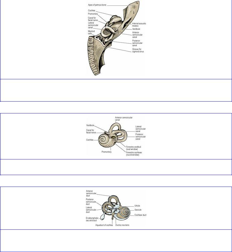
Figure 6.70 Left osseous labyrinth in the temporal bone, from above and behind. The cochlea is at the front, the vestibule in the middle and the semicircular canals at the back.
Figure 6.71 Left osseous labyrinth, from the lateral side.
Figure 6.72 Left membranous labyrinth, from the medial side. The stippled part represents the osseous labyrinth.
Osseous labyrinth
The cavity of the osseous labyrinth is lined by endosteum and opens into the medial wall of the middle ear through the oval window (closed in life by the footpiece of the stapes) and the round window (closed in life by the secondary tympanic membrane) (Figs 6.71 and 6.69). It also opens into the posterior cranial fossa through the aqueduct of the vestibule (Fig. 6.105), closed in life by the endolymphatic duct, and through the aqueduct of the cochlea (Fig. 6.72) through which perilymph is

believed to drain into the cerebrospinal fluid. The source of perilymph is uncertain; it may be derived from cerebro-spinal fluid or as an ultrafiltrate from perilymphatic blood capillaries.
The cochlea is a conical snail-shaped cavity in the petrous bone. It consists of two and three-quarter spiral turns of a tapering canal. The bony canal is of greatest calibre at the basal turn; this part projects laterally, producing the promontory on the medial wall of the middle ear (Fig. 6.69).
The axial conical bony stem around which the canal spirals is the modiolus. The base of the modiolus lies at the fundus of the internal acoustic meatus and its apex lies across the long axis of the petrous bone, pointing towards the middle ear. The apex of the modiolus is overlaid by the blind extremity of the apical turn of the cochlea.
From the modiolus a spiral shelf of bone projects into the canal, like a thread projecting from a screw. This is the spiral lamina. Its projection is widest in the basal and narrowest in the apical turn. The membranous cochlear duct (Figs 6.72 and 6.73) is attached to the spiral lamina and to the outer bony wall of the canal.
Figure 6.73 Transverse section through a single turn of the cochlea.
The bony canal of the cochlea is thus partitioned by the spiral lamina and the membranous cochlear duct, which contains endolymph. The canal on the apical side of the partition is the scala vestibuli, that on the basal side the scala tympani (Fig. 6.73); they contain perilymph. They communicate with each other around the blind apical extremity of the cochlear duct.
The basal turn of the cochlea sees the termination of the spiral lamina. Here the scala tympani is sealed off into a blind end. There are two holes in this cul-de-sac. One leads laterally into the middle ear—the round window—which is closed in life by the secondary tympanic membrane. The other is the beginning of a canal, the aqueduct of the cochlea (perilymphatic duct), which leads down through the substance of the petrous bone and opens into the cochlear canaliculus, below the internal acoustic meatus, in the anterior compartment of the jugular foramen. The aqueduct of the cochlea is patent in life. The arachnoid mater is attached to the margin of its opening, so that perilymph draining down the aqueduct is received into the cerebrospinal fluid in the subarachnoid space.
The modiolus is perforated spirally at its base in the internal acoustic meatus by the branches of the cochlear nerve (Fig. 6.108). These run into the modiolus and fan out spirally towards the base of the spiral lamina. Here the spiral ganglion containing their bipolar cell bodies lies in the spiral canal at the base of the spiral lamina of the modiolus (Fig. 6.73). This is the counterpart of the posterior root ganglion of a spinal nerve (i.e. it contains the cell bodies of the first neuron of a sensory pathway). The spiral ganglion (cochlear nerve) connects the sound receptors in the spiral organ (see below) with the cochlear nuclei in the brainstem.
Figure 6.108 Left internal acoustic meatus with the osseous labyrinth.
The vestibule is a hollow in the petrous bone which contains the membranous saccule and utricle (Figs 6.70 and 6.72). The scala vestibuli of the cochlea opens into the front of the vestibule and the five orifices of the semicircular canals open posteriorly.
The medial wall abuts on the internal acoustic meatus and is perforated by minute foramina for the branches of the vestibular nerve to the saccule, utricle and semicircular canals.
The lateral wall of the vestibule abuts on the middle ear behind the promontory. Here is the opening of the oval window (Fig. 6.71), closed in life by the foot-piece of the stapes and its annular ligament.
The semicircular canals lie in three planes at right angles to each other (Fig. 6.71). Each is about two-thirds of a circle; in length along the curve they measure about 20 mm. Their calibre is 1 mm except at one end, where each is dilated as the ampulla to a calibre of 2 mm.
The anterior (superior) semicircular canal is placed in a vertical plane across the long axis of the petrous bone, convexity upwards, ampulla laterally (Fig. 6.70). Its convexity produces the arcuate eminence on the upper surface of the petrous bone in the middle cranial fossa (see p. 448). It lies highest of the three canals.
The posterior semicircular canal is placed in a vertical plane in the long axis of the petrous bone, convexity backwards, ampulla below. The ampulla is innervated separately by a branch of the vestibular nerve which pierces the foramen singulare in the internal acoustic meatus (Fig. 6.108).
The lateral semicircular canal is placed 30° off the horizontal plane, convexity backwards and laterally, ampulla anteriorly. The ampulla bulges the medial wall of the aditus and epitympanic recess above the facial canal. The lateral semicircular canal lies horizontal if the head nods 30° forwards. The canal opens by each end separately into the back of the vestibule. The anterior and posterior canals open separately at their ampullated ends, but their non-ampullated ends fuse into a common canal. Thus only five openings connect the three canals with the cavity of the vestibule.
The anterior and posterior canals, lying across and along the axis of the petrous bone, are each at 45° with the sagittal plane. Thus the posterior canal of one side lies parallel with the anterior canal of the opposite side.
Membranous labyrinth
The membranous labyrinth is a reduced replica of the hollow bony labyrinth (Fig. 6.72). It consists of one continuous closed cavity containing endolymph. The membranous covering consists of three layers. The outer fibrous layer is vascular and in places adherent to the endosteum of the bony labyrinth forming the stria vascularis which produces endolymph. The intermediate layer is homogeneous like a basal lamina and the inner epithelial layer is elaborated in three places into receptors of sound, static balance and kinetic balance, supplied by the cochlear (hearing) and vestibular (balance) divisions of the eighth nerve.
The cochlear duct is the spiral anterior part of the membranous labyrinth which contains the sound receptors. It is attached to the apical surface of the spiral lamina and to the outer bony walls of the cochlea. It commences at a blind extremity at the apex of the cochlea and after two and three-quarter spiral turns ends in a bulbous extremity in the basal turn of the cochlea. The minute ductus reuniens connects this extremity with the saccule (Fig. 6.72).
Two membranes enclose the duct of the cochlea. It is triangular in cross-section ( Fig. 6.73). One side of the triangle is formed by the basilar membrane which extends in the line of the spiral lamina to the outer bony wall of the cochlea. Throughout its length it supports the spiral organ.
Another side is formed by the delicate vestibular (Reissner's) membrane which passes obliquely across the cochlea on the apical side of the basilar membrane. Connecting the two membranes, and completing the triangle, is the endosteum of the outer wall of the cochlea, which is thickened here to form the spiral ligament.
In the spiral organ (of Corti) the sensory hair cells, the receptors for hearing, are lodged on supporting cells. The ‘hairs’ are modified microvilli (stereocilia) and are overlaid by the tectorial membrane, a sheet of keratin-like protein that projects from beneath the inner attachment of the vestibular membrane (Fig. 6.73). The hair cells are supplied by the dendrites of the spiral ganglion.
The mechanism of the organ of Corti is not fully understood. It is probable that sound vibrations are communicated from the perilymph to the endolymph through the delicate vestibular membrane and cause appropriate fibres in the basilar membrane to bulge. The overlying hair cells are thus stimulated.
The saccule is a fibrous sac that lies in the lower front part of the vestibule, connected to the basal part of the cochlear duct by the very small ductus reuniens. The utricle is a similiar sac in the upper and back part of the vestibule and receives the five openings of the three semicircular ducts (Fig. 6.72). The two sacs lie with their adjacent walls in contact; a small duct leads from each and they unite in a Y-shaped manner to form the endolymphatic duct. This lies in the aqueduct of the vestibule and projects as a blind diverticulum, the endolymphatic sac, beneath the dura mater of the posterior cranial fossa. Endolymph is absorbed by the epithelium of the sac.
The medial wall of the saccule and the floor of the utricle are thickened to form the maculae, the
areas that contain the sensory receptors for static balance. As in the cochlear duct, they are called hair cells (with stereocilia), and are here overlaid by the gelatinous otolithic (statoconial) membrane which contains particles of calcium carbonate (otoliths or statoconia).
The semicircular ducts are only a quarter the calibre of the bony canals except at the ampullae, which they almost fill. Each membranous duct is adherent by its convexity to the wall of the bony canal in which it lies. The ducts open into five orifices in the back of the utricle inside the elliptical recess.
A transverse crest on the medial surface of each ampulla forms the crista, containing the sensory receptors (hair cells) for kinetic balance, here overlaid by the gelatinous cupula.
The hair cells of the maculae of the utricle and saccule, and the cristae of the semicircular ducts, are stimulated by movements of the otolithic membranes or cupulae respectively in response to head movements, e.g. changes in position, speed of change, etc. They are supplied by the vestibular nerve.
Blood supply of the labyrinth
The labyrinthine artery (from the basilar or its anterior inferior cerebellar branch) divides in the internal acoustic meatus into branches which accompany the cochlear and vestibular nerves to the labyrinth. Branches of the stylomastoid artery assist. The veins unite to form a labyrinthine vein which leaves the internal acoustic meatus and joins the inferior petrosal sinus. Various irregular veins penetrate the petrous bone independently to open into the superior petrosal sinus. A small vein lies in each aqueduct; that in the aqueduct of the cochlea joins the inferior petrosal sinus, that in the aqueduct of the vestibule joins the superior petrosal sinus.
Distribution of the eighth nerve
The cochlear nerve enters the front of the inferior part of the fundus of the internal aconstic meatus in spiral fashion to reach the organ of hearing (Fig. 6.108). The lower division of the vestibular nerve supplies the macula of the saccule and, through the foramen singulare, the ampulla of the posterior semicircular duct. The upper division of the vestibular nerve supplies the macula of the utricle and the ampullae of the anterior and lateral semicircular ducts. The cell bodies of the cochlear fibres lie in the spiral ganglion in the base of the spiral lamina. The cell bodies of the vestibular fibres lie in the vestibular ganglion in the depths of the internal acoustic meatus.

Part eighteen. Vertebral column
The vertebral column (spine) forms the central axis of the skeleton. It supports the skull and gives attachment, by way of the ribs, to the thoracic cage and, by way of this cage, to the pectoral girdle and upper limb. By the pelvic girdle it is strongly united to the lower limbs, which serve the double function of support and propulsion. The great strength of the column comes from the size and architecture of the bony elements, the vertebrae, and the ruggedness of the ligaments and muscles that hold them together. This great strength is combined with great flexibility; the column is flexible because it has so many joints so close together. Finally, the vertebral column contains in its cavity the spinal cord, to which it gives protection.
The vertebral column is made up of five parts with individual vertebrae peculiar to each: cervical, thoracic, lumbar, sacral and coccygeal.
In the fetus in utero the column lies flexed in its whole extent, like the letter C. This anterior flexion or concavity is the primary curvature of the column, and it is retained throughout life in the thoracic, sacral and coccygeal parts. After birth secondary extension of the column produces the secondary curvatures with an anterior convexity (i.e. lordosis) in the cervical and lumbar regions, the former associated with muscular support of the head and the latter with that of the trunk (see p. 35).
As the secondary curvatures develop in the neck and lumbar regions the vertebral column is opened out from its original C shape, and elongated into a vertical column characterized by gentle sinuous bends. These bends give a certain resilience to the column, but the actual shock-absorbing factors in the spinal column are the intervertebral discs.
General features of vertebrae
The general features of vertebrae are best exemplified by a thoracic vertebra. It consists of a ventral body and a dorsal vertebral or neural arch; they enclose between them the vertebral foramen (vertebral canal is the collective name given to the whole series of foramina when the vertebrae are strung together as a column). From the neural arch three processes diverge: in the posterior midline, the spinous process or spine, and on either side the transverse processes. That part of the neural arch between spinous process and transverse process is the lamina, that between transverse process and body is the pedicle. The vertical height of the pedicle is less than that of the body, to allow room for passage of the spinal nerve through the intervertebral foramen between the pedicles of adjacent vertebrae (Fig. 6.76). At the junction of lamina and pedicle (i.e. at the root of the transverse process) are articular processes, superior and inferior, which have hyaline cartilage facets for the synovial joints between the neural arches. The direction of the facets determines the nature of the movement possible between adjacent vertebrae.
Each cervical vertebra has a foramen in the transverse process (foramen transversarium or vertebrarterial foramen) and it has no costal facets. Each thoracic vertebra has costal facets on the side of the body. Each lumbar vertebra has neither a foramen in the transverse process nor costal facets. These two features, foramen in the transverse process and presence or absence of costal facets, serve to distinguish cervical, thoracic and lumbar vertebrae.
During its development a vertebra ossifies in three parts, the centrum and the right and left halves of the neural arch. In the thoracic region costal elements develop separately as the ribs, which

articulate with the vertebrae. The centrum is not the same thing as the anatomical body of a vertebra. Part of the neural arch is incorporated into the body of the vertebra, and the neurocentral junction lies anterior to the costal facets on the body of a thoracic vertebra; hence these facets lie on the neural arch, and not on the centrum.
Costal elements develop in association with all vertebrae. But, except in the thoracic region, the costal elements are vestigial and fuse with the neural arches to become incorporated into the vertebrae. The foramen in the transverse process of a cervical vertebra is produced by this fusion (Fig. 6.74). The costal element consists of the anterior bar and tubercle, the intertubercular lamella and the posterior tubercle.
Figure 6.74 Essential characteristics of cervical, thoracic and lumbar vertebrae (as viewed from above).
A cervical rib is due to the elongation of the costal element of C7 vertebra. It presents as either bony elements or fibrous tissue bands, passing down from C7 vertebra to the first rib (Fig. 6.75). The subclavian artery and lowest root (T1) of the brachial plexus become displaced upwards over such a rib or band, and pressure upon the neurovascular structures from below may cause severe symptoms. The pressure produced by a thin fibrous band may do more harm than that due to a smooth bony rib. The presence of a fibrous band may be inferred if the anterior tubercle of C7 vertebra is enlarged. The patient whose radiograph is shown in Figure 6.75 had symptoms on the right side which were due to a fibrous band, and the well-ossified cervical rib on the left side produced no symptoms. When a cervical rib is well developed the brachial plexus is more likely to be prefixed (i.e. its roots are C4– 8), thus preserving the normal nerve to rib relationship.

Figure 6.75 Radiograph of a left cervical rib. On the right the anterior tubercle of C7 vertebra is greatly enlarged; a fibrous band passed from this tubercle to the first rib.
The so-called transverse processes of the lumbar vertebrae are in reality costal elements. The true transverse process is contracted into a small mass of bone which is grooved by the medial branch of the posterior ramus of the spinal nerve. Above the groove lies the small mamillary process (on the superior articular facet) and below the groove is found the tiny accessory tubercle (Figs 6.94 and 6.96).
Figure 6.94 Typical lumbar vertebra, from above.
Figure 6.96 L3 vertebra, from behind.
The five sacral vertebrae are fused into a single bone and so, too, are the five costal elements. The latter produce the lateral mass of the sacrum, lying lateral to the transverse tubercles (lateral crest) on

the back of the sacrum and extending between the anterior sacral foramina on the front of the bone to the side of the sacral vertebral bodies (Figs 6.97 and 6.98). The auricular surface for the sacroiliac joint lies wholly on the lateral mass.
Figure 6.97 Sacrum: pelvic surface.
Figure 6.98 Sacrum from behind. The five pedicles and laminae have been cut through on the left to show the sheaths of dura mater around the nerve roots.
Vertebral joints
Adjacent vertebrae are held together by strong ligaments and small joints. The vertebrae articulate between their bodies and between their neural arches. These joints are very different from each other, and they allow a greater range of movement between the neural arches than between the bodies.
Joints between the bodies
The bodies of adjacent vertebrae are held together by the strong intervertebral disc, and by the anterior and posterior longitudinal ligaments.
A n intervertebral disc is a secondary cartilaginous joint, or symphysis. The upper and lower surfaces of each vertebral body are covered completely by a thin plate of hyaline cartilage. These

plates are united by a peripheral ring, the annulus fibrosus, which has a narrow outer collagenous zone and a wider inner fibrocartilaginous zone. It consists of concentric laminae, the fibres of which lie at 25–45° with the horizontal plane. Alternate layers of the annulus contain fibres lying at right angles to each other. By this means the annulus is able to withstand strain in any direction. Inside the annulus is a bubble of semiliquid gelatinous substance, the nucleus pulposus, derived from the embryonic notochord. (The notochord extended originally as far cranially as the sella turcica of the skull, but it disappears except in the nucleus pulposus of each intervertebral disc and in the apical ligament of the atlas.) The nucleus pulposus in the embryo lies at the centre of the disc. Subsequent growth of the vertebral bodies and discs occurs in a ventral and lateral direction (the spinal cord prevents a corresponding growth dorsally). Thus in the adult and especially in the lumbar region the nucleus pulposus lies nearest to the back of the disc (Fig. 6.76) and if it herniates through the annulus it will be most likely to do so posteriorly and press on the roots of a spinal nerve near the intervertebral foramen, or on the spinal cord itself.
Figure 6.76 Bisected vertebral column (lumbar region). The left half is seen from the right, so showing the inside of the vertebral canal, intervertebral discs in section, and the boundaries of two intervertebral foramina.
The nucleus pulposus accounts for 15% of the whole disc. It contains about 90% water at birth, and this diminishes to about 70% in old age. The water content keeps the nucleus under constant pressure since its mucoprotein (proteoglycan) component has the property of imbibing and retaining water. Imbibition of water by the nucleus accounts for the overnight increase in height of a young adult by 1 cm; when upright during the day, water is squeezed out. In old age there is little height change between night and morning; imbibition of water becomes less and the nucleus more fibrous. In astronauts, who have been relieved of gravity, there may be a height increase of several centimetres.
The relationship of nerve roots to intervertebral discs is of clinical importance, and is best understood by considering the lowest disc—the fifth lumbar or lumbosacral disc—which is the one most frequently herniated or prolapsed (‘slipped disc’), with its nucleus pulposus being extruded posterolaterally. At the level of this fifth lumbar disc, the fifth lumbar nerve root within its dural sheath has already emerged from the intervertebral foramen, hugging the pedicle of L5 vertebra and so is not lying low enough to come in contact with the fifth lumbar disc. The roots that lie behind the posterolateral part of this disc are those of the first sacral nerve, and these are the ones liable to be irritated by a prolapse. Thus the general rule throughout the vertebral column is that when a disc

herniates (usually posterolaterally rather than in the midline) it may irritate the nerve roots numbered one below the disc: S1 nerve by L5 disc; L5 nerve by L4 disc; and C8 nerve by C6 disc (there are 8 cervical nerve roots and 7 cervical vertebrae). These are the commonest clinical examples.
The posterolateral lip, or uncus, on the upper surface of cervical vertebrae 3 to 7 (see p. 432) may appear to form a joint with the side of the vertebra above because a small cavity may develop in this region (the so-called neurocentral, uncovertebral or Luschka's joint). It is disputed as to whether these are synovial joints, or are due to degenerative changes in the adjacent disc.
The anterior longitudinal ligament extends from the basiocciput of the skull and the anterior tubercle of the atlas to the front of the upper part of the sacrum. It is firmly united to the periosteum of the vertebral bodies, but is less so over the intervertebral discs. It is a flat band, broadening gradually as it passes downwards.
The posterior longitudinal ligament extends from the back of the body of the axis to the anterior wall of the upper sacral canal. It narrows gradually as it passes downwards. It has serrated margins, being broadest over the discs to which it is firmly attached, and narrow over the vertebral bodies to which it is more loosely attached in order to give free exit to the basivertebral veins emerging from the backs of the bodies (Fig. 6.10). At the top the ligament is continued above the body of the axis as the tectorial membrane (see p. 426 and Fig. 6.80).
Figure 6.80 Ligaments of the atlanto-occipital and atlantoaxial joints. The posterior part of the occipital bone and the laminae of the upper cervical vertebrae have been removed.
Joints between the arches
The pedicles of adjacent vertebrae are not attached to one another, so leaving a space—the intervertebral foramen—for the emergence of the spinal nerve. All other parts of the neural arch and its processes are joined to their adjacent companions: the articular processes by synovial joints, and the remainder by ligaments, of which the most important are the ligamenta flava and the supraspinous ligament.
The joints between the articular facets of the superior articular processes of one vertebra and the articular facets of the inferior articular processes of the vertebra above are termed the zygapophyseal joints (or simply known as facet joints). They are synovial with a simple capsule which blends medially with a ligamentum flavum. The articular surfaces allow gliding of one on the other; the

direction of the surfaces determines the direction of the possible movements between adjacent vertebrae. The joints have a nerve supply from the nerve of their own segmental level and from the nerve of the segment above. One nerve thus supplies two joints; this may be important when considering nerve root pain which can be referred from facet joints. Although most of the weight transmission by the vertebral column takes place via the vertebral bodies and intervening discs, a small amount does occur through these joints.
The paired ligamenta flava are yellowish from their high content of elastic fibres. They join the contiguous borders of adjacent laminae (Fig. 6.76). They are attached above to the front of the upper lamina and below to the back of the lower lamina. Thus adjacent laminae and ligamenta flava overlap each other slightly like the tiles of a roof. The ligamenta extend from the facet joints to the midline where they partially fuse; small veins connecting the internal and external vertebral venous plexuses may pass between a pair of ligamenta. They are stretched by flexion of the spine; in leaning forward their increasing elongation becomes an increasing antigravity support.
The supraspinous ligaments join the tips of adjacent spinous processes (Fig. 6.76). They are strong bands of white fibrous tissue and are lax in the extended spine. They are drawn taut by full flexion, and then support the spine (no action currents can be obtained from the erector spinae muscles when the spine is fully flexed, as in touching the toes). They are indistinct below the L4 spine where the lumbar fascia is thick. In the neck they are replaced by the ligamentum nuchae (see p. 430).
The interspinous ligaments are relatively weak sheets of fibrous tissue uniting spinous processes along their adjacent borders (Fig. 6.76). They are well developed only in the lumbar region. They fuse with the supraspinous ligaments.
The intertransverse ligaments are similar weak sheets of fibrous tissue joining the transverse processes along their adjacent borders.
Vertebral column
In the normal erect posture the vertebral column supports the head and trunk on the pelvis. (The pelvis is supported by the lower limbs in standing and by its own ischial tuberosities in sitting.) This support is maintained by the bodies of the vertebrae and the intervertebral discs, which thus become progressively larger from above downwards. The curvatures of the spine are produced partly by the wedge-shape of the vertebral bodies, but mostly by the wedge-shape of the intervertebral discs. This is particularly noticeable in the lower part of the spine; L5 vertebra is usually wedge-shaped and the disc between it and the sacrum is very thick anteriorly.
The vertebral canal (see p. 421) becomes progressively smaller from above downwards. It is closed anteriorly by the vertebral bodies, the intervertebral discs and the posterior longitudinal ligament and posteriorly by the laminae and the ligamenta flava. Laterally it is occupied by the pedicles, which are narrower than the height of the vertebral bodies. Thus a series of intervertebral foramina is produced between adjacent pedicles which form the upper and lower boundaries of each foramen. In the thoracic and lumbar regions each intervertebral foramen is bounded in front by the lower part of a body of a vertebra (mainly, in the thoracic region; Fig. 6.92) and the adjacent intervertebral disc (mainly, in the lumbar region; Fig. 6.93), and behind by the facet joint and its capsule. In the cervical region, because the pedicle arises from a little lower down the back of the body, a small part of the
vertebral body below the disc is also included in this anterior boundary (Fig. 6.88). The intervertebral foramina lodge the spinal nerves and posterior root ganglia and give passage to the spinal arteries and veins.
Figure 6.93 T11 and T12 vertebrae, from the left.
Movements of the vertebral column
In general the movements of the spine are simple enough. Flexion and extension, and lateral flexion (abduction) are possible in cervical, thoracic and lumbar regions, though in varying degree in the three parts. Rotation occurs mainly in the thoracic region. Movements of the head occur at the specialized atlanto-occipital and atlantoaxial joints.
Lumbar region
The articular facets lie in an anteroposterior plane; they lock, and greatly limit rotation of the bodies on each other. Flexion and extension are free, and a good deal of lateral flexion is possible.
Thoracic region
The synovial joints between T12 and L1 are lumbar in type (Fig. 6.93); elsewhere the direction of the articular facets on the neural arches is quite different. On any one neural arch the upper facets face backwards and laterally (Fig. 6.74); they lie on the circumference of a circle whose centre lies in the vertebral body. The lower facets face reciprocally forwards and medially. Thus rotation of the bodies on each other is possible, though restricted by the splinting effect of the ribs. As in the lumbar region, flexion and extension occur, as well as ‘lateral flexion’. The thoracic spine is thus the most mobile region of all, but the range of movements is limited by the ribs.
Cervical region
The atlanto-occipital and atlantoaxial joints are specialized for head nodding and head rotation. They are considered below.
The upper articular facets of the other joints face back-wards and upwards; the lower facets face, reciprocally, forwards and downwards. While flexion and extension are free, pure rotation is impossible. Lateral flexion is not a simple movement. The neural arch of the abducted vertebra slides downwards (and therefore backwards) on the concave side and upwards (and therefore forwards) on the convex side, thus inevitably producing slight concomitant rotation.
Special vertebrae and joints

The atlas (Fig. 6.77) lacks a centrum (see p. 422). The vertebral arch has become modified to form a thick lateral mass on each side, joined at the front by a short anterior arch and with a longer posterior arch at the back. The articular facets on the upper and lower surfaces of the lateral mass differ markedly. The upper surface is kidney-shaped and concave for articulation with the occipital condyle, while the lower is round or oval and nearly flat for the lateral atlantoaxial joint. The articular facets are in line with the uncovertebral joints (see p. 424) of the other cervical vertebrae, not with the articular facets on the neural arches. Thus the C1 and C2 nerves send their anterior rami behind, and not in front of, the joints.
Figure 6.77 Superior surface of the atlas. C1 nerve divides into anterior and posterior rami just behind the atlanto-occipital joint (right side), where it lies in the groove beneath the vertebral artery (left side).
The axis is characterized by the dens and a large spinous process (Fig. 6.78). The dens (odontoid process) has an articular facet at the front for the joint with the anterior arch of the atlas. It bears no weight. The weight of the skull is transmitted through the lateral mass of the atlas to the superior articular process of the axis which lies immediately lateral to the dens. The lower articulations of the axis are as for the ordinary cervical vertebrae: body to body with intervening disc and the two uncovertebral joints, and the ordinary articular facets on the neural arch. From the axis downwards the weight of the skull is supported by the vertebral bodies. The bifid spinous process is very large, due to the attachments of muscles of the suboccipital triangle above (Fig. 6.84).
Figure 6.78 Anterior and lateral views of the axis.

Figure 6.84 Suboccipital region and the suboccipital triangle.
The atlanto-occipital and the atlantoaxial joints are adapted to provide freedom of head movement, the former for nodding and lateral flexion, the latter for rotation.
The atlanto-occipital joint is a synovial joint between the convex occipital condyle and the concave facet on the lateral mass of the atlas. Both surfaces are covered with hyaline cartilage. The synovial cavity of the joint is contained in a lax but strong capsule, which is attached to the articular margins of both bones and is innervated by C1 nerve.
The anterior and posterior atlanto-occipital membranes are attached to the upper borders of the respective arches of the atlas and to the outer margins of the foramen magnum (Fig. 6.84). The posterior membrane is deficient at each lateral extremity to allow passage for the vertebral artery and C1 nerve; the lateral margin of the membrane sometimes ossifies, converting the groove for the vertebral artery into a foramen. The membranes are innervated by C1 nerve.
The curved surfaces of the joint are well adapted for head flexion and extension, and allow also for a considerable amount of lateral flexion of the skull on the atlas. In the ordinary erect position the centre of gravity of the skull lies in front of the joint and the head is maintained in position by the tonus of the extensor muscles, notably semispinalis capitis. It is flexed by relaxation of the extensors (i.e. by gravity) and, actively, by longus capitis and the two sternocleidomastoids acting together. The effect of gravity is considerable on account of the weight of the head and, of course, varies with position. Lateral flexion is produced by unilateral contraction of such muscles as sternocleidomastoid, trapezius and splenius capitis. No rotation is possible at the atlanto-occipital joints.
The median atlantoaxial joint is where the dens articulates with the back of the anterior arch of the atlas (Fig. 6.79). The smooth facets seen on the dry bones are covered with hyaline cartilage in life, with a capsule attached to their margins to make this a small synovial joint. The dens is held in position by the transverse ligament; between the two is a relatively large bursa.

Figure 6.79 Atlas and related structures. 1. Dens of axis with apical and alar ligaments. 2. Mucosa of nasopharynx over levator palati. 3. Internal carotid artery and last four cranial nerves (Roman figures). 4. Prevertebral fascia. 5. Superior oblique. 6. Vertebral artery. 7. Posterior ramus of C1. 8. Medulla/spinal cord junction (denticulate ligament, accessory nerve and roots of C1 not labelled). 9. Rectus capitis posterior major. 10. Spinous process of axis. 11. Rectus capitis posterior minor, from posterior arch of atlas. 12. Posterior atlanto-occipital membrane. 13. Spinal dura and arachnoid. 14. Inferior oblique. 15. Tectorial membrane. 16. Transverse band of cruciform ligament. 17. Anterior ramus of C1 giving branch to hypoglossal nerve and ending in longus capitis. 18. Internal jugular vein. 19. Pharyngobasilar fascia. 20. Anterior atlanto-occipital membrane.
On each side there is a lateral atlantoaxial joint (also synovial) between the inferior articular facet of the atlas and the superior articular facet of the axis (Fig. 6.81). The joint surfaces are nearly circular and flat with hyaline cartilage lining, and there is a lax capsule supplied by C2 nerve.
Accessory ligaments connect the axis to the occiput, bypassing the atlas: the tectorial membrane, cruciform ligament, apical ligament and the paired alar ligaments.
The tectorial membrane extends upwards in continuity with the posterior longitudinal ligament (Fig. 6.80). It is attached to the back of the body of the axis and diverges upwards to become attached to the basilar part of the occipital bone above the foramen magnum. It lies in front of the spinal dura mater, which is firmly attached to it (Fig. 6.79).
The transverse ligament is a broad strong band that runs across (and grooves) the back of the dens from its attachment on each side to a tubercle in the ‘hilum’ of the kidney-shaped upper articular facet of the atlas. A weaker longitudinal band runs from the back of the body of the axis to the basiocciput and together with the transverse ligament constitutes a cruciform ligament (Fig. 6.80). This lies in contact with the front of the tectorial membrane. They hold the dens in position; rupture of the ligament and membrane allows the dens to dislocate backwards with fatal pressure on the medulla.
The weak apical ligament joins the apex of the dens to the anterior margin of the foramen magnum, and is a fibrous remnant of the notochord (see p. 23).
The alar ligaments lie obliquely one on either side of the apical ligament. From the sides of the dens they diverge upwards to the margins of the foramen magnum. They are very strong and limit rotation of the head.

Movements at the atlantoaxial joints are simply those of rotation about a vertical axis passing through the dens. The atlas rotates by its anterior arch and transverse limb of the cruciform ligament gliding around the dens and by the lower flat facets on its lateral mass gliding on the superior facets of the axis. The head rotates with the atlas; the curved surfaces of the atlanto-occipital joints do not allow independent rotation of occiput on atlas.
The muscles chiefly responsible for rotation are sterno-cleidomastoid, splenius capitis and the inferior oblique.
The atlantoaxial region of the cervical spine can be visualized in transoral anteroposterior radiographs (Fig. 6.81). The transoral route is also utilized in surgical approaches to this region, with upward retraction of the soft palate and division of the posterior wall of the pharynx.
Figure 6.81 Transoral anteroposterior radiograph.
Blood supply of the vertebrae
The vertebrae are supplied segmentally by the vertebral, ascending and deep cervical, posterior intercostal, lumbar and lateral sacral arteries, which give multiple small branches to the vertebral bodies.
The richly supplied red marrow of the vertebral body drains through its posterior surface by a pair of large basivertebral veins into the internal vertebral venous plexus, which lies inside the vertebral canal, outside the dura (Fig. 6.110). It drains into the external vertebral venous plexus. This intramuscular plexus, which also receives blood from the neural arch, drains into the regional segmental veins (vertebral, posterior intercostal, lumbar and lateral sacral veins), which in turn drain into brachiocephalic veins, superior vena cava, inferior vena cava and internal iliac veins. Venous communication is thus established in the pelvis with veins draining the pelvic viscera, in the abdomen with the renal veins, in the thorax with the azygos venous system (and thereby with the venous drainage of the breast and bronchus), and in the neck with the inferior thyroid veins. In this way, by reflux blood flow through these largely valveless veins, malignant disease may spread from prostate, kidney, breast, bronchus and thyroid gland to the bodies of the vertebrae.
Extensor muscles of the spine
Running along the whole length of the vertebral column from skull to sacrum is a posterior mass of

mainly longitudinal extensor muscles, derived from the outer of the three layers of the body wall and supplied segmentally by posterior rami of spinal nerves. They form a bulge on either side of the midline of the back, often best seen in the lumbar region. In the neck the posteriormost layer is the splenius muscle. Elsewhere the muscles are covered posteriorly by the thoracolumbar fascia.
The deepest muscles are the small interspinales and intertransversales. The remainder form intermediate and superficial masses collectively called transversospinalis and erector spinae, each of which is composed of three groups. Transversospinalis includes the rotatores, multifidus and semispinalis, while erector spinae comprises iliocostalis, longissimus and spinalis.
Deep layer
The interspinales join adjacent borders of spinous processes, alongside the interspinous ligaments. The inter-transversales join adjacent transverse processes; they are best developed in the upper part of the vertebral column.
Transversospinalis (intermediate layer)
As a group these muscles run from transverse processes to spines, hence the name. The rotatores are small, but multifidus and semispinalis form larger muscle masses.
The rotatores are confined to the thoracic spine, the only region where pure rotation occurs. Each extends from the base of a transverse process to the root of the spinous process of the vertebra above (Fig. 6.82).
Figure 6.82 Rotator spinae and levator costae muscles.
Multifidus fibres slope upwards from the back of the sacrum, mamillary processes of lumbar vertebrae, transverse processes of thoracic vertebrae and articular processes of cervical vertebrae to the spinous processes of vertebrae two or three above their level of origin. They commence at the upper part of the sacrum and extend to the upper part of the neck.
Semispinalis lies on the surface of multifidus. Its fibres arise from the transverse processes and slope steeply upwards to the spinous processes. It extends from the lower thoracic region to the skull. Semispinalis thoracis extends from the transverse processes of the lower thoracic vertebrae and each part is inserted into the spinous process six or more vertebrae higher. Semispinalis cervicis arises in continuity at a higher level; the uppermost part of the muscle is inserted into the concavity of the bifid

spinous process of the axis.
Semispinalis capitis is the most powerful part of this layer. It arises from the transverse processes of the upper six thoracic and the articular processes of the lower four cervical vertebrae and is inserted into the occipital bone near the midline, between the superior and inferior nuchal lines (see p. 506). It lies beneath splenius and trapezius and is the chief extensor of the head. It contains a large plexus of veins within and around it.
Erector spinae (superficial layer)
This forms the most powerful muscle group. It commences below, deep to the lumbar fascia, on the back of the sacrum and the inner side of the iliac crest. The thick mass of fibres passes upwards and slightly outwards dividing as it does so into two main bundles, iliocostalis laterally and longissimus medially (Fig. 6.83). The iliocostalis fibres are inserted by shining tendons into the angles of the lower six ribs. From these attachments new muscle bundles arise and each runs up to be attached to the angle of the sixth rib above. From there further fibres run up to reach the transverse processes of the lower four cervical vertebrae. Iliocostalis forms the most lateral part of erector spinae.
Figure 6.83 Parts of the left erector spinae muscle. The most lateral mass is iliocostalis. Most of longissimus (whose lower part is seen adjacent to iliocostalis) has been removed to show various parts of spinalis. Prosection in the Anatomy Museum of the Royal College of Surgeons of England.
The more medial longissimus bundle arising from the sacrum and iliac crest passes up to be inserted into the gutter between transverse processes and ribs; this is longissimus thoracis. At its insertion it is replaced by new fibres on the medial side that pass up to the transverse processes of the cervical vertebrae; this is longissimus cervicis. Longissimus capitis arises from the transverse processes of the lower three or four cervical vertebrae and is inserted into the mastoid process deep to splenius capitis.

The most medial part of erector spinae is the spinalis part. Its fibres run alongside the spinous processes and are small and often indefinite.
Splenius
In the neck, the underlying extensor muscles are bound down by splenius, a flat sheet arising from upper thoracic spinous processes and the supraspinous ligament, and from the ligamentum nuchae. The fibres slope upwards and laterally, deep to trapezius and sternocleidomastoid (Fig. 6.4), and are inserted (as splenius capitis) into the mastoid process and the lateral third of the superior nuchal line, and (as splenius cervicis) into the transverse processes of the upper three or four cervical vertebrae (deep to levator scapulae). The whole muscle is like a bandage that holds down the deeper extensor muscles at the back of the neck. It, like the muscles deep to it, is supplied by posterior rami.
Back of the neck
The back of the neck consists of muscles connecting the skull to the spine and pectoral girdle. In the midline the ligamentum nuchae separates the muscles of the two sides. This is a triangular septum of fibroelastic tissue attached to the external occipital crest, the bifid spines of the cervical vertebrae, the tubercle of the spine of C7 vertebra and the investing layer of deep cervical fascia which encloses the trapezius muscles.
Beneath the trapezius and sternocleidomastoid lies the splenius, and beneath the splenius lie longissimus capitis and semispinalis capitis. When all these are removed the deeper structures of the back of the neck are seen to be divided into upper and lower portions by the prominent backward projection of the massive spinous process of the axis (C2 vertebra). Below this level semispinalis cervicis is seen converging almost vertically upwards to the internal surfaces of the bifid axial spine. Above the spine lie the right and left suboccipital triangles.
Suboccipital triangle
The suboccipital triangle is bounded by rectus capitis posterior major and the superior and inferior oblique muscles (Fig. 6.84). Its floor contains the posterior arch of the atlas and the posterior atlantooccipital membrane. Across the floor runs the vertebral artery, and through the floor emerges the suboccipital (C1) nerve. Across the roof run the greater occipital (C2) nerve and the occipital artery.
Rectus capitis posterior major arises from the outer surface of the bifid spinous process of C2 vertebra and extends obliquely upwards and outwards to be attached to the lateral part of the area below the inferior nuchal line. Its action is to extend the head and rotate it (with the atlas) back towards its own side.
The inferior oblique (obliquus capitis inferior) is attached between the outer surface of the bifid spine of the axis (below rectus capitis posterior major) and the back of the transverse process of the atlas. Its action is to rotate the atlas (and the skull with it) back towards its own side.
The superior oblique (obliquus capitis superior) extends from the upper surface of the transverse process of the atlas to the lateral part of the occipital bone between superior and inferior nuchal lines. It is a lateral flexor of the skull.
Rectus capitis posterior minor is the only muscle attached to the posterior arch of the atlas, from

where it passes vertically upwards to be inserted into the medial part of the area below the inferior nuchal line. It lies deep to rectus capitis posterior major. It weakly extends the head.
All four muscles are supplied by the posterior ramus of C1.
The second part of the vertebral artery (see p. 348) ascends through the foramina in the transverse processes of the upper six cervical vertebrae, anterior to the emerging spinal nerves. It gives a spinal branch into each intervertebral foramen. Its course from C6 to C2 vertebra is vertical. Between the foramina in the transverse processes of the axis and atlas it passes laterally with a pronounced posterior convexity and then loops upwards beside the atlantoaxial joint (Figs 6.84, 6.85, 6.86 and 6.109). This must be to allow for taking up slack during rotation of the atlas on the axis.
Figure 6.85 Vertebral arteriogram: anteroposterior view.
Figure 6.86 Vertebral arteriogram: lateral view.
On emerging from the foramen in the transverse process of the atlas, the third part of the vertebral artery curves backwards and medially behind the lateral mass of the atlas. Here it lies in the floor of the suboccipital triangle before piercing the lateral angle of the posterior atlanto-occipital membrane. It deeply grooves the posterior arch of the atlas before entering the skull through the foramen magnum.
The vertebral veins exist only in the neck, not inside the cranial cavity which the vertebral arteries enter. Blood from the veins in and around semispinalis capitis and from the muscles of the
suboccipital triangle is collected in a plexus of veins that surrounds the second and third parts of the vertebral artery. The thin walls of these veins are adherent to the periosteum of the posterior arch of the atlas and the foramina in the transverse processes. From this plexus two vertebral veins usually emerge, one from the sixth foramen with the vertebral artery and one alone through the foramen in the transverse process of C7 vertebra, and join the brachiocephalic vein at the root of the neck (Fig. 6.9B).
In the groove between artery and bone on the posterior arch of the atlas lies the posterior ramus of the suboccipital (C1) nerve, as it passes backwards to supply the two recti, the two obliques and the upper fibres of semispinalis capitis. The anterior ramus winds round the lateral side of the lateral mass of the atlas between it and the vertebral artery, and passes forwards between rectus capitis lateralis and anterior to join the cervical plexus (Fig. 6.8). Neither branch reaches the skin.
The greater occipital nerve is the long posterior ramus of C2 and emerges below the posterior arch of the atlas. It curls around the lower border of the inferior oblique muscle and passes upwards across the roof of the suboccipital triangle. It pierces semispinalis capitis (first supplying it) and extends up to supply the skin of the scalp up to the vertex.
The occipital artery is a large vessel that passes back along the occipitomastoid suture of the skull deep to digastric and longissimus capitis. It runs across the upper part of the roof of the suboccipital triangle and passes to the scalp. The companion veins form a rich plexus around and within semispinalis capitis.
Part nineteen. Osteology of vertebrae
Cervical vertebrae
A typical cervical vertebra
The broad kidney-shaped body is the same size as, or smaller than, the vertebral foramen (Fig. 6.87). On each side the lateral edge is projected up into a posterolateral lip or uncus, and its lower margin laterally is bevelled reciprocal with this. The anterior surface of the body is concave from above down. Basivertebral veins emerge from the posterior surface.
Figure 6.87 C6 vertebra from above.
The pedicle is attached below the upturned lip on the body. Thus an intervertebral foramen in the neck is bounded in front by both vertebral bodies and the uncovertebral joint and disc between them (Fig. 6.88). Attached to the pedicle and body is the lateral projection of the transverse process, perforated by its foramen. The proximal part of the posterior bar of bone that projects from the pedicle behind the foramen is the true transverse element; it ends in the posterior tubercle, which is part of the costal element, as is the bar of bone that projects from the body in front of the foramen ends and in the anterior tubercle. The anterior tubercles enlarge progressively from C3 to C6. The large C6 anterior tubercle is the carotid tubercle (of Chassaignac) because the common carotid artery can be compressed against it. The anterior and posterior tubercles are joined by the intertubercular lamella, on the concave upper surface of which lies the anterior ramus of the cervical nerve of the same number (Fig. 6.87). The vertebral artery, with its accompanying sympathetic nerve fibres, and the vertebral venous plexus lie in the foramen of the transverse process, and the posterior root ganglion of the nerve of the same number lies behind it (Fig. 6.109).
Figure 6.88 C3 and C4 vertebrae articulated to show the boundaries of the intervertebral foramen.
The laminae enclose a relatively large vertebral foramen, somewhat triangular in cross-section. At

the junction of pedicle and lamina there are superior and inferior articular processes. The upper facets face obliquely up and back; the lower facets face down and forward.
The spinous process is usually bifid.
Atypical cervical vertebrae
The seventh cervical vertebra is often called the vertebra prominens because of its prominent spine. The vertebra is atypical in that its long spine is not bifid but ends in a rounded tubercle, and the foramen in the transverse process does not transmit the vertebral artery. The foramen is small, and contains the posterior vein when the vertebral vein is doubled. The anterior tubercle is very small; the suprapleural membrane is attached to it.
Some of the essential features of the atlas are described on page 425. Its distinctive feature is the lack of a body. The short anterior arch is projected into a tubercle in front (Fig. 6.89), for attachment of the anterior longitudinal ligament and longus colli. The longer posterior arch is grooved by the vertebral artery. The lateral mass carries the weight-bearing articular facets. The lateral mass is projected into the transverse process, which is perforated by the foramen. There are no anterior and posterior tubercles; but the blunt end of the transverse process represents a posterior tubercle and has corresponding muscle attachments: scalenus medius and levator scapulae. The atlas is the widest cervical vertebra, and the tip of its transverse process can be palpated with deep pressure behind the mandibular ramus, 1 cm below and in front of the apex of the mastoid process. The internal jugular vein, crossed by the accessory nerve, lies in front of the transverse process.
Figure 6.89 Atlas, from above. The epiphyseal junction between the ossification centre of the anterior arch and that of the right lateral mass has not fused completely.
Some of the essential features of the axis are described on page 426. The dens (odontoid process) is characteristic, projecting up from the body between a pair of massive weight-bearing lateral masses (Fig. 6.78). The weight is communicated from these shoulders through the body to the body of C3 vertebra.
The transverse process has no anterior and posterior tubercles but ends in a rounded tip, which corresponds with the posterior tubercles of lower cervical vertebrae with equivalent muscle attachments. The foramen in the transverse process is not vertical as in the other vertebrae, but is directed upwards and outwards (Fig. 6.90) to communicate a lateral bend to the vertebral artery to enable it to ascend to the transverse process of the wider atlas (Fig. 6.84). An inferior articular process extends down from the junction of pedicle and lamina; its articular facet faces downwards and forwards as in typical cervical vertebrae (Fig. 6.78). The laminae are thick and rounded and
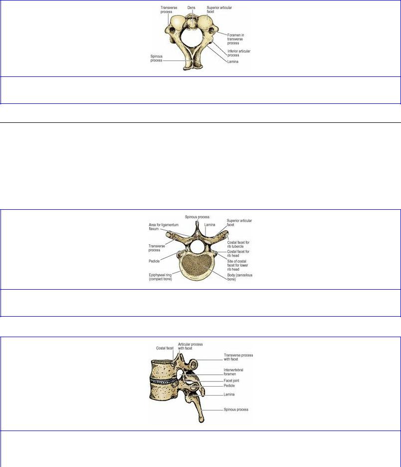
project posteriorly into a massive spinous process (Fig. 6.90), the lower surface of which is grooved and ends in a wide bifurcation; the tip of the spinous process thus resembles an inverted U (Fig. 6.84).
Figure 6.90 Axis, from above.
Thoracic vertebrae
A typical thoracic vertebra
The distinctive feature of a thoracic vertebra is the presence of costal facets (Fig. 6.91). On each side of the body of a typical thoracic vertebra there are a pair of semicircular demifacets (Fig. 6.92). The upper and larger of these is on the body at its junction with the upper border of the pedicle. The smaller lower facet is at the lower border of the body.
Figure 6.91 Typical thoracic vertebra, from above.
Figure 6.92 Two articulated midthoracic vertebrae from the left. The intervertebral foramen is bounded in front by half of the body of the upper vertebra and the intervertebral disc, at the back by

the facet joint, and above and below by pedicles.
The body is concave from above down around its circumference. The posterior surface of the body is concave from side to side, making the body heart-shaped and the vertebral foramen almost circular in outline (Fig. 6.91). Two large foramina open centrally on the back of the body for the basivertebral veins. The upper and lower surfaces of the body have a heart-shaped ring of compact bone at their margins. This is the fused epiphyseal ring of the body, and it encloses a large central area in which cancellous bone reaches the surface.
The pedicle projects back from the upper half of the body. Its upper border is level with the upper surface of the body. The upper border of the pedicle curves up in a slight concavity to a superior articular process. The lower border curves markedly down to the inferior articular process, so making the upper boundary of the intervertebral foramen. This accommodates the thoracic nerve of the same number as the vertebra. The flat laminae slope downwards and backwards from the pedicles to unite in the midline and complete the neural arch. The superior articular process projects up from the junction of pedicle and lamina. It carries an oval articular facet facing backwards and slightly laterally; so the two articular surfaces lie on the arc of a wide circle, permitting rotation of adjacent bodies. The inferior articular facets are reciprocal, facing forwards and somewhat medially.
The spinous processes of successive thoracic vertebrae slope downwards with gradually increasing declivity as far as T7, below which they progressively level out to become almost horizontal at T12. Thus the tips of the upper four spines lie opposite the bodies of vertebrae one lower in the series, the next four spines (those of T5–8) opposite the upper margin of two vertebral bodies lower, and the lowest spines opposite one lower or their own vertebral bodies.
The transverse process projects backwards as well as laterally from the junction of pedicle and lamina. Its anterior surface expands towards the tip, to carry the characteristic costal facet. The upper six costal facets are concave, the lower ones flat, and the rib tubercles with which they articulate have correspondingly rounded or flat facets. Accordingly, when ribs elevate during inspiration, a rotational movement occurs at the upper costotransverse joints and a gliding movement at the lower joints. The primary curvature of the thoracic vertebral column results in the upper thoracic vertebrae leaning forwards, so their transverse processes are tilted up a little. Thus the neck of the first rib slopes backwards and upwards, a feature that enables the side to which it belongs to be determined (see p. 218).
First thoracic vertebra
The body is broad and not heart-shaped. On the body, at its junction with the pedicle, is a large round facet for the single articular surface of the head of the first rib which does not articulate with the body of C7 vertebrae. There is a demifacet for the second rib at the lower border of the body. The pedicle is attached below the upper margin of the body, as in a cervical vertebra, so the body takes part in the formation of the intervertebral foramen above it as well as below it. These are the foramina for the exit of C8 and T1 spinal nerves.
Lower thoracic vertebrae
The tenth, eleventh and twelfth thoracic vertebrae have only single facets on each side of their bodies for articulation with the heads of their same numbered ribs (Fig. 6.93). This facet is at, close to and

somewhat below the upper border of the body of the tenth, eleventh and twelfth vertebrae respectively. The last two vertebrae have no costal facets on their small transverse processes. The twelfth vertebra has some lumbar features: the lower articular facet faces laterally and mamillary processes and accessory tubercles are present.
Lumbar vertebrae
T he bodies of lumbar vertebrae increase in breadth from above down, and this is reflected posteriorly by a progressive widening between the articular processes. Thus in L1 and 2 the four processes as seen from behind make a rectangle set vertically; in L3 they may also make a vertical rectangle, or they may be like those of L4 and make a square; in L5 they make a horizontal rectangle. The body shares with the smaller thoracic vertebrae the characteristics of being concave from above down, of having pedicles attached to its upper half, and of being perforated by a pair of basivertebral veins posteriorly. It differs from the heart-shaped thoracic vertebra in being kidney-shaped. The posterior surface is less concave from side to side, so the vertebral canal is somewhat triangular in cross-section (Fig. 6.94).
The transverse processes are variable in length, but the fourth is usually the longest (Fig. 6.74). The transverse process of the fifth, however, is quite characteristic. Short, massive, triangular, its base is attached to both the pedicle and the lateral side of the body itself (Fig. 6.95).
Figure 6.95 L5 vertebra from above. Compare with Figure 6.94 and note that the transverse process extends well forward on to the side of the body, a feature unique to L5.
The pedicles are stout and form the upper and lower margins of the intervertebral foramina. The laminae do not show such a downward slope as in the thoracic vertebrae. The quadrangular spinous process is roughly horizontal. The upper border is straight but the lower border is concave.
The articular processes are characteristic. The upper pair rise up and carry articular facets that face medially (Fig. 6.94). The articular surfaces are concave from front to back. The lower pair of articular processes project down, face laterally and are convex from front to back.
The transverse processes are fused ribs (costal elements). The true transverse element consists of two small elevations with a groove between them occupied by the medial branch of the posterior ramus of the overlying lumbar nerve. The mamillary process is a convexity projecting back from the margin of the superior articular process; the smaller accessory tubercle lies below this, at the root of the transverse process (Figs 6.94 and 6.96).

The inferior articular processes of the fifth lumbar vertebra face well forwards, and are received into backward-facing facets on the sacrum, and this locking prevents L5 vertebra from sliding forwards down the slope of S1 vertebra. Furthermore, the adjacent bodies are strongly united by the intervertebral disc. Thus, although the sloping lumbosacral joint carries the whole body weight, it is extremely stable. A strongly contracting erector spinae acts as a supporting strap posteriorly. However, if the neural arch is disrupted between the superior and inferior articular processes, i.e. in the so-called pars interarticularis, the body of L5 tends to slip downwards and forwards (spondylolisthesis).
The fifth lumbar vertebra may be fused on one or both sides to the first sacral vertebra, a condition known as ‘sacralization’. More rarely the first sacral vertebra may be partially or completely separate (‘lumbarization’).
Sacrum
Five progressively smaller sacral vertebrae and their costal elements fuse to make this bone, which is triangular in outline and curved with a concavity towards the pelvis. On its lateral aspect it has an auricular surface for articulation with the ilium to make the upper posterior wall of the pelvis. Below the sacroiliac joints the sacrum tapers off down to its apex. The upper surface of the first sacral vertebra forms the base of the sacrum. The body of S1 vertebra is large, and wider transversely; its anterior projecting edge is the sacral promontory. Lateral to the body is the winglike ala of the sacrum on each side, consisting of fused costal elements and transverse processes. The ala is crossed anteriorly by the sympathetic trunk, lumbosacral trunk and obturator nerve, in that order from medial to lateral. In the anatomical position the upper surface of the base slopes downwards and forwards at 30° or more. From here the sacrum is directed backwards before curving down over the pelvic cavity.
Pelvic surface
This concave surface is smooth (Fig. 6.97). Across the midline five diminishing bodies are fused, with four ridges persisting to mark the lines of ossification; these transverse lines represent the intervertebral discs. On each side are the four anterior sacral foramina. The rounded bars of bone between adjacent foramina (costal elements) represent the heads and necks of ribs. The medial boundaries of the anterior sacral foramina are thus formed by the bodies of the sacral vertebrae, but the other three-quarters of their circumference is costal in origin. The rounded bar of bone above the first sacral foramen continues the arcuate line of the ilium to form the posterior part of the pelvic brim (see p. 288).
The mass of bone lateral to the foramina, the lateral mass, is formed by fusion of the costal elements (shafts of ribs) with each other. It is deeply indented by grooves for the anterior rami of the upper four sacral nerves, which pass laterally from the anterior sacral foramina. Piriformis arises from the three ridges (costal elements) that separate the anterior foramina, and from the lateral mass nearby. The sacral anterior primary rami emerging from these foramina lie on piriformis behind its covering fascia (see Fig. 5.56, p. 292). Peritoneum is draped over the front of the upper two bodies and below that level the retroperitoneal rectum lies surrounded by the mesorectum (see p. 293). From the front of the lower sacrum the rectosacral fascia passes downwards and forwards to fuse with the mesorectum 3–5 cm above the anorectal junction. Behind this fascia the median sacral artery and vein lie in the

midline with some lymph nodes. On each side, the sacral sympathetic trunk lies medial to the sacral foramina, and the lateral sacral vessels lie in front of the piriformis fascia.
Dorsal surface
This convex surface is irregular and rough (Fig. 6.98). In the midline it is closed by fusion of adjacent laminae. The gap above the first sacral laminae is closed by the ligamenta flava attached to the laminae of L5 vertebra. The sacral hiatus below, variable in its extent, indicates failure of fusion of the laminae of S5 and often of S4 vertebrae. This hiatus is closed by fibrous tissue forming the superficial sacrococcygeal ligament. Adjacent spinous processes are fused with each other to produce a midline ridge, the median sacral crest, that projects dorsally from the fused laminae. The superior articular process on S1 vertebra carries a backward facing facet for the synovial joint with L5 vertebra. Below this, medial to the posterior foramina is a line of irregular tubercles that represent fusion of adjacent articular processes of the sacral vertebrae. This low ridge forms the intermediate sacral crest and it is projected below, alongside the sacral hiatus, to end in the rounded sacral cornu. Lateral to the superior articular process is a prominent boss of bone which is the transverse process of S1 vertebra. Below this the transverse processes are fused with each other, making a ridge, the lateral sacral crest, lateral to the posterior foramina. It is marked by bosses of bone that represent the tips of the fused transverse processes. The fused costal elements lie lateral to the lateral sacral crest. The gutter between the median and lateral sacral crests is filled by erector spinae, and the posterior layer of the lumbar fascia that covers it is attached to both crests. Between the lateral sacral crest and the auricular surface are deep fossae for the attachment of the posterior sacroiliac ligament.
Sacral canal
This is triangular in cross-section and curves with the sacrum. The sacral canal contains the meninges which extend down to S2 vertebra. From here the filum terminale (pia mater), piercing the dura, runs down to blend with the periosteum on the back of the coccyx. The space around the dura mater and its prolongations is filled with loose fat and the internal vertebral venous plexus. The posterior root ganglia are contained within the sacral canal and the anterior and posterior rami of sacral nerves emerge separately from the anterior and posterior sacral foraminae.
Sex differences are present in the sacrum. The most useful guide is the comparison of the width of the body of S1 vertebra with the width of the lateral mass, or ala. The body is wider than the ala in the male equal to or narrower than the ala in the female (Fig. 6.99). Other differences lie in the curvature of the bone. In the male the anterior surface is gently and uniformly concave; in the female it is flat above and turns forward more prominently below. The auricular surface occupies two and a half vertebrae in the male, but is smaller in the female and may be restricted to two vertebrae.
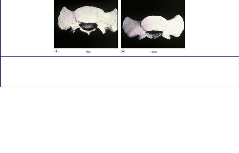
Figure 6.99 Male and female sacrum from above. In the male the body of the first sacral vertebra is broad and the alae narrow; in the female the body and the alae broad are of approximately equal width.
Coccyx
This is contracted into four pieces fused together into a small triangular bone joined by its base to the apex of the sacrum at the sacrococcygeal joint (see p. 325). At each side a lateral sacrococcygeal ligament joins the transverse process of the first piece of the coccyx to the inferolateral angle of the sacrum and completes the foramen for S5 nerve anteriorly; posteriorly the foramen is closed by the sacral and coccygeal cornua connected by the intercornual ligament. The upper surface of the coccyx is in the pelvic floor, the lower surface is in the buttock, beneath the skin of the natal cleft.
Development of vertebrae
The vertebrae develop from the sclerotome parts of the mesodermal somites (see p. 23). The sclerotomes surround the notochord and neural tube in a sheath of mesoderm. A series of hyaline cartilaginous rings appears in the mesodermal sheath. Each ring is formed by fusion of adjacent halves (caudal and cranial) of the original somites. Thus the vertebrae lie not in segments of the body wall, but in the intersegmental planes.
Each ring ossifies in three centres to form the centrum and the two halves of the neural arch of a vertebra. The centre for the centrum is initially double, but the two areas rapidly fuse; failure of one half results in a hemivertebra; a series of hemivertebrae is one cause of congenital scoliosis (lateral curvature of the spine). By the eighth week of fetal life ossification has commenced in the centrum and the two halves of the neural arch. At birth a vertebra consists of three ossifying parts, centrum and two half arches, united by cartilage. The half arches unite in the first year after birth, first in the lumbar, then thoracic and finally cervical regions. The centra unite with the arches first in the cervical region, about the third year, but in the lumbar region this union is not complete until the sixth year. Failure of fusion of the vertebral arches in the midline results in the condition known as spina bifida. When the overlying skin is intact this is qualified as being occult. A more severe developmental defect results in a cerebrospinal fluid-filled sac (meningocoele) or a sac containing a part of the spinal cord and nerve roots (myelomeningocoele) being exposed on the surface. These defects are more common in the lumbar or lumbosacral region.
The vertical cylindrical surface of the body is covered with compact bone, but the cancellous bone on the flat upper and lower surfaces remains covered with a layer of hyaline cartilage. The epiphyses for the body appear as bony rings, upper and lower, soon after puberty. They are ridged and grooved

reciprocally with the margins of the surfaces of the body (Fig. 6.100). Fusion of the epiphyseal ring and body occurs in the early twenties. Soon after puberty secondary centres appear also at the tip of the spinous process (double in the bifid spines of the cervical vertebrae) and at the tips of the transverse processes of all the vertebrae, and in the mamillary processes of T12 and the lumbar vertebrae. These fuse in the early twenties. The costal elements of cervical and lumbar vertebrae do not have a separate bony centre, but ossify by direct extension from the neural arch. An occasional centre in the costal element of C7 or L1 vertebra may lead to the formation of a cervical or lumbar rib. On the other hand the weight-bearing costal elements of the sacrum have primary ossification centres. In general the development of the five segments of the sacrum resembles that of typical vertebrae. After puberty these segments coalesce from below upwards.
Figure 6.100 L5 vertebra from above. The neurocentral junction has not yet completely ossified. The radial grooves around the anterolateral convexity of the body indicate that the epiphyseal ring had not united.
The atlas ossifies in the seventh week of fetal life by a centre in each lateral mass. These extend around the posterior arch and unite at the fourth year. In the meantime a centre in the anterior arch has appeared at the first year. Its junction with the bone of the lateral mass cuts across the anterior part of the upper articular surface; these epiphyses fuse at 7 years. This epiphyseal junction may permanently divide the articular surface (as in Fig. 6.89, right side).
Each half of the neural arch of the axis ossifies from a primary centre in the second month; a pair of centres for the centrum, which soon fuse, ossify in the fourth month. The dens is mainly ossified from a pair of centres which appear in the sixth month and join before birth. Thus at birth the axis is in four parts and these fuse with each other in the first few years thereafter; but although the cartilage between the dens and the body ossifies circumferentially, the centre may remain cartilaginous till old age. A secondary centre for the tip of the dens appears at about 6 years and unites with the rest of the dens at about 12 years. As in the case of a typical vertebra, a secondary centre for the lower surface of the body appears at puberty and fuses in the early twenties.

Part twenty. Cranial cavity and meninges
The interior of the cranium is lined with dura mater, the surface of the brain is covered with pia mater. Between the two, in contact with the dura mater, lies the arachnoid mater, which is connected to the pia by many fine filamentous processes (hence the name arachnoid: spider-like). These three tissue layers constitute the meninges. The cranial meninges are described here; their continuations around the spinal cord as the spinal meninges are considered on page 453.
Pia mater
The pia mater invests the brain and spinal cord as periosteum invests bone. Like periosteum it contains blood vessels, and nowhere does any structure intervene between pia mater and the underlying nervous tissue. It closely invests the surface of the central nervous system to the depths of the deepest fissures and sulci. It is made of thin vascular fibrous tissue and can be stripped away from the brain surface. It is prolonged out over the cranial nerves and spinal nerve roots to fuse with their epineurium, and it is invaginated into the substance of the brain by the entering cerebral arteries. The arteries lie loose in these sheaths of pia, surrounded by a narrow perivascular space containing cerebrospinal fluid.
The region between the pia and the arachnoid is the subarachnoid space, filled with cerebrospinal fluid.
Arachnoid mater and subarachnoid space
The arachnoid mater consists of an impermeable delicate membrane that everywhere is supported by the inner surface of the inner layer of the dura mater with only a thin film of tissue fluid between them in the subdural space. Vessels and nerves pierce the dura and arachnoid mater both at the same place; they cross the subdural space, but do not run along between the two membranes.
In certain areas the arachnoid herniates through little holes in the dura mater into the venous sinuses. Such herniae are the arachnoid villi; through their walls the cerebrospinal fluid ‘oozes’ back into the blood. The arachnoid villi are most numerous in the superior sagittal sinus and its laterally projecting blood lakes. In the child the villi are discrete; as age progresses they become aggregated into visible clumps, the arachnoid granulations (Pacchionian bodies) (see Fig. 7.16, p. 474). These latter leave indentations on the inner table of the cranial vault mainly alongside the superior sagittal sinus, at the site of the blood lakes.
Between the base of the brain and the base of the skull several larger spaces exist as a result of the incongruities in the contours of bone and brain. These spaces form the subarachnoid cisterns.
The cerebellomedullary cistern (cisterna magna) is the largest. It occupies the angle between the undersurface of the cerebellum and the posterior surface of the medulla. Cerebrospinal fluid flows into it from the midline aperture (foramen of Magendie) in the roof of the fourth ventricle (see p. 482). The lateral part of the cistern contains the vertebral artery and its posterior inferior cerebellar branch on each side (see Fig. 7.19, p. 475). The cisterna magna can be tapped in the midline by a needle passed above the posterior arch of the atlas, through the posterior atlanto-occipital membrane and spinal dura.
The pontine cistern lies between the clivus (see p. 510) and the front of the pons and medulla. Cerebrospinal fluid flows into it from the lateral apertures (foramina of Luschka) of the fourth ventricle (see p. 483). The cistern contains the basilar artery and its pontine and labyrinthine branches and the fifth to twelfth cranial nerves.
T he interpeduncular cistern lies between the dorsum sellae of the sphenoid and the cerebral peduncles; it is roofed in by the floor of the third ventricle (mamillary bodies and posterior perforated substance). The floor of the cistern, on the dorsum sellae, is formed by the arachnoid membrane passing across, in contact with the dura mater, between the right and left temporal lobes. The cistern contains the terminal branches of the basilar artery (including the posterior part of the circle of Willis), the stalk of the pituitary gland, and the third and fourth cranial nerves (see Fig. 7.19, p. 475).
The chiasmatic cistern lies above the optic chiasma, beneath the rostrum of the corpus callosum (see Fig. 7.3A, p. 457). It contains the anterior communicating artery.
Dura mater
The dura mater consists of an outer endosteal layer, and an inner meningeal layer. The two layers are united except where they separate to enclose the venous sinuses of the dura. The outer layer is the periosteum which invests the surface of any bone, and blood vessels pass through it to supply the bone. Around the margins of every foramen in the skull it lies in continuity with the periosteum on the outer surface of the cranial bones (pericranium). It is not prolonged into the dura mater of the vertebral canal nor is it evaginated by any cranial nerve. The inner layer consists of a dense, strong fibrous membrane, which is really the dura mater proper. Over the vault of the skull the fused layers are easily stripped away from the bone as a single sheet, a fact which makes removal of the vault relatively easy. Over the base of the skull the fused layers are so firmly attached that they can only be stripped off with difficulty. Although theoretically structures such as the middle meningeal vessels (see p. 442) lie between the two layers, they appear to be on the outer surface of this single sheet (as when the bone of the skull vault is removed leaving the dura intact), and so are usually described as being extradural. The vessels are thicker than the outer layer and so bulge out to make impressions on the bone with a negligible amount of intervening tissue. Haemorrhage from these vessels is described as extradural (see p. 447).
Folds of the inner layer project into the cranial cavity. One such fold, the tentorium cerebelli, roofs in the posterior cranial fossa; another forms the falx cerebri, lying in the midline between the two cerebral hemispheres. The falx cerebelli and diaphragma sellae are smaller derivatives of the inner dural layer. These fibrous flanges or septa minimize rotary displacement of the brain. Concussion is caused more readily by rotary movement of the brain rather than by mass displacement of the head. At the foramen magnum the inner layer leaves the outer layer and is projected down the vertebral canal as the spinal dura mater (see p. 453). The inner layer is likewise evaginated around the cranial nerves and spinal nerve roots.
The tentorium cerebelli is a flange of the inner layer which projects from the margins of the transverse sinuses and the margins of the superior petrosal sinuses (Fig. 6.101). It is attached to the posterior clinoid processes, along the upper borders of the petrous temporal bones, and horizontally along the inner surface of each side of the skull to the internal occipital protuberance; this is the

attached margin of the tentorium. Its upper and lower layers are separated at their bony attachments by the superior petrosal and transverse sinuses, but elsewhere are intimately fused with each other. The free margin of the tentorium is U-shaped and lies at a higher level than the bony attachment. The large central gap is the tentorial notch (incisure), through which passes the upper part of the brainstem (midbrain). The membrane slopes concavely upwards as it converges from the attached to the free margin, in conformity with the shape of the upper surface of the cerebellum and the undersurface of the posterior part of the cerebral hemisphere. The concave free margin is traceable forwards to the anterior clinoid process on each side. At the medial end of the superior petrosal sinus it overlies the attached margin, and from this point forwards to the anterior clinoid process it lies as a ridge of dura mater on the roof of the cavernous sinus. To the medial side of the ridge is the concave triangular roof of the cavernous sinus (see p. 443), which is pierced by the third and fourth nerves.
Figure 6.101 Cranial fossae. The dura mater has been removed on the left side.
The midline attachment of the falx suspends the tentorium and the straight sinus lies in the midline at the junction of the two.
The falx cerebri is a sickle-shaped flange of the inner layer in the midline between the cerebral hemispheres. Its anterior margin is attached to the crista galli of the ethmoid bone and to the cavity of the foramen caecum (see p. 508), into which it projects like a peg, an enlarged Sharpey's fibre. The posterior margin is attached to the upper surface of the tentorium cerebelli in the midline, from the attached to the free margin of the tentorium; here its layers separate to enclose the straight sinus. Its convex upper border is attached alongside the midline to the whole length of the concave inner surface of the skull, from the foramen caecum to the internal occipital protuberance. Its two layers are separated a short distance above the foramen caecum to accommodate the superior sagittal sinus, which becomes progressively broader from this point to the internal occipital protuberance. The concave lower border of the falx cerebri is free and contains the inferior sagittal sinus within its two layers; this border lies just above the corpus callosum (see Fig. 7.15, p. 473). Between superior and inferior sagittal sinuses the two layers of the falx are firmly united to form a strong inelastic
membrane.
The falx cerebelli is a low elevation of the inner layer in the midline of the posterior cranial fossa, extending from the internal occipital protuberance along the internal occipital crest to the posterior margin of the foramen magnum. It lodges the small occipital sinus between its layers, and it projects a little into the sulcus between the cerebellar hemispheres.
The diaphragma sellae is a horizontal sheet of the inner layer that forms a roof for the pituitary fossa (see p. 448). The dura of the floor of the fossa is prolonged up the sides of the fossa, to be attached to the middle and posterior clinoid processes, forming an intervening flange between the fossa and the cavernous sinus. From this the dura extends medially to form the diaphragma, which is perforated centrally for the passage of the pituitary stalk, and is continuous laterally with the roof of the cavernous sinus.
Blood supply
The inner layer of the dura mater requires very little blood to nourish it. The outer layer, on the other hand, is richly supplied, with the adjacent bone. In the supratentorial part it is supplied by the middle meningeal artery. The dura is supplied, in the anterior cranial fossa, by meningeal branches of the ophthalmic and anterior and posterior ethmoidal arteries and a branch of the middle meningeal artery, in the middle cranial fossa by the middle and accessory meningeal arteries and by meningeal branches of the internal carotid and ascending pharyngeal arteries and in the posterior cranial fossa by meningeal branches of the vertebral artery (Fig. 6.109). All these meningeal arteries are chiefly distributed to bone.
The middle meningeal artery, a branch of the maxillary, arises in the infratemporal fossa and passes upwards between the two roots of the auriculotemporal nerve to enter the foramen spinosum. It thus enters the middle cranial fossa, accompanied by its own plexus of sympathetic nerves. It is accompanied throughout all its ramifications by veins which lie between it and bone. It courses laterally on the floor of the middle cranial fossa and turns upwards and forwards on the greater wing of the sphenoid, where it divides into anterior and posterior branches.
The frontal (anterior) branch courses up towards the pterion (see p. 504) and then curves back to ascend towards the vertex, lying over the precentral gyrus. Haemorrhage from the vessel thus causes pressure on the motor area. In the region of the pterion the artery frequently lies in a tunnel in the parietal bone for a centimetre or more.
The parietal (posterior) branch courses horizontally backwards, on a groove in the squamous part of the temporal bone, and ramifies over the posterior part of the skull. It lies alongside the superior temporal gyrus; haemor-rhage here may cause contralateral impaired hearing through pressure on the auditory area (see p. 463).
The main purpose of the middle meningeal artery is to supply the bones of the vault of the skull; it does not supply the brain. These bones receive very little blood from the vessels of the scalp; scalping produces no necrosis of the underlying bones. Only where the bones give attachment to muscles (temporal fossa and suboccipital region) is any substantial supply received from the exterior.
Much of the blood from the marrow is drained by large diploic veins, which emerge on the exterior.

Other diploic veins drain into the venous sinuses, especially the superior sagittal. The remaining blood drains into the middle meningeal veins.
The middle meningeal veins are sinuses in the dura mater and accompany the branches of the artery. They lie between the artery and the bone, grooving the latter. Some converge to two veins which leave the skull through the foramen spinosum and foramen ovale to join the pterygoid plexus. Some join the sphenoparietal and superior sagittal sinus.
The surface marking of the middle meningeal artery as it enters the skull lies just above the midpoint of the zygomatic arch. The frontal branch can be surgically approached through a burr hole made in the region of the pterion, about 3 cm above the zygomatic arch and 3 cm behind the zygomatic process of the frontal bone; a position that is above the midpoint of the zygomatic arch and behind the palpable frontozygomatic suture. The posterior branch runs backwards parallel with and above the upper border of the zygomatic arch and the supramastoid crest; it is usually exposed vertically above the mastoid process on a level horizontal with the upper margin of the orbit.
Nerve supply
Most of the supratentorial part of the dura mater is supplied from the ophthalmic division of the trigeminal nerve; the tentorial nerves course up and back from the anterior end of the cavernous sinus to supply the falx, the dura of the vault, and the upper surface of the tentorium cerebelli.
The anterior cranial fossa is supplied by the anterior and posterior ethmoidal nerves and receives some twigs from the maxillary nerve. The middle fossa is supplied, in its anterior portion by a branch of the maxillary nerve, and in its posterior part by the meningeal branch of the mandibular nerve (nervus spinosus). The posterior fossa is supplied by meningeal branches of the vagus and hypoglossal nerves; these are C1 and C2 fibres carried by the cranial nerves. They innervate the undersurface of the tentorium cerebelli and the upper part of the bony fossa. The dura around the foramen magnum is supplied directly by the second and third cervical nerves. Clinical experience suggests that this cervical supply extends farther afield than the posterior cranial fossa; cervical spondylosis, for example, may be associated with supra-orbital pain, presumably referred from spinal nerves.
Venous sinuses of the dura mater
All the venous sinuses, except the inferior sagittal and straight sinuses, lie between the inner and outer layers of the dura. They receive all the blood from the brain, and with the above exceptions they receive blood also from the adjacent bone. Several of them have important communicating branches (emissary veins) with veins outside the skull. Like all vascular channels the venous sinuses are lined by endothelium. They do not contain valves.
The superior sagittal sinus lies between the two layers of the falx cerebri along the convexity of its attached margin. It commences just above the foramen caecum and grows progressively larger as it passes back to the internal occipital protuberance. It grooves the bones along the midline of the vault of the skull. Three or four lakes of blood project laterally from it, between the inner dura and the endosteum; into these lakes arachnoid granulations project to return cerebrospinal fluid to the bloodstream (see Fig. 7.16, p. 474).
The superior sinus does not drain the frontal pole of the hemisphere, but receives veins from the upper and posterior parts of both medial and lateral surfaces of both hemispheres. These superior cerebral veins enter the sinus obliquely, against the flow of the bloodstream (see Fig. 7.15, p. 473). The superior sagittal sinus turns at the internal occipital protuberance, generally to the right, and becomes the transverse (lateral) sinus.
The inferior sagittal sinus begins some little distance above the crista galli and lies between the folds of the free margin of the falx cerebri. It drains the lower parts of the medial surface of each hemisphere. At the attachment of falx cerebri and tentorium cerebelli it flows into the straight sinus (see Fig. 7.15, p. 473).
The straight sinus lies between the folds of the fibrous dura at the junction of falx cerebri and tentorium cerebelli. It commences anteriorly by receiving the inferior sagittal sinus and the great cerebral vein (of Galen) (see p. 474). The straight sinus also receives veins from the adjoining occipital lobes and from the upper surface of the cerebellum. It slopes down steeply and ends at the internal occipital protuberance by turning into the transverse (lateral) sinus, generally the left (see Fig. 7.15, p. 473).
The transverse sinus commences at the internal occipital protuberance and runs laterally between the two layers of the attached margin of the tentorium cerebelli (Fig. 6.101). It courses horizontally forwards, grooving the occipital bone and the mastoid angle of the parietal bone. Reaching the junction of petrous and mastoid parts of the temporal bone it curves downwards, deeply grooving the inner surface of the mastoid bone, as the sigmoid sinus. One sinus is larger than the other, namely that which receives the superior sagittal sinus; this is usually the right.
The two transverse sinuses communicate at their commencement at the internal occipital protuberance (confluence of the sinuses). Each receives tributaries from the nearby surfaces of cerebral and cerebellar hemispheres and, at its termination at the commencement of the sigmoid sinus, the superior petrosal sinus enters.
T he surface marking of the transverse sinus is a horizontal line from the external occipital protuberance to the top of the mastoid process, that is, at the upper limit of the neck muscles where they are attached to the skull, along the superior nuchal line.
The sigmoid sinus commences as the termination of the transverse sinus, deeply grooving the inner surface of the mastoid part of the petrous bone. It curves downwards and then forwards to the posterior margin of the jugular foramen, through which it passes, and expands into the superior jugular bulb, from which emerges the internal jugular vein (see p. 343). The sigmoid sinus is connected with the exterior in its upper part by the mastoid emissary vein which joins the posterior auricular vein, and in its lower part by a vein which passes through the posterior condylar foramen (when present) to join the suboccipital plexus of veins.
As the superior petrosal sinus drains into the termination of the transverse sinus, it could be said that the sigmoid sinus receives the superior petrosal sinus at its upper end and the occipital sinus at its lower end. Cerebellar veins drain to it, and it receives veins also from the mastoid air cells. Thrombophlebitis in these veins may lead to cerebellar abscess from mastoid infection.

The occipital sinus runs downwards from the beginning of the transverse sinus to the foramen magnum, skirts the margin of the foramen and drains into the sigmoid sinus. Along the attachment of the falx cerebelli this sinus is often a single trunk. Around the margins of the foramen magnum a pair of sinuses communicate with the veins outside the spinal dura (the internal vertebral plexus). The occipital sinus receives tributaries from the cerebellum and medulla and drains the choroid plexus of the fourth ventricle.
The basilar plexus consist of a network of veins, lying between the two layers of the dura, on the clivus (see p. 510). They connect the two inferior petrosal sinuses and receive veins from the lower part of the pons and from the front of the medulla. Thrombosis in this plexus is therefore usually fatal.
No veins accompany the vertebral and basilar arteries; the vertebral vein itself commences outside the skull below the occipital bone (see p. 430).
Cavernous sinus
The cavernous sinus lies alongside the body of the sphenoid bone in the middle cranial fossa. Each contains the internal carotid artery (Figs 6.102 and 6.103) and transmits some cranial nerves; each receives blood from three sources (orbit, vault bones, and cerebral hemisphere); each drains by the superior and inferior petrosal sinuses to the transverse sinus and internal jugular vein respectively, and each is connected to the pterygoid plexus by emissary veins.
Figure 6.102 Pituitary fossa, from above.

Figure 6.103 Pituitary fossa and cavernous sinuses in coronal section. Reproduced from Williams: Gray's Anatomy 38th edition, 1999, Churchill Livingstone, with permission.
The cavernous sinus lies in a space between the periosteum of the body of the sphenoid (outer layer of the dura mater) and a fold of the inner layer of the dura, which forms the upper part of the medial wall, the roof and the lateral wall of the sinus (Fig. 6.103). Medially the roof is continuous with the diaphragma sellae. At its lower edge the lateral wall is continued laterally as the inner layer of dura across the middle cranial fossa (Fig. 6.101). Anteriorly the roof is attached to the anterior and middle clinoid processes of the sphenoid bone; it is perforated between these processes by the emerging internal carotid artery. Posteriorly the roof has a triangular depression between the attached edge of the tentorium cerebelli (to the posterior clinoid process) and the ridge raised by the free edge of the tentorium, as it extends forwards to the anterior clinoid process. The third and fourth cranial nerves invaginate the roof here (Fig. 6.101), and then run forwards in the lateral wall (i.e. between the dura and the endothelial lining) above the ophthalmic and maxillary branches of the fifth nerve. Further back the lateral wall is medial to the anterior parts of the trigeminal ganglion and the trigeminal (Meckel's) cave (see p. 450). The rest of the cave and ganglion are posteroinferior to the lateral wall. The floor of the sinus is a narrow strip of periosteum along the base of the greater wing of the sphenoid.

Figure 6.104 Longitudinal section through the right orbit and middle cranial fossa, viewed from the right (lateral) side.
The cavernous sinus extends from the apex of the orbit back to the apex of the petrous temporal bone. Each end is pointed, so that the sinus is spindle-shaped in lateral view. It is about 2 cm long and 1 cm wide. Despite its name, the cavernous sinus is usually a plexus of veins and not a trabeculated venous space, like the corpora cavernosa of the penis.
Medial to the sinus lies the fibrous lateral wall of the pituitary fossa and the body of the sphenoid, with the sphenoid air sinus within it (Fig. 6.103). The air sinus lies towards the front of the pituitary fossa at a lower level; its extent is very variable and tends to increase posteriorly with advancing years. Lateral to the cavernous sinus lies the medial surface of the temporal lobe of the hemisphere. Superiorly, the emerging internal carotid artery lies in contact with the forepart of the roof of the sinus as the artery passes backwards a little before turning up towards the anterior perforated substance of the brain (Fig. 6.102). Further back and somewhat above the roof lies the uncus of the temporal lobe.
The contents of the cavernous sinus include the structures lying within the cavity (the internal carotid artery and sixth nerve) and those embedded within the lateral wall (the third and fourth nerves and the ophthalmic and maxillary branches of the fifth nerve) (Fig. 6.103).
The internal carotid artery curves upwards from the foramen lacerum to enter the posterior part of the sinus and runs forwards within the sinus, deeply grooving the body of the sphenoid and the base of its greater wing. The artery then curves upwards again to pierce the roof of the sinus, medial to the anterior clinoid process, and turns backwards (Figs 6.102 and 6.105). The artery is accompanied by a plexus of postganglionic sympathetic fibres from the superior cervical ganglion.

Figure 6.105 Part of the posterior and middle cranial fossae seen from behind after sectioning the skull coronally through the mastoid process. All dura mater has been removed except for that over the superior and inferior petrosal sinuses, the dura around the opening of the trigeminal cave and the cut edge of the tentorium cerebelli at its attachment to the petrous temporal bone.
The abducens nerve (see p. 451) enters the back of the cavernous sinus after passing over the apex of the petrous part of the temporal bone. It may, however, enter the inferior petrosal sinus beside the clivus and run along this sinus into the cavernous sinus within which it runs forwards on the inferolateral side of the internal carotid artery (Fig. 6.105). At the anterior end of the sinus it enters the superior orbital fissure (Fig. 6.49).
The oculomotor nerve (see p. 449) enters the roof between the free and attached margins of the tentorium cerebelli (Fig. 6.101), and then passes forward in the lateral wall. As it does so it inclines downwards medial to the other nerves, namely, the trochlear nerve and the branches of the ophthalmic nerve (Fig. 6.104), and at the anterior end of the sinus it breaks into its superior and inferior divisions which enter the superior orbital fissure. In its course it picks up sympathetic fibres from the internal carotid plexus; these are for the smooth muscle part of levator palpebrae superioris.
The trochlear nerve (see p. 450) enters the roof of the sinus behind the oculomotor nerve, alongside the ridge raised by the free margin of the tentorium, and courses horizontally forwards in the lateral wall of the sinus to enter the superior orbital fissure at a higher level and lateral to the oculomotor nerve.
The trigeminal ganglion, in its anterior part, lies forward of Meckel's cave. Its mandibular division passes downwards to the foramen ovale and does not come in contact with the lateral wall of the cavernous sinus (Fig. 6.101). The maxillary division runs horizontally forwards in the lateral wall and leaves the middle fossa through the foramen rotundum. The ophthalmic division runs forwards above the maxillary division and divides into its three branches towards the anterior end of the lateral wall; these enter the orbit through the superior orbital fissure. At the anterior end of the sinus the ophthalmic division gives off its tentorial branches to the dura mater (see p. 442). In its course through the sinus the ophthalmic division picks up sympathetic fibres from the internal carotid plexus; these eventually enter the long ciliary nerves to reach the dilator pupillae muscle.
Veins of the cavernous sinus
Although it is customary to think of venous blood as entering the cavernous sinus at the front and
leaving from behind and below, blood can flow in either direction in the sinus, depending on local venous pressures. There are no valves in the cavernous sinus or its connected veins.
The superior ophthalmic vein passes back directly into the anterior end of the sinus at the superior orbital fissure. The inferior ophthalmic vein drains into both the pterygoid plexus and the cavernous sinus.
The superficial middle cerebral vein traverses the subarachnoid space and drains into the sinus by piercing its roof near the emerging carotid artery. Some inferior cerebral veins also drain through the roof of the sinus.
The sphenoparietal sinus runs just beneath the edge of the lesser wing of the sphenoid, lying between the two layers of the dura mater, and enters the sinus through its roof.
The superior petrosal sinus leaves the top of the posterior end and, bridging the groove made by the underlying trigeminal nerve, runs back along the upper border of the petrous bone between the two layers at the attached margin of the tentorium cerebelli (Fig. 6.105). It enters the termination of the transverse sinus at the commencement of the sigmoid sinus.
The inferior petrosal sinus is larger and empties the bulk of the blood from the cavernous sinus. It leaves the posterior end of the sinus beneath the petrosphenoid ligament, a fibrous band stretched between the apex of the petrous part of the temporal bone and the side of the dorsum sellae (Fig. 6.105); it is occasionally partly ossified. The abducens nerve may enter the sinus and pass through it into the cavernous sinus. The inferior petrosal sinus runs down between the two layers of the dura along the suture between the apex of the petrous bone and the side of the clivus (occipital bone), enters the anterior compartment of the jugular foramen medial to the glossopharyngeal nerve, and joins the internal jugular vein as its first (highest) tributary.
The cavernous sinus communicates with the pterygoid venous plexus by emissary veins which usually pass through the foramen ovale and the foramen lacerum. When the venous foramen (of Vesalius) is present medial to the foramen ovale (Fig. 6.102), it too transmits a vein.
The cavernous sinuses communicate with each other through the intercavernous sinuses, which form a small plexus that lies between the two layers of the dura of the floor of the pituitary fossa. An anterior and a posterior intercavernous sinus lie also in the diaphragma sellae.
The cavernous sinus is in venous connection with the skin of part of the face whence infection may produce thrombosis (see p. 355). By the superficial middle cerebral vein such thrombosis can spread to the cerebral hemisphere. The ‘danger area of the face’ comprises the upper lip and nose and medial part of the cheek. It lies between the two veins which communicate with the cavernous sinus, namely, the angular vein (via superior ophthalmic vein) and the deep facial vein (via pterygoid plexus and emissary veins). Thrombosis of the cavernous sinus causes ophthalmoplegia from ocular nerve interruption. Spread of thrombosis to the inferior petrosal sinus and medullary veins is usually fatal.
Rupture of the internal carotid artery within the cavernous sinus, following a fracture of the skull base, produces a pulsating exophthalmos (bulging eyeball) from suffusion of the ophthalmic veins with arterial blood.
Extradural, subdural and subarachnoid haemorrhage
Fractures of the side of the skull may rupture the middle meningeal artery (especially its frontal branch) causing extradural haemorrhage which leads to the formation of a haematoma between the bone of the skull and the dura. The resultant swelling may cause (apart from the clinical features of raised intracranial pressure, namely headache, drowsiness and a slow pulse) pressure on the cerebral hemisphere in the region of the motor area (see p. 463), giving contralateral hemiparesis (commencing usually in the face and then spreading to the upper limb), and the medial edge of the temporal lobe may be displaced over the free edge of the tentorium, compressing the oculomotor nerve and causing dilatation of the pupil on the injured side.
Subdural haemorrhage may be caused by rupture of a superior cerebral vein as it crosses the subdural space to enter the superior sagittal sinus; the venous blood escapes into the (potential) space between the dura and arachnoid. There may be similar pressure symptoms to those caused by extradural haemorrhage, but because venous rather than arterial blood is involved they are slower to develop and less severe (chronic subdural haematoma). As the haematoma tends to be located near the vertex the contralateral hemiparesis usually commences in the lower limb.
Subarachnoid haemorrhage is usually caused by rupture of arteries that lie within the space, such as aneurysms of the arterial circle at the base of the brain (see p. 471). This causes blood to contaminate the cerebrospinal fluid.

Part twenty-one. Cranial fossae
Anterior cranial fossa
The anterior cranial fossa is formed by the frontal bone, the cribriform plate of the ethmoid, the lesser wings and anterior part of the body of the sphenoid. The floor of the fossa roofs in the orbits, ethmoidal sinuses and the nose.
The gyri and sulci of the undersurface of the frontal lobes cause grooves and ridges on the orbital parts of the frontal bone. From the nose up to 20 olfactory nerve filaments (on each side) perforate the dura and arachnoid mater over the cribriform plate and pass upwards through the subarachnoid space to enter the olfactory bulb, from which the olfactory tract passes back on the inferior surface of the frontal lobe (see Fig. 7.4, p. 459).
The anterior ethmoidal nerve and artery have a very short course in the fossa. Coming in from the orbit they lie beneath the dura at the edge of the cribriform plate, where they run forwards in a slit and pass down into the nose at the side of the crista galli.
The posterior border of the lesser wing of the sphenoid lies at the boundary between the anterior and middle cranial fossae. The sphenoparietal sinus runs between the two layers of the dura at this edge, which fits into the stem of the lateral cerebral sulcus (see p. 456); a branch of the middle meningeal artery crosses over the sinus to reach the anterior cranial fossa.
Middle cranial fossa
The middle cranial fossa consists of a median part, formed by the sphenoid body, and right and left lateral parts, formed by the cerebral surfaces of the greater wings of the sphenoid and the squamous and petrous parts of the temporal bones. The median part (Fig. 6.102) contains the pituitary gland, optic nerves and chiasma, and the intercavernous sinuses. The right and left lateral parts each contain the cavernous sinus, the third to the sixth cranial nerves and trigeminal ganglion, internal carotid artery, middle and accessory meningeal vessels, and the greater and lesser petrosal nerves. The temporal lobes lie on the right and left lateral parts.
The superior border of the petrous temporal bone lies at the boundary between the middle and posterior cranial fossae; the superior petrosal sinus lies here between the two layers of the dura.
Pituitary gland
The pituitary gland (hypophysis cerebri) lies in its fossa (sella turcica) with the diaphragma sellae above it. Intercavernous sinuses lie in the diaphragma and in the dural floor of the fossa. On each side is a flange of dura mater separating the gland from the upper part of the cavernous sinus. The pia and arachnoid blend with the capsule of the gland. Below the fossa lies the body of the sphenoid bone containing the sphenoidal air sinuses. When small the sinuses lie anteroinferior to the fossa, but when large one or both extend back beneath the fossa.
The pituitary stalk slopes downwards and forwards to the perforation in the diaphragma, connecting the posterior part of the gland to the hypothalamus (see p. 470). The upper, or infundibular, part of the stalk is hollowed out (continuous with the third ventricle). The optic chiasma lies above the diaphragma sellae, anterosuperior to the pituitary stalk. A pituitary tumour, rising upwards, presses
on the lower anterior part of the chiasma and the medial sides of the optic nerves (so causing hemianopia of the temporal fields; see p. 500).
Structure. The gland is a composite structure consisting essentially of two main parts, the anterior and posterior lobes; these terms are not entirely synonymous with the terms adenohypophysis and neurohypophysis, which indicate different developmental origins. The adenohypophysis, developed from an ectodermal saccule (Rathke's pouch) of the stomodeum (see p. 24), consists of the pars anterior (distalis), pars tuberalis and the rudimentary pars intermedia. The neurohypophysis, developed as a neuroectodermal downgrowth from the floor of the third ventricle, consists of the pars posterior (nervosa), the infundibular stalk and the median eminence (see p. 470). The pars anterior and the pars posterior form the anterior and posterior lobes respectively.
The pars anterior accounts for 75% of the whole gland. It is highly vascular, and its cells consist of 50% chromophobes, 35% acidophils and 15% basophils. The acidophils (eosinophils) secrete growth hormone and prolactin, with the basophils producing ACTH, TSH, FSH and LH (adrenocorticotropic, thyroid-stimulating, follicle-stimulating and luteinizing hormones). The pars tuberalis is a small extension of the pars anterior along the pituitary stalk, but its cells are different and their function not known. The pars anterior lies in front of a narrow cleft (the remains of the pharyngeal downgrowth), behind which is the small pars intermedia. It consists of a number of colloid-filled vesicles and secretes MSH (melanocyte-stimulating hormone). Its cells contain endorphins.
The pars posterior consists largely of about 100 000 unmyelinated nerve fibres whose cell bodies are in the supraoptic and paraventricular nuclei of the hypothalamus. Neurosecretory material manufactured in the cell bodies (mainly oxytocin from the paraventricular cells and vasopressin— antidiuretic hormone—from the supraoptic cells) migrates at the rate of about 3 mm/day along their axons which run in the pituitary stalk and end in the pars nervosa. Both hormones are combined with a carrier protein, neurophysin. Scattered among the nerve fibres are the rather scanty pituicytes which resemble astrocytes.
Blood supply and the hypophyseal portal system. A single inferior and several superior hypophyseal arteries arise from the internal carotid artery. Venous blood enters the adjacent cavernous and intercavernous sinuses. In addition to these vessels there is a hypothalamohypophyseal portal venous system connecting the hypothalamus and neurohypophysis with the adenohypophysis. These fine vessels form a plexus on the stalk and pars anterior. Into them in the upper part of the stalk are secreted the various hypophysiotropic hormones (hormone-releasing factors) derived from cells of the median eminence and possibly other parts of the hypothalamus. The portal vessels then transport these substances to their target cells which are the chromophils of the anterior lobe. Thus the anterior lobe cells are controlled by hypothalamic messengers delivered to them by a portal venous system. Reverse blood flow in this system and venous drainage from the hypophysis to the hypothalamus has also been suggested. There is no blood–brain barrier (see p. 472) in the posterior pituitary or median eminence.
Surgical approach. The gland can be approached through the anterior cranial fossa by elevating the frontal lobe of the brain, the subfrontal approach, or by the trans-sphenoidal approach, entering the pituitary fossa from below through a sphenoidal air sinus. This is entered either via the ethmoidal air

cells after raising the periosteum from the medial wall of the orbit, or by elevating the nasal mucosa from the nasal septum and removing the septum (the nasal cavity itself is not entered).
The internal carotid artery emerges from the roof of the cavernous sinus medial to the anterior clinoid process and curves immediately backwards, lying on the roof of the sinus before curving upwards lateral to the optic chiasma. At the anterior perforated substance it divides into its terminal branches (Fig. 6.106). The curve of the internal carotid artery in and above the cavernous sinus as seen in a lateral carotid arteriogram (like a U on its side, opening backwards) is commonly called the carotid siphon (Fig. 6.107).
Figure 6.106 Right carotid arteriogram: anteroposterior view.
Figure 6.107 Right carotid arteriogram: lateral view.
T he ophthalmic artery branches from the internal carotid immediately above the roof of the cavernous sinus. The internal carotid arteries come from below and laterally, the optic nerves come from above and medially (i.e. from the chiasma), and this is the relationship of nerve and artery in the optic canal (Fig. 6.53).
The optic nerve passes forward, down and laterally from the chiasma to the optic canal. Clad only in pia mater, it receives its sleeve of arachnoid and dura mater at the optic canal. Its intracranial part, in the chiasmatic cistern, is supplied by branches of the anterior cerebral artery that run down from the chiasma.
The oculomotor nerve leaves the medial side of the crus of the cerebral peduncle (see Fig. 7.18, p. 475). The nerve passes forwards between the posterior cerebral and superior cerebellar arteries (see Fig. 7.19, p. 475), just below the free margin of the tentorium cerebelli, below the posterior communicating artery; aneurysms may damage the nerve here. It crosses the interpeduncular cistern and enters the roof of the cavernous sinus in the middle fossa.
The trochlear nerve, after emerging from the dorsal surface of the brainstem below the inferior colliculus (see Fig. 7.19, p. 475), curls around the cerebral peduncle and runs forward between the posterior cerebral and the superior cerebellar arteries, lateral to the oculomotor nerve in the interpeduncular cistern, just below the free margin of the tentorium cerebelli (i.e. in the posterior fossa). It enters the middle fossa just behind the oculomotor nerve and enters the roof of the cavernous sinus, near where the free and attached margins of the tentorium cerebelli cross each other.
The abducens nerve enters the middle cranial fossa by passing over the apex of the petrous temporal and then runs round the lateral side of the ascending part of the internal carotid artery (Fig. 6.105) as it enters the cavernous sinus.
The posterior communicating artery, joining the internal carotid and posterior cerebral artery in the circle of Willis, lies in the interpeduncular cistern, above and lateral to the pituitary gland (see Fig. 7.19, p. 475).
Trigeminal ganglion
The trigeminal ganglion lies beneath the dura mater in the floor of the middle cranial fossa alongside the cavernous sinus, and occupies the trigeminal impression, a small fossa on the front of the apex of the petrous temporal bone posterolateral to the foramen lacerum (Figs 6.101 and 8.4, p. 529). The trigeminal nerve leaves the pons in the posterior fossa and runs forwards to cross the upper border of the petrous bone, upon which it leaves a shallow groove some 5 mm wide; it is accompanied by the small motor root, which lies below the sensory root. They pass beneath the superior petrosal sinus at this point (Fig. 6.105). As they do so, with their covering of pia, they evaginate a diverticulum of the inner layer of the dura and the arachnoid, the trigeminal cave (of Meckel), which passes forwards with them to lie in the trigeminal impression beneath the dural floor of the middle cranial fossa, the sensory root now enlarged to form the ganglion, with the motor root still below it. The evaginated inner layer of dura and the arachnoid fuse with the pia mater at the middle of the trigeminal ganglion, so the sensory and motor roots of the nerve and the posterior half of the ganglion are bathed in cerebrospinal fluid (Fig. 6.104).
Blood supply. The accessory meningeal artery enters through the foramen ovale and runs up along the mandibular division to reach the ganglion, a similar arrangement to that in the spinal nerves. Small ganglionic branches are also given off by the internal carotid artery in the cavernous sinus.
The surgical approach to the sensory root and posterior half of the ganglion, in Meckel's cave, traverses the subarachnoid space. The anterior half of the ganglion and the three divisions of the trigeminal nerve lie in front of Meckel's cave. The upper part of the anterior half of the ganglion, with the ophthalmic and maxillary divisions, lies in the fibrous lateral wall of the cavernous sinus; the lower part of the ganglion and the mandibular division lie in the middle fossa between the otherwise fused two layers of dura mater. An extradural approach is therefore possible across the floor of the

middle fossa by stripping the dura from the bone, avoiding entry into the subarachnoid space.
Transient facial palsy sometimes follows the latter approach. The explanation is thought to be that in stripping up the dura from the floor of the middle fossa tension is exerted on the greater petrosal nerve (which lies below the trigeminal ganglion; see below) and therefore on the geniculate ganglion. Subsequent oedema here causes pressure on the motor fibres of the facial nerve with paralysis of the muscles until the oedema subsides and the nerve recovers.
The mandibular division of the trigeminal nerve passes laterally to descend through the foramen ovale (Fig. 6.101). It is joined just below the foramen ovale by the small motor root to form the (mixed) mandibular nerve, a similar arrangement to that of the spinal nerves in the intervertebral foramina.
The maxillary nerve passes forwards within the inner layer of dura to leave the skull through the foramen rotundum (Fig. 6.102). The branches of the ophthalmic nerve pass forwards likewise, to leave the skull through the medial end of the superior orbital fissure.
The greater petrosal nerve (from the nervus intermedius part of the facial, see p. 498) emerges from its hiatus in the petrous bone (see Fig. 8.4, p. 507) and runs obliquely forwards, between the two layers of the dura mater and beneath the trigeminal ganglion to the foramen lacerum. Here it is joined by the deep petrosal nerve, a branch from the carotid plexus of sympathetic nerves. The two join to form the nerve of the pterygoid canal (Vidian nerve). This nerve enters the posterior end of the pterygoid canal at the front of the foramen lacerum and runs along the canal, through the body of the sphenoid bone, to join the pterygopalatine ganglion (see p. 370).
The lesser petrosal nerve (from the glossopharyngeal) leaves its hiatus in the petrous bone (see Fig. 8.4, p. 507) and runs forwards, lateral to the greater petrosal nerve, between the two layers of the dura mater to emerge through the foramen ovale to join the otic ganglion (see p. 366).
The middle meningeal artery is considered on page 442.
Posterior cranial fossa
The posterior cranial fossa lies behind the clivus (see p. 510) and in front of the squamous part of the occipital bone which extends laterally to where the fossa is bounded anterolaterally by the petrous and mastoid parts of the temporal bone. It lodges the convexities of the cerebellar hemispheres as well as the pons and medulla oblongata. In it the fifth to twelfth cranial nerves inclusive pierce the dura mater (Fig. 6.109).

Figure 6.109 Posterior cranial fossa and the vertebral canal opened from behind.
The trigeminal nerve leaves the middle of the anterolateral surface of the pons by two roots, a large sensory and a small motor (see Fig. 7.18, p. 475). They lie close together. The motor root emerges somewhat above and medial to the sensory root, but spirals to enter the mouth of Meckel's cave below it. The sensory root itself shows a spiral arrangement of its fibres. At the junction with the pons the mandibular fibres lie superior, the ophthalmic inferior with the maxillary fibres between, but in Meckel's cave the mandibular fibres lie most laterally and the ophthalmic fibres most medially.
The abducens nerve leaves the brainstem near the ventral midline at the junction of the pons and the pyramid of the medulla (see Fig. 7.18, p. 475) and runs upwards through the pontine cistern. It enters the dura mater of the clivus some distance above its origin from the brainstem, and runs thence upwards between the two layers of the dura, passing over the apex of the petrous bone, to enter the posterior end of the cavernous sinus (see also p. 445). The relatively long intracranial course of this delicate nerve renders it particularly vulnerable to increase of intracranial pressure; paralysis of the lateral rectus may be an early sign in such cases.
The facial and vestibulocochlear nerves, with the intervening nervus intermedius part of the facial nerve, leave the lateral end of the junction of pons and medulla (see Fig. 7.6, p. 461) and pass upwards to enter the internal acoustic meatus, accompanied by the labyrinthine artery. This vessel is a branch of the basilar artery, or arises from the anterior inferior cerebellar artery.
The internal acoustic meatus is a foramen directed laterally in the posterior surface of the obliquely set petrous bone. Its fundus consists of a plate of bone divided by a horizontal crest into an upper and lower semicircle (Fig. 6.108). The facial nerve and its nervus intermedius part pierce the front of the upper part, the cochlear nerve the front of the lower part (by many branches in spiral arrangement). The vestibular nerve pierces the plate posteriorly, by upper and lower divisions that lie behind the facial nerve foramen (separated by a vertical bar of bone) and the spiral cochlear foramina respectively. Each division of the vestibular nerve is connected to the vestibular ganglion deep in the meatus. Behind the vestibular area is a single foramen (the foramen singulare) for the passage of the branch of the inferior division to the posterior semicircular duct. The labyrinthine artery divides in the meatus and its branches accompany the nerves through the bony plate.
The subarcuate fossa lies lateral to the internal acoustic meatus (Fig. 6.105). It is a very shallow fossa against which the flocculus of the cerebellum lies.
Further laterally on the posterior surface of the petrous bone is the orifice of the aqueduct of the vestibule, a narrow slit overhung by a sharp scale of bone (Fig. 6.105). The endolymphatic sac (see p. 421) hangs down from this slit beneath the inner layer of the dura.
The glossopharyngeal, vagus and accessory nerves arise from the side of the medulla oblongata as a series of rootlets lying vertically between the olive and the inferior cerebellar peduncle (see Fig. 7.6, p. 461). The three nerves run laterally across the occipital bone and pass through the jugular foramen (Fig. 6.109).
The spinal root of the accessory nerve enters the posterior fossa through the foramen magnum (Fig. 6.109). It arises by a series of rootlets that emerge from the lateral surface of the upper five or six segments of the cervical cord posterior to the denticulate ligament. These rootlets unite into a single trunk that passes forwards over the top of the ligament and ascends lateral to the vertebral artery to unite with the cranial root medial to the jugular foramen.
The jugular foramen is divided by two transverse septa of the inner layer of dura into three compartments. These septa may ossify. The glossopharyngeal nerve and inferior petrosal sinus share the anterior compartment, vagus and accessory nerves lie in the middle compartment, while the large posterior compartment is occupied by the termination of the sigmoid sinus. The inferior border of the petrous bone shows a deep notch immediately below the internal acoustic meatus (Fig. 6.105). This notch is indented by the inferior ganglion of the glossopharyngeal nerve; the aqueduct of the cochlea (see p. 419) opens into the depths of the notch and by this means perilymph drains into the subarachnoid space. The groove made by the inferior petrosal sinus is seen to enter the jugular foramen medial to the notch.
The hypoglossal nerve leaves the medulla by a vertical series of rootlets between the pyramid and the olive (see Fig. 7.18, p. 475). The rootlets unite into two roots which enter the hypoglossal canal separately, divided from each other by a septum of dura mater which occasionally ossifies.
The arteries in the posterior fossa comprise the two vertebral and the basilar arteries with their branches. After piercing the posterior atlanto-occipital membrane the vertebral arteries give off meningeal branches which enter the posterior fossa between the two layers of the dura mater at the foramen magnum (Fig. 6.109).
The vertebral artery then pierces the spinal dura mater and arachnoid and gives off the small posterior spinal artery (which may arise instead from the posterior inferior cerebellar branch). This fourth part of the vertebral artery runs forward in front of the highest denticulate ligament, in front of or between the rootlets of the hypoglossal nerve. It gives off the anterior spinal artery and the posterior inferior cerebellar artery and spirals up to meet its opposite fellow at the lower border of the pons to form the basilar artery. The posterior and anterior spinal arteries pass downwards through the foramen magnum on the spinal cord (see p. 491). The posterior inferior cerebellar artery is perhaps the most tortuous artery in the body. Its coils insinuate themselves between the rootlets of the hypoglossal, accessory and vagus nerves and the vessel is distributed to the cerebellum and medulla (see pp. 481 and 486).
The basilar artery runs up in front of the pons. It is not responsible for the ventral median groove in the pons; indeed, the artery is usually curved to one side of the midline (see Fig. 7.19, p. 475). It gives off the anterior inferior cerebellar artery and many pontine branches. The labyrinthine artery arises from the anterior inferior cerebellar or directly from the basilar trunk. The basilar artery ends at the upper border of the pons by dividing into the posterior cerebral arteries (see p. 472), immediately after giving off the superior cerebellar branches.