

Chapter 1. Introduction to regional anatomy
Part one. Tissues and structures
Skin
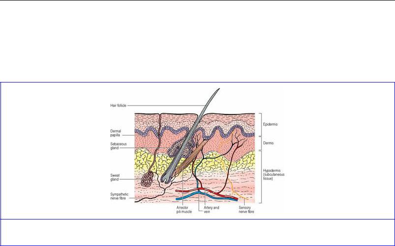
Skin consists of two components: epidermis and dermis (Fig. 1.1). The surface epithelium of the skin is the epidermis and is of the keratinized stratified squamous variety. The various skin appendages— sweat glands, sebaceous glands, hair and nails—are specialized derivatives of this epidermis, which is ectodermal in origin. The deeper dermis is mesodermal in origin and consists mainly of bundles of collagen fibres together with some elastic tissue, blood vessels, lymphatics and nerve fibres.
Figure 1.1 Structure of the skin and subcutaneous tissue.
The main factor determining the colour of skin is the degree of pigmentation produced by melanocytes in the basal layer of the epidermis. Melanocyte numbers are similar in all races. In darker skins the melanocytes produce more pigment. Melanins vary in colour from yellows to browns and blacks.
Sweat glands are distributed all over the skin except on the tympanic membranes, lip margins, nipples, inner surface of prepuce, glans penis and labia minora. The greatest concentration is in the thick skin of the palms and soles, and on the face. Sweat glands are coiled tubular structures that extend into the dermis and subcutaneous tissue. They are supplied by cholinergic fibres in sympathetic nerves. Apocrine glands are large, modified sweat glands confined to the axillae, areolae, periumbilical, genital and perianal regions; their ducts open into hair follicles or directly on to the skin surface. Their odourless secretion acquires a smell through bacterial action. They enlarge at puberty and undergo cyclic changes in relation to the menstrual cycle in females. They are supplied by adrenergic fibres in sympathetic nerves.
Sebaceous glands are small saccular structures in the dermis, where they open into the side of hair follicles. They also open directly on to the surface of the hairless skin of the lips, nipples, areolae, inner surface of prepuce, glans penis and labia minora. There are none on the palms or soles. They are particularly large on the face. Androgens act on these glands which have no motor innervation.
Hair and nails are a hard type of keratin; the keratin of the skin surface is soft keratin. Each hair is formed from the hair matrix, a region of epidermal cells at the base of the hair follicle, which extends

deeply into the dermis and subcutaneous tissue. As the cells move up inside the tubular epidermal sheath of the follicle they lose their nuclei and become converted into the hard keratin hair shaft. Melanocytes in the hair matrix impart pigment to the hair cells. The change with age is due to decreasing melanocyte activity. An arrector pili muscle attached to the connective tissue of the base of the follicle passes obliquely to the upper part of the dermis. Contraction of this smooth muscle, with a sympathetic innervation, makes the hair ‘stand on end’, and squeezes the sebaceous gland that lies between the muscle and the hair follicle. Hair follicles are richly supplied by sensory nerves.
Nails consist of nail plates lying on nail beds on the dorsum of the terminal segment of fingers and toes. Compacted keratin-filled squames form the nail plate, which develops from epidermal cells deep to its proximal part. Here the nail plate is overlapped by the skin of the proximal nail fold. Blood vessels and sensory nerve endings are plentiful in the nail bed.
The arteries of the skin are derived from a tangential plexus in the subcutaneous connective tissue. Branches from this plexus form a subpapillary network in the dermis (Fig. 1.1). The veins have a similar arrangement to the arteries and arteriovenous anastomoses are abundant. From a meshwork of lymphatic capillaries in the papillary layer of the dermis, lymphatics pass inwards and then run centrally with the blood vessels. Cutaneous nerves carry afferent somatic fibres, mediating general sensation, and efferent autonomic (sympathetic) fibres, supplying smooth muscle of blood vessels, arrector pili muscles and sweat glands. Both free sensory nerve endings and several types of sensory receptors are present in the skin.
The proportionate surface area of the skin over different regions of the body can be estimated by the ‘rule of nines’ and this is useful in assessing the need for fluid replacement after burns. This rule is a guide to the size of body parts in relation to the whole: head 9%; upper limb 9%; lower limb 18%; front of thorax and abdomen 18%; back of thorax and abdomen 18%.
Tension lines of the skin, due to the patterns of arrangement of collagen fibres in the dermis, run as shown in Figure 1.2. They are often termed relaxed skin tension lines because they coincide with fine furrows present when the skin is relaxed. Wrinkle lines are caused by the contraction of underlying muscles; they do not always correspond to tension lines. Flexure lines over joints run parallel to tension lines. The cleavage lines originally described by Langer in 1861 on cadavers do not entirely coincide with the lines of greatest tension in the living. Incisions made along skin tension lines heal with a minimum of scarring (Fig. 1.3).
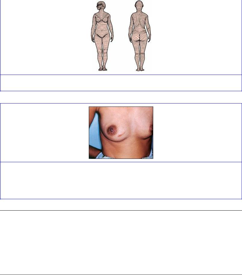
Figure 1.2 Tension lines of the skin, front and back.
Figure 1.3 An incision over the medial part of the right breast has crossed tension lines and resulted in excess scar formation. An incision at the lower margin of the areola along a tension line has healed with minimal scarring. The tubercles in the areola are due to the presence of large sebaceous glands.
Superficial fascia
The skin is connected to the underlying bones or deep fascia by a layer of loose areolar connective tissue. This layer, usually referred to as superficial fascia, is of variable thickness and fat content. Flat sheets of muscles are also present in some regions. These include both skeletal muscles (platysma, palmaris brevis) and smooth muscles (subareolar muscle of the nipple, dartos, corrugator cutis ani). The superficial fascia is most distinct on the lower abdominal wall where it differentiates into two layers. Strong connective tissue bands traverse the superficial fasica binding the skin to the underlying aponeurosis of the scalp, palm and sole.
Deep fascia
The limbs and body wall are wrapped in a membrane of fibrous tissue, the deep fascia. It varies widely in thickness. In the iliotibial tract of the fascia lata, for example, it is very well developed, while over the rectus sheath and external oblique aponeurosis of the abdominal wall it is so thin as to be scarcely demonstrable and is usually considered to be absent. In other parts, such as the face and the ischioanal fossa, it is entirely absent. Where deep fascia passes directly over bone it is always
anchored firmly to the periosteum and the underlying bone is described as being subcutaneous. In the neck, as well as the investing layer of deep fascia, there are other deeper fascial layers enclosing neurovascular structures, glands and muscles. Intermuscular septa are laminae of deep fascia which extend between muscle groups. Transverse thickenings of deep fascia over tendons, attached at their margins to bones, form retinaculae at the wrists and ankles and fibrous sheaths on the fingers and toes.
Ligaments
Ligaments are composed of dense connective tissue, mainly collagen fibres, the direction of the fibres being related to the stresses which they undergo. In general ligaments are unstretchable, unless subjected to prolonged strain. A few ligaments, such as the ligamenta flava between vertebral laminae and the ligamentum nuchae at the back of the neck, are made of elastic fibres, which enables them to stretch and regain their original length thereafter. Ligaments are usually attached to bone at their two ends.
Tendons
Tendons have a similar structure to collagenous ligaments, and attach muscle to bone. They may be cylindrical, or flattened into sheet-like aponeuroses. Tendons have a blood supply from vessels which descend from the muscle belly and anastomose with periosteal vessels at the bony attachment.
Synovial sheaths
Where tendons bear heavily on adjacent structures, and especially where they pass around loops or pulleys of fibrous tissue or bone and change the direction of their pull, they are lubricated by being provided with a synovial sheath. The parietal layer of the sheath is attached to the surrounding structures, the visceral layer is fixed to the tendon, and the two layers glide on each other, lubricated by a thin film of synovial fluid secreted by the lining cells of the sheath. The visceral and parietal layers join each other at the ends of their extent. Usually they do not enclose the tendon cylindrically; it is as though the tendon was pushed into the double layers of the closed sheath from one side. In this way blood vessels can enter the tendon to reinforce the longitudinal anastomosis. In other cases blood vessels perforate the sheath and raise up a synovial fold like a little mesentery—a vinculum—as in the flexor tendons of the digits (see Fig. 2.47C, p. 90).
Cartilage
Cartilage is a type of dense connective tissue in which cells are embedded in a firm matrix, containing fibres and ground substance composed of proteoglycan molecules, water and dissolved salts. There are three types of cartilage. The most common is hyaline cartilage which has a bluewhite translucent appearance. Costal, nasal, most laryngeal, tracheobronchial, articular cartilage of typical synovial joints and epiphyseal growth plates of bones are hyaline cartilage.
Fibrocartilage is like white fibrous tissue but contains small islands of cartilage cells and ground substance between collagen bundles. It is found in intervertebral discs, the labrum of the shoulder and hip joints, the menisci of the knee joints and at the articular surface of bones which ossify in membrane (squamous temporal, mandible and clavicle). Both hyaline cartilage and fibrocartilage tend to calcify and they may even ossify in old age.

Elastic cartilage has a matrix that contains a large number of yellow elastic fibres. It occurs in the external ear, auditory (Eustachian) tube and epiglottis. Elastic cartilage never calcifies.
Fibrocartilage has a sparse blood supply, but hyaline and elastic cartilage have no capillaries, their cells being nourished by diffusion through the ground substance.
Muscle
There are three kinds of muscle—skeletal, cardiac and smooth—although the basic histological classification is into two types: striated and non-striated. This is because both skeletal and cardiac muscle are striated, a structural characteristic due to the way the filaments of actin and myosin are arranged. The term striated muscle, however, is usually taken to mean skeletal muscle. Smooth muscle, also known as visceral muscle, is non-striated. Smooth muscle also contains filaments of actin and myosin, but they are arranged differently. The terms ‘muscle cell’ and ‘muscle fibre’ are synonymous. Smooth muscle fibres have a single nucleus, cardiac muscle fibres have one or two nuclei and skeletal muscle fibres are multinucleated.
Smooth muscle consists of narrow spindle-shaped cells, usually lying parallel. They are capable of slow but sustained contraction. In tubes that undergo peristalsis they are arranged in longitudinal and circular fashion (as in the alimentary canal and ureter). In viscera that undergo a mass contraction without peristalsis (such as urinary bladder and uterus) the fibres are arranged in whorls and spirals rather than demonstrable layers. Contractile impulses are transmitted from one cell to another at sites called nexuses or gap junctions, where adjacent cell membranes lie unusually close together. Innervation is by autonomic nerves.
Cardiac muscle consists of broader, shorter cells that branch. Cardiac muscle is less powerful than skeletal muscle, but is more resistant to fatigue. Part of the boundary membranes of adjacent cells make very elaborate interdigitations with one another to increase the surface area for impulse conduction. The cells are arranged in whorls and spirals; each chamber of the heart empties by mass contraction. Although the heart has an intrinsic impulse generating and conduction system, the rate and force of contraction are influenced by autonomic nerves.
Skeletal muscle consists of long, cylindrical non-branching fibres. Individual fibres are surrounded by a fine network of connective tissue, the endomysium. Parallel groups of fibres are surrounded by less delicate connective tissue, the perimysium, to form muscle bundles or fasciculi. Thicker connective tissue, the epimysium, envelops the whole muscle. Neurovascular structures pass along the sheaths.
The orientation of individual skeletal muscle fibres is either parallel or oblique to the line of pull of the whole muscle. The range of contraction is long with the former arrangement, while the latter provides increased force of contraction. Sartorius is an example of a muscle with parallel fibres.
Muscles with an oblique disposition of fibres fall into several patterns:
•Unipennate muscles, where all the fibres slope into one side of the tendon, giving a pattern like a feather split longitudinally (e.g. flexor pollicis longus).
•Bipennate muscles, where muscle fibres slope into the two sides of a central tendon, like an
intact feather (e.g. rectus femoris).
• Multipennate muscles, which take the form of a series of bipennate masses lying side by side (e.g. subscapularis), or of a cylindrical muscle within which a central tendon forms. Into the central tendon the sloping fibres of the muscle converge from all sides (e.g. tibialis anterior).
The attachment of a muscle, where there is less movement, is generally referred to as its origin, and the attachment, where there is greater movement, as its insertion. These terms are relative; which end of the muscle remains immobile and which end moves depends on circumstances and varies with most muscles. Simple usage of ‘attachment’ for both sites of fixation of a muscle avoids confusion and inaccuracy.
Movements are the result of the coordinated activity of many muscles, usually assisted or otherwise by gravity. Bringing the attachments of a muscle (origin and insertion) closer together is what is conventionally described as the ‘action’ of a muscle (isotonic contraction, shortening it). If this is the desired movement the muscle is said to be acting as a prime mover, as when biceps is required to flex the elbow. A muscle producing the opposite of the desired movement—triceps in this example— is acting as an antagonist; it is relaxing but in a suitably controlled manner to assist the prime mover. Two other classes of action are described: fixators and synergists. Fixators stabilize one attachment of a muscle so that the other end may move, e.g. muscles holding the scapula steady are acting as fixators when deltoid moves the humerus. Synergists prevent unwanted movement; the long flexors of the fingers pass across the wrist joint before reaching the fingers, and if finger flexion is the required movement, muscles that extend the wrist act as synergists to stabilize the wrist so that the finger flexors can act on the fingers. A muscle that acts as a prime mover for one activity can of course act as an antagonist, fixator or synergist at other times. Muscles can also contract isometrically, with increase of tension but the length remaining the same, as when the rectus abdominis contracts prior to an anticipated blow on the abdomen. Many muscles can be seen and felt during contraction, and this is the usual way of assessing their activity, but sometimes more specialized tests such as electrical stimulation and electromyography may be required.
Muscles have a rich blood supply. Arteries and veins usually pierce the surface in company with the motor nerves. From the muscle belly vessels pass on to supply the adjoining tendon. Lymphatics run back with the arteries to regional lymph nodes.
Embedded among the ordinary skeletal muscle cells are groups of up to about 10 small specialized muscle fibres that constitute the muscle spindles. The spindle fibres are held together as a group by a connective tissue capsule and are called intrafusal fibres (lying within a fusiform capsule), in contrast to ordinary skeletal muscle fibres which are extrafusal. Spindles act as a type of sensory receptor, transmitting to the central nervous system information on the state of contraction of the muscles in which they lie.
Skeletal muscle is supplied by somatic nerves through one or more motor branches which also contain afferent and autonomic fibres. The efferent fibres in spinal nerves are axons of the large α anterior horn cells of the spinal cord which pass to extrafusal fibres, and axons of the small γ cells which supply the spindle (intrafusal) fibres. The motor nuclei of cranial nerves provide the axons for those skeletal muscles supplied by cranial nerves.

The nerves supplying the ocular and facial muscles (third, fourth, sixth and seventh cranial nerves) contain no sensory fibres. Proprioceptive impulses are conveyed from the muscles by local branches of the trigeminal nerve. The spinal part of the accessory nerve and the hypoglossal nerve likewise contains no sensory fibres. Proprioceptive impulses are conveyed from sternoclei-domastoid and trapezius by branches of the cervical plexus, and from the tongue muscles probably by the lingual nerve (trigeminal).
Bone
Bone is a type of vascularized dense connective tissue with cells embedded in a matrix composed of organic materials, mainly collagen fibres, and inorganic salts rich in calcium and phosphate.
Macroscopically, bone exists in two forms: compact and cancellous. Compact bone is hard and dense, and resembles ivory. It occurs on the surface cortex of bones, being thicker in the shafts of long bones, and in the surface plates of flat bones. The collagen fibres in the mineralized matrix are arranged in layers, embedded in which are osteocytes. Most of these lamellae are arranged in concentric cylinders around vascular channels (Haversian canals), forming Haversian systems or osteons, which usually lie parallel to each other and to the long axis of the bone. Haversian canals communicate with the medullary cavity and each other by transversely running Volkmann's canals containing anastomosing vessels. Cancellous bone consists of a spongework of trabeculae, arranged not haphazardly but in a very real pattern best adapted to resist the local strains and stresses. If for any reason there is an alteration in the strain to which cancellous bone is subjected there is a rearrangement of the trabeculae. The moulding of bone results from the resorption of existing bone by phagocytic osteoclasts and the deposition of new bone by osteoblasts. Cancellous bone is found in the interior of bones and at the articular ends of long bones. The organization of cancellous or trabecular bone is also basically lamellar but in the form of branching and anastomosing curved plates. Blood vessels do not usually lie within this bony tissue and osteocytes depend on diffusion from adjacent medullary vessels.
The medullary cavity in long bones and the interstices of cancellous bone are filled with red or yellow marrow. At birth all the marrow of all the bones is red, active haemopoiesis going on everywhere. As age advances the red marrow atrophies and is replaced by yellow, fatty marrow, with no power of haemopoiesis. This change begins in the distal parts of the limbs and gradually progresses proximally. By young adult life red marrow remains only in the ribs, sternum, vertebrae, skull bones, girdle bones and the proximal ends of the femur and humerus; these tend to be sites of deposition of malignant metastases.
The outer surfaces of bones are covered with a thick layer of vascular fibrous tissue, the periosteum, and the nutrition of the underlying bone substance depends on the integrity of its blood vessels. The periosteum is osteogenic, its deeper cells differentiating into osteoblasts when required. In the growing individual new bone is laid down under the periosteum, and even after growth has ceased the periosteum retains the power to produce new bone when it is needed, e.g. in the repair of fractures. The periosteum is united to the underlying bone by collagen (Sharpey's) fibres, particularly strongly over the attachments of tendons and ligaments. Periosteum does not, of course, cover the articulating surfaces of the bones in synovial joints; it is reflected from the articular margins and blends with the capsule of the joint.
The single-layered endosteum that lines inner bone surfaces (marrow cavity and vascular canals) is also osteogenic and contributes to new bone formation.
One or two nutrient arteries enter the shaft of a long bone obliquely and are usually directed away from the growing end. Within the medullary cavity they divide into ascending and descending branches. Near the ends of bone they are joined by branches from neighbouring vessels and from periarticular arterial anastomoses. Cortical bone receives blood supply from the periosteum and from muscular vessels at their attachments. Veins are numerous and large in the cancellous red marrow bones (e.g. the basivertebral veins). Lymphatics are present, but scanty; they drain to the regional lymph nodes of the part.
Subcutaneous periosteum is supplied by the nerves of the overlying skin. In deeper parts the local nerves, usually the branches to nearby muscles, provide the supply. Periosteum in all parts of the body is very sensitive. Other nerves, probably vasomotor in function, accompany nutrient vessels into bone.
Bone develops by two main processes, intramembranous and endochondral ossification (ossification in membrane and cartilage). In general the bones of the vault of the skull, the face and the clavicle ossify in membrane, while the long bones of the skeleton ossify in cartilage.
I n intramembranous ossification, osteoblasts simply lay down bone in fibrous tissue; there is no cartilage precursor. The bones of the skull vault, face and the clavicle develop in this way and the growth in the thickness of other bones (subperiosteal ossification) is also by intramembranous ossification.
I n endochondral ossification a pre-existing hyaline cartilage model of the bone is gradually destroyed and replaced by bone. The majority of the bones of the skeleton, including the long bones, are formed in this way. The cartilage is not converted into bone; it is destroyed and then replaced by bone.
During all the years of growth there is constant remodelling with destruction (by osteoclasts) and replacement (by osteoblasts), whether the original development was intramembranous or endochondral. Similarly endochondral ossification, subperiosteal ossification and remodelling occurs in the callus of fracture sites.
The site where bone first forms is the primary centre of ossification, and in long bones is in the middle of the shaft (diaphysis), the centre first appearing about the eighth week of intrauterine life. The ends of the bone (epiphyses) remain cartilaginous and only acquire secondary ossification centres much later, usually after birth. The growing end of the diaphysis is the metaphysis, and the adjacent epiphyseal cartilage is the epiphyseal plate. When ossification occurs across the epiphyseal plate, the diaphysis and epiphysis fuse and bone growth ceases. The more actively growing end of a bone starts to ossify earlier and is the last to fuse with the diaphysis.
In the metaphysis the terminal branches of the nutrient artery of the shaft are end arteries, subject to the pathological phenomena of embolism and infarction; hence osteomyelitis in the child most commonly involves the metaphysis. The cartilaginous epiphysis has, like all hyaline cartilage, no blood supply. As ossification of the cartilaginous epiphysis begins, branches from the periarticular
vascular plexus penetrate to the ossification centre. They have no communication across the epiphyseal plate with the vessels of the shaft. Not until the epiphyseal plate ossifies, at cessation of growth, are vascular communications established. Now the metaphysis contains no end arteries and is not subject to infarction from embolism; therefore osteomyelitis no longer has any particular site of election in the bone.
Sesamoid bones
Sesame seed-like sesamoid bones are usually associated with certain tendons where they glide over an adjacent bone. They may be fibrous, cartilaginous or bony nodules, or a mixture of all three, and their presence is variable. The only constant examples are the patella, which is by far the largest, and the ones in the tendons of adductor pollicis, flexor pollicis brevis and flexor hallucis brevis. In the foot they can also occur in the peroneus longus tendon over the cuboid, the tibialis anterior tendon against the medial cuneiform and the tibialis posterior tendon opposite the head of the talus. A sesamoid bone in the lateral head of gastrocnemius (the fabella) is not associated with a tendon. The reasons for the presence of sesamoids are uncertain. Sometimes they appear to be concerned in altering the line of pull of a tendon (patella in the quadriceps tendon) or with helping to prevent friction (as in the peroneus longus tendon moving against the cuboid bone).
Joints
Union between bones can be in one of three ways: by fibrous tissue; by cartilage; or by synovial joints.
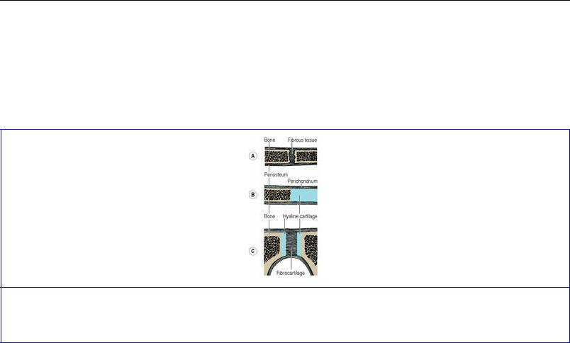
Fibrous joints occur where bones are separated only by connective tissue (Fig. 1.4A) and movement between them is negligible. Examples of fibrous joints are the sutures that unite the bones of the vault of the skull and the syndesmosis between the lower ends of the tibia and fibula.
Figure 1.4 Fibrous and cartilaginous joints in section: A fibrous joint; B primary cartilaginous joint; C secondary cartilaginous joint.
Cartilaginous joints are of two varieties, primary and secondary. A primary cartilaginous joint (synchondrosis) is one where bone and hyaline cartilage meet (Fig. 1.4B). Thus all epiphyses are primary cartilaginous joints, as are the junctions of ribs with their own costal cartilages. All primary cartilaginous joints are quite immobile and are very strong. The adjacent bone may fracture, but the

bone–cartilage interface will not separate.
A secondary cartilaginous joint (symphysis) is a union between bones whose articular surfaces are covered with a thin lamina of hyaline cartilage (Fig. 1.4C). The hyaline laminae are united by fibrocartilage. There may be a cavity in the fibrocartilage, but it is never lined with synovial membrane and it contains only tissue fluid. Examples are the pubic symphysis and the joint of the sternal angle (between the manubrium and the body of the sternum). An intervertebral disc is part of a secondary cartilaginous joint, but here the cavity in the fibrocartilage contains a gel (p. 423).
A limited amount of movement is possible in secondary cartilaginous joints, depending on the amount of fibrous tissue within them. All symphyses occur in the midline of the body.
Typical synovial joints, which include all limb joints, are characterized by six features: the bone ends taking part are covered by hyaline cartilage and surrounded by a capsule enclosing a joint cavity, the capsule is reinforced externally or internally or both by ligaments, and lined internally by synovial membrane, and the joint is capable of varying degrees of movement. In atypical synovial joints the articular surfaces of bone are covered by fibrocartilage.
The synovial membrane lines the capsule and invests all non-articulating surfaces within the joint; it is attached round the articular margin of each bone. Cells of the membrane secrete a hyaluronic acid derivative which is responsible for the viscosity of synovial fluid, whose main function is lubrication. The viscosity varies, becoming thinner with rapid movement and thicker with slow. In normal joints the fluid is a mere film. The largest joint of all, the knee, only contains about 0.5 mL.
The extent to which the cartilage-covered bone-ends make contact with one another varies with different positions of the joint. When the surfaces make the maximum possible amount of contact, the fully congruent joint is said to be close-packed (as in the knee joint in full extension). In this position the capsule and its reinforcing ligaments are at their tightest. When the surfaces are less congruent (as in the partly flexed knee), the joint is loose-packed and the capsule looser, at least in part.
Intra-articular fibrocartilages, discs or menisci, in which the fibrous element is predominant, are found in certain joints. They may be complete, dividing the joint cavity into two, or incomplete. They occur characteristically in joints in which the congruity between articular surfaces is low, e.g. the temporomandibular, sternoclavicular and knee joints.
Fatty pads are found in some synovial joints, occupying spaces where bony surfaces are incongruous. Covered in synovial membrane, they probably promote distribution of synovial fluid. The Haversian fat pad of the hip joint and the infrapatellar fold and alar folds of the knee joint are examples.
Mucous membranes
A mucous membrane is the lining of an internal body surface that communicates with the exterior directly or indirectly. This definition must not be taken to imply that all mucous membranes secrete mucus; many parts of the alimentary and respiratory tracts do, but most of the urinary tract does not. Mucous membranes consist of two and sometimes three elements: always an epithelium and an underlying connective tissue layer, the lamina propria, which in much of the alimentary tract contains a thin third component of smooth muscle, the muscularis mucosae. The whole mucous membrane, often called ‘mucosa’, usually lies on a further connective tissue layer, the submucous layer or

submucosa. The epithelium of a mucous membrane varies according to the site and functional needs, e.g. stratified squamous in the mouth, columnar in the intestine, ciliated in the trachea.
Serous membranes
A serous membrane (serosa) is the lining of a closed body cavity—pericardial, pleural and peritoneal —and consists of connective tissue covered on the surface by a single layer of flattened mesothelial cells (derived from the mesoderm of the coelomic cavity). The part of the serosa that lines the wall of the cavity (the parietal layer of pericardium, pleura or peritoneum) is directly continuous with the same membrane that covers or envelops the mobile viscera within the cavity (the visceral layer). The peritoneal, pericardial and pleural cavities are potential slit-like spaces between the visceral layer and the parietal layer. The two layers slide readily on each other, lubricated by a film of tissue fluid. There are no glands to produce a lubricating secretion. The serous membranes are usually very adherent to the viscera. The parietal layer is attached to the wall of the containing cavity by loose areolar tissue and in most places can be stripped away easily.
The parietal layer of all serous membranes is supplied segmentally by spinal nerves. The visceral layer has an autonomic nerve supply.
Blood vessels
Blood vessels are of three types: capillaries; arteries; and veins.
Capillaries are the smallest vessels. Their walls consist only of flattened endothelial cells. Capillaries form an anastomotic network in most tissues. Certain structures, such as the cornea of the eye and hyaline cartilage, are devoid of capillaries.
Arteries conduct blood from the heart to the capillary bed, becoming progressively smaller, and as they do so, give way to arterioles which connect with the capillaries. Arterial walls have three layers. The tunica intima is very thin and comprises the endothelial lining, little collagenous connective tissue and an internal elastic lamina. Surrounding this layer is the tunica media consisting mainly of elastic connective tissue fibres and smooth muscle in varying amounts. The aorta and major arteries have a large proportion of elastic tissue which enables them to regain their original diameter after the expansion that follows cardiac contraction. Smaller arteries have less elastic tissue and more muscle. The tunica media of arterioles is almost entirely composed of smooth muscle. The outermost layer of the arterial wall is the tunica adventitia, which has an external elastic lamina surrounded by collagenous connective tissue.
Veins collect blood from the capillaries. They generally have a thinner wall and a larger diameter than their corresponding arteries. Veins have the same three layers in their walls as arteries, but a distinct internal elastic lamina is absent and there is much less muscle in the media. Peripheral limb veins are often double, as venae comitantes of their arteries. In the proximal parts of limbs venae comitantes unite into a single large vein. Many veins in the limbs and the neck have valves which prevent reflux of blood. These valves usually have two cup-shaped cusps formed by an infolding of the tunica intima. These cusps are apposed to the wall as long as the flow is towards the heart; when blood flow reverses, the valves close by assuming their cup-shaped form. On the cardiac side of a valve the vein wall is expanded to form a sinus. In general, there are no valves in the veins of the

thorax and abdomen; testicular veins have valves.
Anastomoses between arteries are either actual or potential. In the former instance arteries meet end to end, such as the labial branches of the two facial arteries. A potential anastomosis is by terminal arterioles. Given sufficient time these arterioles can dilate to convey adequate blood, but with sudden occlusion of a main vessel the anastomosis is inadequate to immediately nourish the affected part, as in the case of the coronary arteries.
In many cases there is no precapillary anastomosis between adjacent arteries. Such vessels are endarteries, and here interruption of arterial flow necessarily results in gangrene or infarction. Examples are found in the liver, spleen, kidney, lung, medullary branches of the central nervous system, the retina and the straight branches of the mesenteric arteries.
Arteriovenous anastomoses are short-circuiting channels between terminal arterioles and primary venules which occur in many parts of the body. They are plentiful in the skin, where they may have a role in temperature regulation.
Sinusoids are wide capillaries which have a fenestrated or discontinuous endothelium. They are numerous in the liver, spleen, adrenal medulla and bone marrow.
Blood vessels are innervated by efferent autonomic fibres which regulate the contraction of the smooth muscle in their walls. These nerves act on muscular arteries and especially on arterioles. Their main effect is vasoconstriction and increase in vascular tone, mediated by adrenergic sympathetic fibres. In some areas sympathetic cholinergic fibres inhibit muscle activity and cause vasodilatation. Circulating hormones and factors such as nitric oxide also act on vessel wall muscle. On account of the thickness of their walls, large vessels have their own vascular supply through a network of small vessels, the vasa vasorum.
Lymphatics
Not all the blood entering a part returns by way of veins; much of it becomes tissue fluid and returns by way of lymphatic vessels. Lymphatic capillaries are simple endothelial tubes. Larger collecting channels have walls similar to those of veins, but the specific tunics, or layers, are less distinct. They differ from veins in having many more valves. In general superficial lymphatics (i.e. in subcutaneous tissues) follow veins, while deep lymphatics follow arteries.
Clinical spread of disease (e.g. infection, neoplasm) by lymphatics does not necessarily follow strictly anatomical pathways. Lymph nodes may be bypassed by the disease process. If lymphatics become dilated by obstruction their valves may be separated and reversal of lymph flow can then occur. Lymphatics communicate with veins freely in many parts of the body; the termination of the thoracic duct in the neck may be ligated with impunity, for lymph finds its way satisfactorily into more peripheral venous channels.
Lymphoid tissue
The defence mechanisms of the body include phagocytosis, which is a non-specific engulfing process, and the immune response, which is a specific reaction to microorganisms and foreign proteins (antigens). The immune response may occur in two ways: (1) by the humoral antibody
response, with production of antibodies which are protein molecules that circulate in the blood and attach themselves to the foreign protein so that the combination of antigen and antibody can be destroyed by phagocytosis; and (2) by the cell-mediated immune response, with the production of specific cells that circulate in the blood and destroy the antigen or stimulate its phagocytosis. Two types of lymphocyte produce these reactions: T cells are responsible for cell-mediated immunity and B cells for humoral antibody production. The B cells become transformed into plasma cells which produce the antibody molecules (the immunoglobulins: IgG; IgM; IgA; IgE; and IgD).
All lymphocytes arise from common stem cells in bone marrow (in the embryo from yolk sac, liver and spleen). Some of them circulate to and settle in the thymus, where they proliferate. After release into the bloodstream as T cells they colonize the spleen, lymph nodes and lymphoid follicles at other sites by passing through the postcapillary venules of those structures. Other stem cells become B cells and colonize lymphoid follicles without passing through the thymus. The T cells are so named because they depend on the thymus for their development; cell-mediated immunity thus depends on this organ. The B cells acquire their name from the bursa of Fabricius in birds, for it was in chickens that this organ (a diverticulum of the cloaca) was first found to be the source of humoral antibodies. The main types of T cell are cytotoxic T, helper T and regulatory T cells. B cells can either form plasma cells or become B memory cells.
The lymphoid organs consist of the thymus, lymph nodes and spleen. All are encapsulated and have an internal connective tissue framework to support the cellular elements. In all except the thymus the characteristic structural feature is the lymphoid nodule or follicle, which is typically a spherical collection of lymphocytes with a pale central area, the germinal centre. Unencapsulated lymphoid tissue occurs in mucosa-associated lymphoid tissue (MALT) in the mucosa and submucosa of the alimentary, respiratory and genitourinary tracts. Gut-associated lymphoid tissue (GALT) and bronchus-associated lymphoid tissue (BALT) are categories of MALT. Waldeyer's peripharyngeal lymphoid ring of tonsils (palatine, lingual, nasopharyngeal and tubal) and Peyer's patches in the ileum are areas of organized mucosa-associated lymphoid tissue (O-MALT). The overlying epithelium of these sites is able to sample antigens in the lumen and translocate them to the underlying lymphoid aggregation.
In the thymus the lymphocytes are not concentrated in rounded follicles but form a continuous dense band of tissue at the outer region or cortex of the lobules into which the organ is divided. The inner (paler) regions of the lobules form the medulla which has fewer lymphocytes and contains the characteristic thymic corpuscles (of Hassall); these are remnants of the epithelium of the third pharyngeal pouches from which the thymus developed.
In a typical lymph node the rounded follicles of lymphocytes are concentrated at the periphery (cortex). Lymphocytes, not collected into follicles, are also present in the paracortical areas and medullary region. B lymphocytes are found in the follicles and medulla; T lymphocytes in the paracortical areas and in the cortex between follicles. Several afferent lymph vessels enter through the capsule of the node and open into the subcapsular sinus. From here radial cortical sinuses drain to medullary sinuses which are confluent with the efferent vessel draining the node at the hilum, where blood vessels enter and leave. The thymus, spleen and the O-MALT aggregations, such as the tonsils, do not have afferent lymphatics.
The (palatine and pharyngeal) tonsils possess lymphoid follicles similar to those of lymph nodes, but while the nodes have a capsule of connective tissue the tonsils have, on their inner surfaces, a covering of mucous membrane that dips down deeply to form the tonsillar crypts.
The lymphoid follicles of the spleen are found in its white pulp, which is scattered in the red pulp that constitutes most of the substance of the spleen and contains large numbers of venous sinuses. In the white pulp T lymphocytes form periarteriolar sheaths. The sheaths are enlarged in places by lymphoid follicles with B lymphocytes in the germinal centres. These follicles are visible to the naked eye on the cut surface of the spleen as whitish nodules up to 1 mm in diameter.
Apart from lymphocytes, all lymphoid organs and organized lymphoid tissue contain macrophages, which are part of the mononuclear phagocyte system of the body.

Part two. Nervous system
The nervous system is divided into the central nervous system, which consists of the brain and spinal cord, and the peripheral nervous system composed of cranial and spinal nerves and their associated ganglia. The central and peripheral parts each have somatic and autonomic components; the somatic are concerned with the innervation of skeletal muscle (along efferent pathways) and the transmission of sensory information (along afferent pathways), and the autonomic are concerned with the control of cardiac muscle, smooth muscle and glands (also involving efferent and afferent pathways). The term autonomic nervous system is applied collectively to all autonomic components.
Neurons and nerves
The structural and functional unit of the nervous system is the nerve cell or neuron. It consists of a cell body containing the nucleus, and a variable number of processes commonly called nerve fibres. A single cytoplasmic process, the axon (often very long), conducts nerve impulses away from the cell body, and may give off many collaterals and terminal branches to many different target cells. Other multiple cytoplasmic processes, the dendrites (usually very short), expand the surface area of the cell body for the reception of stimuli.
Pathways are established in the nervous system by communications between neurons at synapses, which are sites on the cell body or its processes where chemical transmitters enable nerve impulses to be handed on from one neuron to another. Transmission between neurons and cells outside the nervous system, for example muscle cells (neuromuscular junctions), is also effected by neurotransmitters. The small number of ‘classic’ transmitters such as acetylcholine and noradrenaline (norepinephrine) has been vastly supplemented in recent years by many substances. These include monoamines, amino acids, nitric oxide and neuropeptides.
Cell bodies with similar function show a great tendency to group themselves together, forming nuclei within the central nervous system and ganglia outside it. Similarly processes from such aggregations of cell bodies tend to run together in bundles, forming tracts within the central nervous system and nerves outside the brain and spinal cord.
Apart from neurons the nervous system contains other cells collectively known as neuroglial cells (neuroglia or glia), which have supporting and other functions but which do not have the property of excitability or conductivity possessed by neurons. The main types of neuroglial cell are astrocytes and oligodendrocytes, which like neurons are developed from ectoderm of the neural tube. A third type of neuroglial cell is the microglial cell (microglia) which is the phagocytic cell of the nervous system, corresponding to the macrophage of connective tissue, and is derived from mesoderm.
Nerve fibres may be myelinated or unmyelinated. In the central nervous system myelin is formed by oligo-dendrocytes, and in peripheral nerves by Schwann cells (neurolemmocytes). In myelinated fibres, the regions where longitudinally adjacent Schwann cells or oligodendrocyte processes join one another are the nodes (of Ranvier). The white matter of the nervous system is essentially a mass of nerve fibres and is so called because of the general pale appearance imparted by the fatty myelin, in contrast to grey matter which is darker and consists essentially of cell bodies.
Peripheral nerve fibres have been classified in relation to their conduction velocity, which is
generally proportional to size, and function:
• Group A—Up to 20 μm diameter, subdivided into: α:12–20 μm. Motor and proprioception (Ia and Ib) β:5–12 μm. Touch, pressure and proprioception (II) γ:5–12 μm. Fusimotor to muscle spindles (II) δ:1–15 μm. Touch, pain and temperature (III)
•Group B—Up to 3 μm diameter. Myelinated. Preganglionic autonomic
•Group C—Up to 2 μm diameter. Unmyelinated. Postganglionic autonomic, and touch and pain (IV).
The widest fibres tend to conduct most rapidly. Unfortunately, as can be seen from the above, it is not possible to make a precise prediction of function from mere size. Thus the largest myelinated fibres may be motor or proprioceptive and the smallest, whether myelinated or unmyelinated, are autonomic or sensory.
Spinal nerves
There are 31 pairs of spinal nerves: 8 cervical, 12 thoracic, 5 lumbar, 5 sacral and 1 coccygeal. Each spinal nerve is formed by the union of an anterior (ventral) and a posterior (dorsal) root which are attached to the side of the spinal cord by little rootlets. The union takes place within the intervertebral foramen through which the nerve emerges immediately distal to the swelling on the posterior root, the posterior root ganglion; most of these are also within the foramen. The anterior root of every spinal nerve contains motor (efferent) fibres for skeletal muscle; those from T1 to L2 inclusive and from S2 to S4 also contain autonomic fibres. The anterior root also contains a small number of unmyelinated afferent pain fibres which have ‘doubled back’ from their cells of origin in the posterior root ganglion to enter the spinal cord by the anterior root instead of by the posterior root. The posterior root of every nerve contains sensory (afferent) fibres whose cell bodies are in the posterior root ganglion. These are unipolar neurons, having a single process that bifurcates to pass to peripheral receptors and the central nervous system. Unlike in autonomic ganglia there are no synapses in posterior root ganglia.
Immediately after its formation the mixed spinal nerve divides into a larger anterior and a smaller posterior ramus. The great nerve plexuses—cervical, brachial, lumbar and sacral—are formed from anterior rami; posterior rami do not form plexuses.
Connective tissue binds the fibres of spinal nerves together to form the single nerve. Delicate loose connective tissue, the endoneurium, lies between individual fibres. Rounded bundles of fibres, or fascicles, are surrounded by the perineurium, a condensed layer of collagenous connective tissue. Fascicles are bound together into a single nerve by a layer of loose but thicker connective tissue, the epineurium. In the largest nerve, the sciatic, only about 20% of the cross-sectional area is nerve, so 80% is connective tissue, but in smaller nerves the amount of neural tissue is proportionally greater. The larger nerves have their own nerves, the nervi nervorum, in their connective tissue coverings.
Peripheral nerve trunks in the limbs are supplied by branches from local arteries. The sciatic nerve in
the buttock and the median nerve at the elbow each have a large branch from the inferior gluteal and common interosseous arteries respectively. Elsewhere, however, regional arteries supply nerves by a series of longitudinal branches which anastomose freely within the epineurium, so that nerves can be displaced widely from their beds without risk to their blood supply.
General principles of nerve supply
Once the nerve supply to a part is established in the embryo it never alters thereafter, unlike the vascular supply. However far a structure may migrate in the devel-oping fetus it always drags its nerve with it. Conversely, the nerve supply to an adult structure affords visible evidence of its embryonic origin.
Skeletal muscles are innervated from motor neuron ‘pools’—groups of motor nerve cell bodies in certain cranial nerve nuclei of the brainstem and anterior horns of the spinal cord. The pool supplying any one muscle overlaps the pools of another, e.g. the anterior horn cells of spinal cord segments C5 and C6 that supply deltoid are intermixed with cells of the same segments supplying subscapularis and other muscles. The only exceptions to the overlapping of neuronal pools are the brainstem nuclei of the fourth and sixth cranial nerves, as they are the only motor nerve cell groups supplying only one muscle (superior oblique and lateral rectus of the eye respectively).
Nerve supply of the body wall
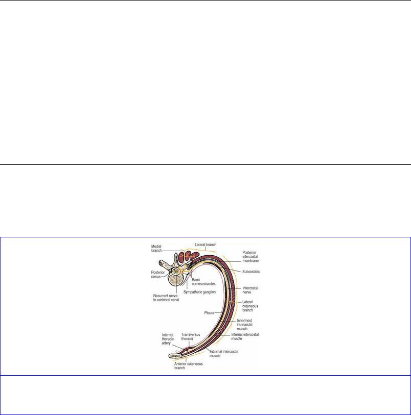
The body wall is supplied segmentally by spinal nerves (Fig. 1.5). The posterior rami pass backwards and supply the extensor muscles of the vertebral column and skull, and to a varying extent the skin that overlies them. The anterior rami supply all other muscles of the trunk and limbs and the skin at the sides and front of the neck and body.
Figure 1.5 Course of a typical intercostal nerve along the neurovascular plane of the body wall, between the middle and innermost of the three muscle layers.
Posterior rami
In the trunk, all the muscles of the erector spinae and transversospinalis groups that lie deep to the thoracolumbar fascia, and the levator costae muscles of the thorax are supplied by the posterior rami

of spinal nerves (Fig. 1.6). In the neck, splenius and all muscles deep to it are similarly supplied.
Figure 1.6 Distribution of posterior rami. On the right, the cutaneous distribution is shown (medial branches down to T6, to clear the scapula, and lateral branches below this); the stippled areas of skin are supplied by anterior rami. On the left, the muscular distribution is shown, to erector spinae and to splenius and the muscles deep to it.
Each posterior ramus divides into a medial and a lateral branch (Fig. 1.5). Both branches of the posterior rami supply muscle, but only one branch, either medial or lateral, reaches the skin. In the upper half of the thorax the medial branches, and in the rest of the body the lateral branches, of the posterior rami provide the cutaneous branches (Fig. 1.6).
C1 has no cutaneous branch, and the posterior rami of the lower two nerves in the cervical and lumbar regions of the cord likewise fail to reach the skin. All 12 thoracic and five sacral nerves reach the skin. No posterior ramus ever supplies skin or muscle of a limb.
Anterior rami
The anterior rami supply the prevertebral flexor muscles segmentally by separate branches from each nerve (e.g. longus capitis and colli, scalene muscles, psoas, quadratus lumborum, piriformis). The anterior rami of the lower four cervical and the first thoracic nerves supply muscles in the upper limb via the brachial plexus. The anterior rami of the 12 thoracic nerves and L1 supply the muscles of the body wall segmentally. Each intercostal nerve supplies the muscles of its intercostal space, and the lower six nerves pass beyond the costal margin to supply the muscles of the anterior abdominal wall. The first lumbar nerve (iliohypogastric and ilioinguinal nerves) is the lowest spinal nerve to supply the anterior abdominal wall. Muscles supplied by anterior rami below L1 are no longer in the body wall; they have migrated into the lower limb.
C2, 3 and 4 supply skin in the neck by branches of the cervical plexus. C5, 6, 7 and 8 and T1 supply
skin of the upper limb via the brachial plexus.
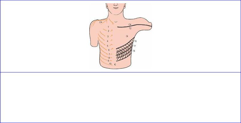
In the trunk the skin is supplied in strips or zones in regular sequence from T2 to L1 inclusive. The intercostal nerves each have a lateral branch to supply the sides and an anterior terminal branch to supply the front of the body wall (Fig. 1.5). The lower six thoracic nerves pass beyond the costal margin obliquely downwards to supply the skin of the abdominal wall (Fig. 1.7).
Figure 1.7 Overlap of dermatomes on the body wall. On the right side, the supraclavicular and thoracic nerves are shown. On the left, the anterior axial line is indicated; this marks the boundary on the chest wall between skin supplied by the cervical plexus and by intercostal nerves. Adjacent dermatomes overlap and thereby, for instance, the dermatomes of T6 and T8 meet each other, completely covering T7, explaining why division of a single intercostal nerve does not give rise to anaesthesia on the trunk.
Neurovascular plane
The nerves of the body wall, accompanied by their segmental arteries and veins, spiral around the walls of the thorax and abdomen in a plane between the middle and deepest of the three muscle layers (see p. 181 and Fig. 1.5). In this neurovascular plane the nerves lie below the arteries as they run around the body wall. But the nerves cross the arteries posteriorly alongside the vertebral column and again anteriorly near the ventral midline, and at these points of crossing the nerve always lies nearer the skin. The spinal cord lies nearer the surface of the body than the aorta, and as a result the spinal nerve makes a circle that surrounds the smaller arterial circle. The arterial circle is made of the aorta with its intercostal and lumbar arteries, completed in front by the internal thoracic and the superior and inferior epigastric arteries. As a part of the same arterial pattern the vertebral arteries pass up to the cranial cavity. The spinal nerves, as they emerge from the intervertebral foramina, pass laterally behind the vertebral artery in the neck, behind the posterior intercostal arteries in the thorax, behind the lumbar arteries in the abdomen and behind the lateral sacral arteries in the pelvis. The anterior terminal branches of the spinal nerves similarly pass in front of the internal thoracic and the superior and inferior epigastric arteries (Fig. 1.5).
The sympathetic trunk runs vertically within the arterial circle. From the base of the skull to the coccyx the sympathetic trunk lies anterior to the segmental vessels (vertebral, posterior intercostal, lumbar and lateral sacral arteries).

Sympathetic fibres
Every spinal nerve without exception, from C1 to the coccygeal, carries postganglionic (unmyelinated, grey) sympathetic fibres which ‘hitch-hike’ along the nerves and accompany all their branches. They leave the spinal nerve only at the site of their peripheral destination. They are in the main vasoconstrictor in function, though some go to sweat glands in the skin (sudomotor) and to the arrectores pilorum muscles of the hair roots (pilomotor). In this way the sympathetic system innervates the whole body wall and all four limbs. This is chiefly for the function of temperature regulation. The visceral branches of the sympathetic system have a different manner of distribution (see p. 19).
Nerve supply of limbs
The body wall has been seen to be supplied segmentally by spinal nerves (Fig. 1.5). A longitudinal strip posteriorly is supplied by posterior rami, a lateral strip by the lateral branches of the anterior rami, and a ventral strip by the anterior terminal branches of the anterior rami. In the fetus the limb buds grow out from the lateral strip supplied by the lateral branches of the anterior rami and these lateral branches, by their anterior and posterior divisions, form the plexuses for supply of the muscles and skin of the limbs. The posterior divisions supply extensor muscles and the anterior divisions supply flexor muscles. Both divisions supply skin of the limbs.
Each limb consists of a flexor and an extensor compartment, which meet at the preaxial and postaxial borders of the limb. These borders are marked out approximately by veins. In the upper limb the cephalic vein lies at the preaxial and the basilic vein at the postaxial border. In the lower limb, extension and medial rotation, which replace the early fetal position of flexion, have complicated the picture. The great saphenous vein marks out the preaxial and the small saphenous vein the postaxial borders of the limb.
The spinal nerves entering into a limb plexus come from enlarged parts of the cord, the cervical enlargement for the brachial plexus and the lumbar enlargement for the lumbar and sacral plexuses. The enlargements are produced by the greatly increased number of motor neurons in the anterior horns at these levels (see p. 487).
On account of the way nerve fibres become combined and rearranged in plexuses, any one spinal nerve can contribute to more than one peripheral nerve and peripheral nerves can receive fibres from more than one spinal nerve. It follows that the area of skin supplied by any one spinal nerve or spinal cord segment is not the same as the area supplied by a peripheral spinal nerve. Two kinds of skin maps or charts are therefore required, one showing segmental innervation and the other showing peripheral nerves. The segmental supplies are reviewed below; the peripheral nerves of the upper and lower limbs are summarized on pages 91 and 162.
Segmental innervation of the skin
The area of skin supplied by a single spinal nerve is called a dermatome. On the trunk, adjacent dermatomes overlap considerably, so that interruption of a single spinal nerve produces no anaesthesia (Fig. 1.7); the same applies to the limbs, except at the axial lines. The line of junction of two dermatomes supplied from discontinuous spinal levels is demarcated by an axial line, and such axial lines extend from the trunk on to the limbs. In the upper limb (Fig. 1.8) the anterior axial line

runs from the sternal angle across the second costal cartilage and down the front of the limb almost to the wrist. The dermatomes lie in orderly numerical sequence when traced distally down the front and proximally up the back of the anterior axial line (C5, 6, 7, 8 and T1) and these dermatomes are supplied by the nerves of the brachial plexus. In addition, skin has been ‘borrowed’ from the neck and trunk to clothe the proximal part of the limb (C4 over the deltoid muscle, T2 for the axilla).
Figure 1.8 Approximate dermatomes and axial lines of the right upper limb. See text for explanation.
Considerable distortion occurs to the dermatome pattern of the lower limb (Fig. 1.9) for two reasons. Firstly the limb, from the fetal position of flexion, is medially rotated and extended, so that the anterior axial line is caused to spiral from the root of the penis (clitoris) across the front of the scrotum (labium majus) around to the back of the thigh and calf in the midline almost to the heel. Secondly, a good deal of skin is ‘borrowed’ from the trunk on the cranial side (from T12, L1, 2 and 3). As in the upper limb, the dermatomes can be traced in numerical sequence down in front and up behind the anterior axial line (L1, 2, 3, 4, 5 and S1, 2, 3).
Figure 1.9 Approximate dermatomes and axial lines of the right lower limb. See text for explanation.

A practical application of the anterior axial line arises in spinal analgesia. A ‘low spinal’ (caudal) anaesthetic anaesthetizes the skin of the posterior two-thirds of the scrotum or labium majus (S3), but to anaesthetize the anterior one-third of the scrotum or labium L1 must be involved, an additional seven spinal segments higher up.
It must be remembered that a single chart cannot indicate individual variations or the differing findings of several groups of investigators, and that such charts are a compromise between the maximal and minimal segmental areas which experience has shown can occur. Original charts, such as those made by Sherrington, Head and Foerster, are being modified by the continuing accumulation of new information. Thus T1 nerve, for example, is not usually considered to supply any thoracic skin but has sometimes been considered to do so, and L5 and S1 have been reported to extend to buttock skin although this is not usually expected. It is probable that posterior axial lines do not exist, but evidence for anterior axial lines is more convincing. Difficulty in investigation arises in the main from the blurring of patterns due to overlap from adjacent dermatomes. A chart of dermatomes must therefore be interpreted with flexibility. The following summary offers selected guidelines that are clinically useful:
C1 |
No skin supply |
C2 |
Occipital region, posterior neck and skin over parotid |
C3 |
Neck |
C4 |
Infraclavicular region (to manubriosternal junction), shoulder and above scapular spine |
C5 |
Lateral arm |
C6 |
Lateral forearm and thumb |
C7 |
Middle fingers |
C8 |
Little finger and distal medial forearm |
T1 |
Medial arm above and below elbow |
T2 |
Medial arm, axilla and thorax |
T3 |
Thorax and occasional extension to axilla |
T4 |
Nipple |
T7 |
Subcostal angle |
T8 |
Rib margin |
T10 |
Umbilicus |
T12 |
Lower abdomen, upper buttock |
L1 |
Suprapubic and inguinal regions, penis, anterior scrotum (labia), upper buttock |
L2 |
Anterior thigh, upper buttock |
L3 |
Anterior and medial thigh and knee |
L4 |
Medial leg, medial ankle and side of foot |
L5 |
Lateral leg, dorsum of foot, medial sole |
S1 |
Lateral ankle, lateral side of dorsum and sole |
S2 |
Posterior leg, posterior thigh, buttock, penis |
S3 |
Sitting area of buttock, posterior scrotum (labia) |
S4 |
Perianal |
|
|
S5 and Co Behind anus and over coccyx.
Segmental innervation of muscles
Most muscles are supplied equally from two adjacent segments of the spinal cord. Muscles sharing a common primary action on a joint irrespective of their anatomical situation are all supplied by the same (usually two) segments. Their opponents, sharing the opposite action, are likewise all supplied by the same (usually two) segments and these segments usually run in numerical sequence with the former. For a joint one segment more distal in the limb the spinal centre lies en bloc one segment lower in the cord.
Thus there are in effect spinal centres for joint movements, and these centres tend to occupy continuous segments in the cord. The upper one or two segments innervate one movement, and the lower one or two innervate the opposite movement (although sometimes the same segment may innervate both movements, but of course from different anterior horn cells). Thus the spinal centre for the elbow is in C5, 6, 7, 8 segments; biceps, brachialis and brachioradialis (the prime flexors of the elbow) are supplied by C5, 6 and triceps (the prime extensor of the elbow) is supplied by C7, 8.
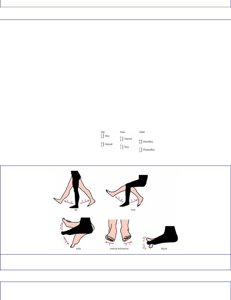
The segments mainly responsible for the various limb joint movements are summarized in Figures 1.10 and 1.11. Flexion/extension at the hip, knee and ankle are the easiest to remember, for each movement involves two segments in logical sequence for each joint, and for each more distal joint the
segments concerned are one segment lower:
Figure 1.10 Segmental innervation of movements of the lower limb.
Figure 1.11 Segmental innervation of movements of the upper limb.
The above pattern enables the segmental innervation of a muscle to be determined, e.g.:
•iliacus (flexes hip) L2, 3
•biceps femoris (flexes knee) L5, S1
•soleus (plantarflexes ankle) S1, 2.
The above are simple flexion–extension movements and, indeed, cover all kneeand ankle-moving muscles. At the hip, however, movements other than flexion and extension are possible, but all are innervated by the same four segments. Thus:
•adduction or medial rotation (same as flexion) L2, 3
•abduction or lateral rotation (same as extension) L4, 5.
For inversion and eversion of the foot the formulae are:
•invert foot L4
•evert foot L5, S1.
Tibialis anterior and tibialis posterior invert the foot and both are innervated by L4 segment. Tibialis anterior is also a dorsiflexor and L4, 5 (from the formula already given for dorsiflexion) is its correct segmental supply. Tibialis posterior, however, lies deep among the plantar flexors of the ankle (S1, 2), but its main action is inversion of the foot (it is the principal invertor) and, although it assists plantar flexion, its segmental innervation is L4, 5.
The upper limb movements, with the segments involved (Fig. 1.11), are as follows:
Shoulder |
Abduct and laterally rotate C5 |
|
Adduct and medially rotate C6, 7, 8 |
Elbow |
Flex C5, 6 |
|
|
|
|

|
Extend C7, 8 |
Forearm |
Supinate C6 |
|
Pronate C7, 8 |
Wrist only |
Flex C6, 7 |
|
Extend C6, 7 |
Fingers and thumb |
Flex C7, 8 |
(long tendons) |
Extend C7, 8 |
Hand |
T1. |
(intrinsic muscles) |
|
In the upper limb the two-and-two segment pattern is not as regular as in the lower limb, probably because in the upper limb much more precise movements are constantly being employed, and the spinal centres have broken up into separate nuclei to control these. Thus, below the elbow the plan does not conform to the basic pattern of four spinal segments for each joint. Flexion and extension share the same two segments; these are C6, 7 for the wrist and C7, 8 for the digits. But the rule holds that the more distal joints are innervated from lower centres in the cord.
As a guide to the level of spinal cord injury it is useful to be aware of a muscle and a movement for which a particular spinal cord segment is mainly responsible:
C4 Diaphragm. Respiration
C5 Deltoid. Abduction of the shoulder
C6 Biceps. Flexion of the elbow. Biceps jerk (see below)
C7 Triceps, Extension of the elbow. Triceps jerk (see below)
C8 Flexor digitorum profundus and extensor digitorum. Finger flexion and extension
T1 Abductor pollicis brevis representing small hand muscles. Abduction of the thumb
T7–12 Anterior abdominal wall muscles. Guarding. Abdominal reflex (see below)
L1 Lowest fibres of internal oblique and transversus abdominis. Guarding
L2 Psoas major. Flexion of the hip
L3 Quadriceps femoris. Extension of the knee. Knee jerk (see below)
L4 Tibialis anterior and posterior. Inversion of the foot
L5 Extensor hallucis longus. Extension of the great toe
S1 Gastrocnemius. Plantarflexion of the foot. Ankle jerk (see below)
S2 Small muscles of the foot
S3 Perineal muscles. Bladder (parasympathetic). Anal reflex (see below).
It is important to note that the term ‘root’ as used in root injuries may be taken to mean either the nerve root proper, i.e. from the side of the spinal cord to the intervertebral foramen, or the roots of the plexuses, i.e. anterior rami distal to the foramen. In lesions of the nerve roots proper, sweating in the distribution of the appropriate nerves is normal, but in more peripheral lesions sweating is reduced, because the postganglionic sympathetic fibres from the sympathetic trunk join the roots of plexuses distal to the nerve roots proper (Fig. 1.12C).

Figure 1.12 Examples of spinal reflex pathways: A the two neurons of a stretch reflex (tendon jerk), which is monosynaptic; B a multisynaptic reflex arc—only one interneuron is shown but there may be several; C the three neurons of a sympathetic reflex, the body of the preganglionic cell is in the lateral horn of the spinal cord and that of the postganglionic cell in a sympathetic ganglion (the preganglionic fibre runs in the white ramus communicans, the more distal connection of the ganglion, and the postganglionic fibre in the proximal grey ramus); D the fusimotor neuron loop; the γ efferent neuron, under the influence of higher centres, stimulates the muscle spindle from which afferent fibres pass back to the spinal cord to synapse with the α motor neuron.
Spinal reflexes
What is commonly called the ‘knee jerk’ and similar tendon reflexes are typical examples of spinal myotatic or stretch reflexes (deep tendon reflexes). They illustrate the simplest kind of reflex pathway and involve only two neurons with one synapse (monosynaptic reflex arc, Fig. 1.12A); indeed the tendon reflexes are the only examples of monosynaptic reflex arcs, for all other reflexes involve two or more synapses (multisynaptic, Fig. 1.12B, C).
Tapping the tendon momentarily stretches the spindles within the muscle and this stimulates the afferent (Ia) fibres of the nerve endings surrounding the intrafusal fibres, which pass into the spinal cord by the posterior nerve root. These afferents synapse directly with the α motor neurons of the anterior horn whose axons form the efferent side of the arc, so causing the extrafusal fibres to contract and produce the ‘jerk’ at the joint.
For most practical purposes the segments mainly concerned with the reflexes most commonly tested may be taken as: biceps jerk—C6; triceps jerk—C7; knee jerk—L3; ankle jerk—S1.
Diminution or absence of the jerk usually indicates some kind of interruption of the arc or muscular defect, but exaggeration of the tendon reflexes is taken as evidence of an upper motor neuron lesion due to alterations in the supraspinal control of the anterior horn cells which are rendered unduly

excitable. In this case the γ motor neurons of the anterior horn are stimulated by such fibres as the reticulospinal and vestibulospinal. The pathway (Fig. 1.12D) is from the γ motor neuron to the intrafusal muscle fibres of the spindle, then from the afferent fibres of the spindle to the α motor neuron and so to the extrafusal fibres. This is the γ reflex loop or fusimotor neuron loop.
In addition to the above deep tendon reflexes, there are superficial skin reflexes which are multisynaptic. Those most commonly tested are the plantar, abdominal and anal reflexes.
Firm stroking of the lateral surface of the sole of the foot (as with the end of a key) to elicit the plantar reflex normally causes plantarflexion of the great toe and probably of the other toes as well. Extension of the great toe—the extensor response (Babinski's sign)—indicates an upper motor neuron lesion. In infants under 1 year old the extensor response is the normal response; only with myelination of the corticospinal tracts during the second year does the normal plantar reflex become flexor.
The abdominal reflex is elicited by lightly stroking across each quadrant of the anterior abdominal wall. Normally there is contraction of the underlying muscles, but the reflexes are absent in upper motor neuron lesions. Patients with paraplegia who are lying down may exhibit Beevor's sign: when trying to lift the shoulders, the umbilicus is displaced upwards, due to weakness of the muscles below the umbilicus.
The anal reflex (‘anal wink’) is a visible contraction of the external anal sphincter following pinprick of the perianal skin and depends on intact sacral segments of the cord (mainly S3).
Autonomic nervous system
The motor part of the somatic nervous system is concerned with the innervation of skeletal muscle. The cell bodies are either in the motor nuclei of cranial nerves or the anterior horn cells of the spinal cord, and the nerve fibres which leave the central nervous system run uninterruptedly to the muscles, ending as motor endplates on the muscle fibres. The motor part of the autonomic nervous system is concerned with the innervation of cardiac and smooth muscle and glands, and the great difference between this and the somatic system is that the pathway from nerve cells in the central nervous system to the target organ is interrupted by synapses in a ganglion. There are thus two sets of neurons, which are logically called preganglionic and postganglionic. The preganglionic cell bodies are always within the central nervous system. If sympathetic, they are in the lateral horn cells of all the thoracic and the upper two lumbar segments of the spinal cord; this is the thoracolumbar part of the autonomic nervous system (the ‘thoracolumbar outflow’). If parasympathetic, they are in certain cranial nerve nuclei and in lateral horn cells of sacral segments of the spinal cord; this is the craniosacral part of the autonomic nervous system (the ‘craniosacral outflow’).
The postganglionic cell bodies are in ganglia in the peripheral nervous system. If sympathetic, the ganglia are either in the sympathetic trunk or in autonomic plexuses situated in the abdomen and pelvis (such as the coeliac ganglia). If parasympathetic, the ganglia are usually within the walls of the viscera concerned, while in the head there are four ganglia which are some little distance from the structures innervated.
Sympathetic nervous system
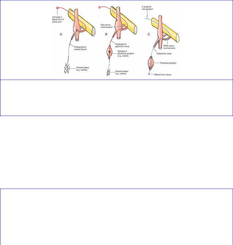
Having reached a sympathetic trunk ganglion, the incoming preganglionic fibres have one of three possible synaptic alternatives. The most common is for them to synapse with cell bodies in a trunk ganglion, either in the one they entered (Fig. 1.13A) or to run up or down the trunk to some other trunk ganglion. The second alternative is to leave the trunk ganglion without synapsing and to pass to a ganglion in an autonomic plexus for synapse (Fig. 1.13B). The third possibility (which applies only to a small number of fibres) is that they leave the trunk (without synapsing) to pass to the suprarenal gland, where certain cells of the medulla can be regarded as modified ganglion cells.
Figure 1.13 Visceral connections of sympathetic ganglia: A efferent pathway with synapse in a sympathetic trunk ganglion; B efferent pathway with synapse in a peripheral ganglion; C afferent pathway for pain fibres, passing through the trunk ganglion and into the spinal nerve by the white ramus communicans.
Because there is no sympathetic outflow from the cervical part of the cord, nor from the lower lumbar and sacral parts, those preganglionic fibres which are destined to synapse with cell bodies whose fibres are going to run with cervical nerves must ascend in the sympathetic trunk to cervical ganglia, and those for lower lumbar and sacral nerves must descend in the trunk to lower lumbar and sacral ganglia.
The segmental levels of the preganglionic cell bodies concerned with the innervation of the different regions of the body (via postganglionic neurons) are indicated in Figure 1.14. In general the body is represented upright from head to perineum but with overlaps and individual variations.

Figure 1.14 Spinal cord levels of sympathetic preganglionic cells. There may be considerable individual variations, especially for the upper limb.
The sympathetic trunk extends alongside the vertebral column from the base of the skull to the coccyx. Theoretically there is a ganglion for each spinal nerve, but fusion occurs, especially in the cervical region where the upper four unite to form the superior cervical ganglion, the fifth and sixth form the middle cervical ganglion, and the seventh and eighth fuse as the inferior cervical ganglion (and often with the first thoracic ganglion as well to form the cervicothoracic or stellate ganglion). Elsewhere there is usually one ganglion less than the number of nerves: 11 thoracic; 4 lumbar; and 4 sacral.
The fibres from the lateral horn cells of each segment of the spinal cord leave in the anterior nerve root (with the axons of anterior horn cells) to reach the spinal nerve and its anterior ramus. The connecting links from here to the sympathetic trunk and its ganglia are the rami communicantes. There are normally two rami; the white ramus communicans is the more distal of the two, and this is the one containing the preganglionic fibres (which are myelinated, hence called white). The other, the grey ramus communicans, contains efferent postganglionic fibres (which are unmyelinated, hence grey). The fibres in the grey ramus are those that are distributed via the branches of the spinal nerve to blood vessels, sweat glands and arrector pili muscles (i.e. they are vasomotor, sudomotor and pilomotor). Every spinal nerve receives a grey ramus. All the thoracic and the upper two lumbar nerves have both white and grey rami connecting them to sympathetic ganglia. But the cervical, lower lumbar and sacral nerves do not have white rami; the ganglia they are connected with receive preganglionic fibres from the thoracolumbar outflow through the chain. Because of the fusion of ganglia, the superior cervical ganglion gives off four grey rami, and the other cervical ganglia two each. Occasionally rami (both grey and white) may be duplicated.
Each sympathetic trunk ganglion has a collateral or visceral branch, usually called a splanchnic nerve in the thoracic, lumbar and sacral regions, but in the cervical region called a cardiac branch because it proceeds to the cardiac plexus. The visceral branches generally arise high up and descend steeply to form plexuses for the viscera (Fig. 1.13). Thus cardiac branches arise from the three cervical ganglia to descend into the mediastinum to the cardiac plexus, which is supplemented by fibres from upper thoracic ganglia. From the fifth and lower thoracic ganglia three splanchnic nerves pierce the diaphragm to reach the coeliac plexus and other pre-aortic plexuses, which are also joined by lumbar splanchnic nerves from the upper lumbar ganglia. Fibres from these plexuses, and
splanchnic nerves from the lower lumbar ganglia, descend to the superior hypogastric plexus and thence to the left and right inferior hypogastric (pelvic) plexuses.
The sympathetic visceral plexuses thus formed are joined by parasympathetic nerves: vagus to the coeliac plexus; and pelvic splanchnics (S2–4) to the inferior hypogastric plexuses. The mixed visceral plexuses reach the viscera by direct branches and by branches that hitch-hike along the relevant arteries.
In addition to the visceral branches, which supply not only the smooth muscle and glands of viscera but also the blood vessels of those viscera, all trunk ganglia give off vascular branches to adjacent large blood vessels. The cervical ganglia give branches to the carotid and vertebral arteries, including (from the superior cervical ganglion) the internal carotid nerve, running upwards on the artery of that name to form the internal carotid plexus on the artery as it enters the skull. The thoracic and lumbar ganglia give filaments to the various aortic plexuses and from there to aortic branches including the common iliac arteries, continued along the internal and external iliac arteries as far as the proximal part of the femoral artery. Branches from the sacral ganglia pass to the lateral sacral arteries. Limb vessels get their sympathetic innervation mainly from nerve fibres that run with the adjacent peripheral nerves before passing to the vessels; the fibres do not run long distances along the vessels themselves. Thus the nerve filaments to the vessels of the tip of a finger or toe run not with the digital arteries but with the digital nerves, and only leave the nerves near the actual site of innervation.
Afferent sympathetic fibres
Many afferent fibres hitch-hike along sympathetic efferent pathways. Some form the afferent limb for unconscious reflex activities; others are concerned with visceral pain. All have their cell bodies in the posterior root ganglia of spinal nerves (not in sympathetic ganglia), at approximately the same segmental level as the preganglionic cells (Fig. 1.14). The afferent fibres reach the spinal nerve via the white ramus communicans (Fig. 1.13C) and then join the posterior root ganglion, from which central processes enter the spinal cord by the posterior nerve root (like any other afferent fibres). Visceral pain fibres enter the posterior horn of the spinal cord, and thereafter the pain pathway is the same as that for spinal nerve pain fibres. Others concerned with reflex activities may synapse with interneurons in the cord or ascend to the hypothalamus and other higher centres by pathways that are not defined.
Sympathectomy
For the control of excessive sweating and vasoconstriction in the extremities of the limbs, parts of the sympathetic trunk with appropriate ganglia can be removed to abolish the normal sympathetic influence. In upper thoracic ganglionectomy for the upper limb the second and third thoracic ganglia with their rami and the intervening part of the trunk are resected; alternatively, the trunk is divided below the third ganglion and the rami communicantes to the second and third ganglia are severed. The first thoracic ganglion is not removed, as the preganglionic fibres for the upper limb do not usually arise above T2 level (see above), and its removal would result in Horner's syndrome (see p. 408). Upper thoracic ganglionectomy is described further on page 211.
For lumbar sympathectomy the third and fourth lumbar ganglia and the intervening trunk are removed. The first lumbar ganglion should be preserved otherwise ejaculation may be compromised. Lumbar

sympathectomy is described further on page 282.
Parasympathetic nervous system
Although all parts of the body receive a sympathetic supply, the distribution of parasympathetic fibres is wholly visceral and not to the trunk or limbs. However, not all viscera are so innervated: the suprarenal glands and the gonads appear to have only a sympathetic supply.
The preganglionic fibres of cranial origin have their cell bodies in the accessory (Edinger–Westphal) oculomotor nucleus, the superior and inferior salivatory nuclei of the seventh and ninth cranial nerves respectively, and the dorsal motor nucleus of the vagus. The fibres of the first three nuclei synapse with cells in the four parasympathetic ganglia, described below; the vagal fibres synapse with postganglionic cell bodies in the walls of the viscera supplied (heart, lungs and gut).
The preganglionic fibres of sacral origin arise from cells in the lateral grey horn of sacral segments 2–4 of the spinal cord, and constitute the pelvic splanchnic nerves. Leaving the anterior rami of the appropriate sacral nerves near the anterior sacral foramina, they pass forwards to enter into the formation of the inferior hypogastric plexuses. From there they run to pelvic viscera and to the hindgut as far up as the splenic flexure, and synapse around postganglionic cell bodies in the walls of these viscera.
Cranial parasympathetic ganglia
The four ganglia—ciliary, pterygopalatine, submandibular and otic—are very similar in plan. Each has parasympathetic, sympathetic and sensory roots, and branches of distribution. The roots and branches are described in general terms below and illustrated in Figure 1.15; the topographical details of each ganglion are dealt with in the regions concerned.
Figure 1.16 An embryo at the beginning of the second week. The trophoblast has differentiated into an inner layer of cells with single nuclei (the cytotrophoblast) and an outer layer with multiple nuclei but without distinct cell boundaries (the syncytiotrophoblast).
The parasympathetic root carries the preganglionic fibres from the cells of origin in a brainstem nucleus. This is the essential functional root of the ganglion; its fibres synapse in it, whereas the fibres of all other roots simply pass through the ganglion without synapse.
The sympathetic root contains postganglionic fibres from the superior cervical ganglion, whose preganglionic cell bodies are in the lateral grey horn of cord segments T1–3.

The sensory root contains the peripheral processes of cell bodies in the trigeminal ganglion.
The branches of each ganglion carry the postganglionic parasympathetic fibres to the particular structure(s) requiring this kind of localized motor innervation: ciliary muscle and sphincter pupillae from the ciliary ganglion, salivary glands from the submandibular and otic ganglia, and lacrimal, nasal and palatal glands from the pterygopalatine ganglion. The other fibres in the branches are sympathetic fibres to the same structures (mainly for their blood vessels) and afferent fibres.
Ciliary ganglion (see p. 403)
Parasympathetic root. From the Edinger–Westphal part of the oculomotor nucleus by a branch from the nerve to the inferior oblique muscle from the inferior division of the oculomotor nerve.
Sympathetic root. From the superior cervical ganglion by branches of the internal carotid plexus.
Sensory root. From a branch of the nasociliary nerve, with cell bodies in the trigeminal ganglion.
Branches. Short ciliary nerves to the eye.
Pterygopalatine ganglion (see p. 370)
Parasympathetic root. From the superior salivary nucleus by the nervus intermedius part of the facial nerve, the greater petrosal nerve and the nerve of the pterygoid canal.
Sympathetic root. From the superior cervical ganglion by the internal carotid plexus, the deep petrosal nerve and the nerve of the pterygoid canal.
Sensory root. From branches of the maxillary nerve, with cell bodies in the trigeminal ganglion.
Branches. To the lacrimal gland via the zygomatic and lacrimal nerves, and to mucous glands in the nose, nasopharynx and palate via maxillary nerve branches. A few fibres (not shown in Fig. 1.15) are taste fibres from the palate, which run in the greater petrosal nerve and have cell bodies in the geniculate ganglion of the facial nerve.
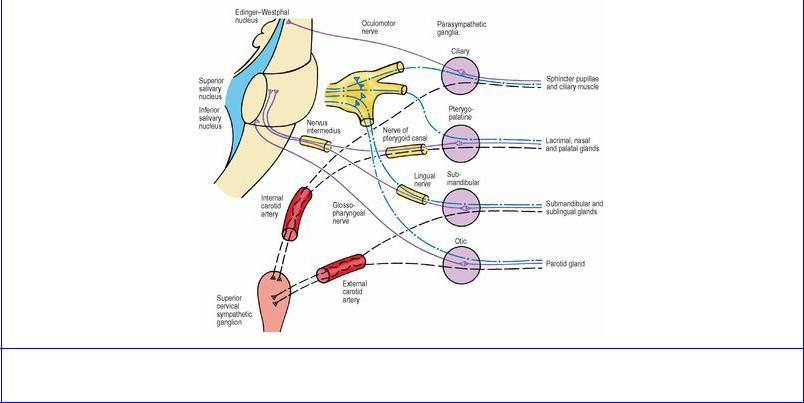
Figure 1.15 Connections of the four parasympathetic ganglia of the head and neck.
Submandibular ganglion (see p. 338)
Parasympathetic root. From the superior salivary nucleus by the nervus intermedius part of the facial nerve, the chorda tympani and the lingual nerve.
Sympathetic root. From the superior cervical ganglion by fibres running with the facial artery.
Sensory root. From a branch of the lingual nerve, with cell bodies in the trigeminal ganglion.
Branches. To the submandibular and sublingual glands via branches of the lingual nerve.
Otic ganglion (see p. 366)
Parasympathetic root. From the inferior salivary nucleus by the glossopharyngeal nerve, its tympanic branch, the tympanic plexus and the lesser petrosal nerve.
Sympathetic root. From the superior cervical ganglion by fibres running with the middle meningeal artery.
Sensory root. From the auriculotemporal nerve with cell bodies in the trigeminal ganglion.
Branches. To the parotid gland via filaments of the auriculotemporal nerve.
Unlike the other three ganglia, the otic ganglion has an additional somatic motor root, from the nerve to the medial pterygoid; the fibres pass through (without synapse) to supply the tensor tympani and tensor palati muscles.
Parasympathetic afferent fibres
As in the sympathetic nervous system, afferent fibres often accompany the parasympathetic supply to various structures. Such fibres that run with the glossopharyngeal and vagus nerves have their cell bodies in the inferior ganglia of those nerves, and their central processes pass to the nucleus of the
tractus solitarius, through which there are connections with other parts of the brainstem and higher centres for the reflex control of respiration, heart rate, blood pressure and gastrointestinal activity.
The pelvic splanchnic nerves also carry afferent fibres. Their cell bodies are in the posterior root ganglia of the second to fourth sacral nerves and the central processes enter the cord by the posterior nerve roots. Some make local synaptic connections, e.g. for bladder reflexes, but others are pain fibres from pelvic viscera, which often seem to use both sympathetic and parasympathetic pathways for pain transmission, e.g. from bladder and rectum.

Part three. Embryology
The development of most of the organs and systems is touched upon in the text descriptions of the regions concerned. Here a very brief account of some important features of early development is included, to provide a background for the later notes.
Early development
For the first 8 weeks of the 40-week human gestation period the developing organism is an embryo; after that time it is a fetus. By the end of the embryonic period most organs have differentiated, and the changes during the fetal period are essentially those of maturation. Many but not all congenital defects are initiated in the embryo rather than the fetus.
The fertilized ovum or zygote undergoes repeated cell divisions (cleavage) to produce a mass of cells, the morula, which travels along the uterine tube towards the uterus. Further division enlarges the morula and a fluid-filled cavity (the extraembryonic coelom) appears in it; the whole structure is now a blastocyst. At this stage implantation into the uterine mucosa takes place, about 6 days after fertilization. The outer layer of cells of the blastocyst, the trophoblast, is destined to become placental. The remainder of the cells are concentrated at one end of the blastocyst to form the inner cell mass or embryoblast, attached to the inner aspect of the trophoblast.
At the beginning of the second week after fertilization, the embryoblast differentiates into two layers, a layer of columnar cells (the epiblast) and a layer of cuboidal cells (the hypoblast). Two cavities then appear, the amniotic cavity, which is related to the epiblast, and the yolk sac, which is related to the hypoblast (Fig. 1.16). The two cavities are surrounded by the extraembryonic coelom, except where the embryoblast is connected to the trophoblast by the connecting stalk, which later develops into the umbilical cord.
A primitive streak appears on the amniotic aspect of the embryoblast towards what will become the caudal end of the embryo (Fig. 1.17A). The streak indicates the site of a groove at the cephalic end of which is a primitive pit with slightly raised margins; this is the primitive node. In the third week, epiblast cells in the region of the primitive streak invaginate, displacing the hypoblast, and spread bilaterally and cephalad forming two new layers, the mesoderm next to the epiblast and the endoderm adjacent to the hypoblast. The remaining epiblast cells form the ectoderm. From the primitive pit a rod of cells invaginate directly cephalad between ectoderm and endoderm; this is the notochord, which extends as far as the buccopharyngeal membrane, where ectoderm and endoderm remain in contact. Similarly at the caudal end of the primitive streak, ectoderm and endoderm are in apposition at the cloacal membrane.
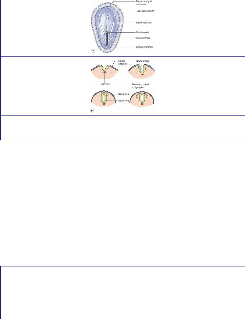
Figure 1.17 A Dorsal view of an embryo at the beginning of the third week; B formation of neural tube and neural crest.
An indentation of the ectoderm overlying the notochord forms the neural groove (Fig. 1.17B). Its edges unite to form the neural tube, which becomes depressed below the surface. In due course, the brain and spinal cord develop from the neural tube. Some of the cells derived from the edges of the groove become isolated between the tube and the overlying ectoderm to form the neural crest. Its cells are destined to migrate and contribute to the development of several structures, including posterior root ganglia of spinal nerves, corresponding ganglia of cranial nerves, autonomic ganglia, neuroglia, Schwann cells, meninges, bones of the skull and face, sclera and choroid of the eye, dentine and cementum of teeth, parafollicular cells of the thyroid, chromaffin cells of the suprarenal medulla and melanocytes.
Alongside the notochord and neural tube the mesoderm lies in three longitudinal strips (Fig. 1.18). That nearest the midline is the paraxial mesoderm; it becomes segmented in cephalocaudal sequence into masses of cells called mesodermal somites. The somites produce: (1) the sclerotome, medially, which surrounds the neural tube and notochord, producing the vertebrae and ribs; and (2) the dermomyotome or muscle plate, laterally, which produces the muscles of the body wall and the dermis of the skin.

Figure 1.18 Cross-section through an embryo at the end of the third week.
The intermediate strip of mesoderm is the intermediate cell mass. From its lateral side in cephalocaudal sequence develop successively the pronephros, mesonephros and metanephros and their associated ducts—the progenitors of the urinary and genital systems. Its medial side gives rise to the gonad and the cortex of the suprarenal gland.
The most lateral strip of mesoderm is the lateral plate. Very early the embryo begins to curl up, a result of the more rapid growth of the dorsal (ectodermal) surface. The embryo becomes markedly convex towards the amniotic cavity and correspondingly concave towards the yolk sac. As the lateral plate curls around to enclose the yolk sac its mesoderm becomes split into two layers by a space that appears within it. The space is the beginning of the intraembryonic coelom or body cavity. The inner layer is the splanchnic (visceral) mesoderm. It encloses the yolk sac in an hourglass constriction; the part of the yolk sac outside persists in the umbilical cord as the vitellointestinal duct; the part inside the embryo becomes the alimentary canal. The outer layer of the lateral plate is the somatic (parietal) mesoderm. Into it the paraxial myotomes migrate in segments to produce the flexor and extensor muscle layers of the body wall. The coelomic cavity at first includes pleural and peritoneal spaces in one continuum; they become separated later. The pleura and peritoneum are thus mesodermal in origin.
The limb buds grow from the lateral plate mesoderm and their muscles develop in situ. Although the lateral plate mesoderm is unsegmented, the motor fibres that grow into it from the spinal cord limb plexuses arrange their distribution in a segmental pattern.
The septum transversum consists of the mass of mesoderm lying on the cranial aspect of the coelomic cavity. Its cranial part contains the pericardial cavity, the walls of which develop into the pericardium and part of the diaphragm. It is invaded by muscles from cervical myotomes, mainly the fourth; they produce the muscle of the diaphragm. The septum transversum later descends, taking the heart with it, to the final position of the diaphragm. The caudal part of the septum transversum is invaded by the developing liver, which it surrounds as the ventral mesogastrium.
The folding of the embryo is impeded to some extent at the tail end by the presence of the connecting stalk, which later becomes the umbilical cord. The greatest amount of folding occurs at the head end of the embryo. By the end of the first fortnight the forebrain capsule is folded down over the pericardium, and a mouth pit, the stomodeum, shows as a dimple between the two. Within the body of the embryo the gut cavity extends headwards dorsal to the pericardium, as far forwards as the buccopharyngeal membrane, which closes the bottom of the mouth pit. The buccopharyngeal

membrane breaks down and disappears in the fourth week and its former site cannot be made out with certainty in the later embryo or adult. Cranial to the site of the membrane the mouth pit is lined with ectoderm; this includes the region of all the mandibular and maxillary teeth, and the anterior twothirds of the tongue. Rathke's pouch arises from this ectoderm and forms the anterior lobe of the pituitary gland. Caudal to the buccopharyngeal membrane is the pharynx, lined with endoderm and lying dorsal to the pericardium.
Pharyngeal arches and pouches
Mesodermal condensations develop in the side walls of the primitive pharynx to form the pharyngeal arches and they grow around towards each other ventrally, where they fuse in the midline. In this way a series of six horseshoe-shaped arches (also called branchial arches) comes to support the pharynx (Fig. 1.19). Deep grooves appear on the surface of the embryo at the intervals between the arches; these are the pharyngeal (or branchial) clefts. The fifth arch is rudimentary and only four clefts are visible. Outpouchings develop from the lining of the pharynx in between the arches and opposite the clefts: the pharyngeal (or branchial) pouches. The fourth and fifth pouches share a common opening into the lumen of the pharynx. In each arch a central bar of cartilage forms and muscle differentiates from the mesoderm around it. An artery and cranial nerve are allocated to the supply of each arch and its derivatives. Vascular patterns are very changeable during development, but a nerve supply, once established, remains constant and knowledge of the nerve supply of a muscle enables its pharyngeal arch origin to be determined.
Figure 1.19 Floor of the developing pharynx. The pharyngeal (branchial) arches are numbered. The foramen caecum lies in the midline between the first and second arches.
First (mandibular) arch
Chondrification in the mesoderm of the first arch produces Meckel's cartilage. The dorsal end of Meckel's cartilage produces the incus and malleus, and the anterior ligament of the malleus. The sphenomandibular ligament (Fig. 1.20) is a remnant of the fibrous perichondrium of Meckel's cartilage. The lingula at the mandibular foramen develops from the cartilage. The mandible starts ossifying in membrane lateral to Meckel's cartilage, and the rest of the cartilage becomes incorporated in the developing mandible. Some time after birth the cartilage disappears.

Figure 1.20 Derivatives of the first arch cartilage.
Ectodermal and endodermal derivatives of this arch are the mucous membrane and glands (but not the muscle) of the anterior two-thirds of the tongue. The muscles of mastication (masseter, temporal and pterygoids), the mylohyoid and anterior belly of digastric, and the two tensor muscles (tensor palati and tensor tympani) develop from the first arch and are supplied by the mandibular nerve, which is the nerve of this arch. Part of the artery of the first arch persists as the maxillary artery.
Other arches
The skeletal and muscular derivatives of the remaining arches can be summarized as follows.
Second (hyoid) arch
Skeletal derivatives: stapes; styloid process; stylohyoid ligament; lesser horn; and superior part of body of hyoid bone (Fig. 1.21).
Figure 1.21 Derivatives of the second and third arch cartilages.
Muscular derivatives: muscles of facial expression (including buccinator and platysma); stapedius; stylohyoid; posterior belly of digastric—all supplied by the facial, the nerve of the second arch.
Third arch
Skeletal derivatives: greater horn and inferior part of body of hyoid bone (Fig. 1.21).
Muscular derivatives: stylopharyngeus—supplied by the glossopharyngeal, the nerve of the third arch.
Fourth and sixth arches
Skeletal derivatives: thyroid; cricoid; epiglottic and arytenoid cartilages.
Muscular derivatives: intrinsic muscles of larynx; muscles of pharynx; levator palati—all supplied by laryngeal and pharyngeal branches of the vagus, the nerve of these arches.
Lateral derivatives of the pharyngeal pouches
Except for the first, each pouch grows laterally into a dorsal and a ventral diverticulum.
First pouch. This is the only pouch in which the endoderm remains in close apposition to the ectoderm of the corresponding cleft, namely at the tympanic membrane, where the mesoderm separating them is minimal. In the other pouches the ectoderm and endoderm are finally widely separated. The endoderm of the first pouch is prolonged laterally, via the auditory tube, to form the middle ear and mastoid antrum (the second pouch gives a contribution to the middle ear; see below). The first pharyngeal cleft becomes deepened to form the external acoustic meatus.
Second pouch. The dorsal part assists the first pouch in the formation of the tympanic cavity, taking what may be its pretrematic nerve (the tympanic branch of the glossopharyngeal) with it; the word ‘trema’ means a cleft. The ventral part of the pouch develops the tonsillar crypts and the supratonsillar fossa from its endoderm, the surrounding mesoderm contributing the lymphatic tissue of the palatine tonsil. The nerve supply of these derivatives is the glossopharyngeal.
Third pouch. Dorsally the inferior parathyroid gland (termed parathyroid III) and ventrally the thymic rudiment grow from this pouch. The latter progresses caudally and joins with that of the other side to produce the bilobed thymus gland. In its descent the thymic bud draws parathyroid III in a caudal direction, so that ultimately the latter lies inferior to parathyroid IV, which is derived from the fourth pouch. From this thymic bud the medulla of the thymus, including the thymic (Hassall's) corpuscles, is derived; the lymphocytes of the cortex migrate from bone marrow.
Fourth pouch. The superior parathyroid glands (parathyroid IV) are derived from the endodermal lining of this pouch.
Fifth pouch. This forms the ultimobranchial body, from which are derived the parafollicular (C) cells of the thyroid gland which produce calcitonin.
Cervical sinus
Concurrently with the growth of the above derivatives from the endoderm of the pouches a change takes place externally in the overlying ectoderm. The only pharyngeal cleft to persist is the first, which forms the external ear. The second arch increases in thickness and grows caudally, over the third, fourth and sixth arches, covering the second, third and fourth clefts and meeting skin caudal to these. During this process a deep groove is formed, which becomes a deep pit, the cervical sinus. The lips of the pit then meet and fuse and the imprisoned ectoderm disappears. Persistence of this ectoderm gives rise to a branchial cyst. Persistence of the deep pit is termed a branchial sinus. A rare branchial fistula results from breaking down of the tissues between the floor of the pit and the side wall of the pharynx (endoderm). The track of the fistula runs from the region of the palatine tonsil, between the external and internal carotid arteries, and reaches the skin anterior to the lower end of sternocleidomastoid.
Ventral derivatives of the floor of the pharynx
The tongue, thyroid gland and larynx are derived from the floor of the mouth (Fig. 1.19).
Buds from the first, third and fourth arches form the stroma of the tongue, the epithelium being derived

from the ectoderm of the stomodeum and the endoderm of the cranial end of the pharynx. Occipital myotomes migrate forwards to provide the musculature, carrying their nerve (hypoglossal) supply with them.
The thyroglossal duct originates from the endoderm of the floor of the pharynx at the foramen caecum (Fig. 1.19) in the region of the developing tongue, and then passes caudally in front of the hyoid bone, behind which it forms a recurrent loop (Fig. 1.22). The thyroid gland buds from the duct's distal end, which itself may give rise to the pyramidal lobe. Other remnants of the duct may persist as accessory thyroid glands or form thyroglossal cysts (Fig. 1.23). Rupture of a cyst gives rise to a thyroglossal sinus (‘fistula’). Removal of retrohyoid duct remnants usually requires excision of a segment from the middle of the hyoid bone. Failure of descent of the thyroglossal duct may result in the development of a lingual thyroid.
Figure 1.22 Course of the thyroglossal duct. The thyroid gland, larynx and trachea have been drawn to a smaller scale than the tongue, mandible and hyoid bone.
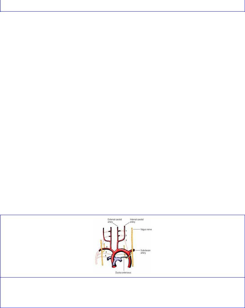
Figure 1.23 Thyroglossal cyst.
In the ventral wall of the pharynx a laryngotracheal groove appears. The cephalic end of this gutter is limited by the furcula, a ridge in the shape of a wish-bone (Fig. 1.19). The ridges which limit the gutter grow towards each other and, by their fusion, convert the gutter into a tube. While maintaining its connection with the pharynx at its cranial end, the larynx, the rest of the tube, the trachea, separates from the oesophagus and buds out into the bronchi and lungs at its caudal end. Failure of proper separation of the trachea and oesophagus results in a tracheo-oesophageal fistula. The furcula persists at the aperture of the larynx, whose cartilages, including that of the epiglottis, derive from the underlying fourth and sixth pharyngeal arches.
Branchial arch arteries
From the cephalic end of the primitive heart tube (see p. 30) a ventral aorta divides right and left into two branches which curve back caudally as the two dorsal aortae. As the branchial arches develop, a vessel in each arch joins the ventral to the dorsal aortae. Thus six aortic arches are to be accounted for. Caudal to this region, the two dorsal aortae fuse to become a single vessel; proximal to the fusion, a part of the right dorsal aorta subsequently disappears.
Parts of the first and second arch arteries form the maxillary and stapedial arteries respectively; the latter does not persist after birth. They are branches of the external carotid artery. The third remains as the common carotid and part of the internal carotid arteries. The fourth on the right contributes to the subclavian artery, on the left to the arch of the aorta. The fifth disappears entirely. By the time the sixth artery appears the upper bulbar part of the heart tube has been divided into aorta and pulmonary trunk and it is to the pulmonary trunk that the sixth arch arteries are connected ventrally. Dorsally they communicate with the dorsal aortae. The dorsal part of the sixth arch artery disappears on the right side but persists on the left as the ductus arteriosus, which thus connects the left pulmonary artery to the arch of the aorta (Fig. 1.24). This explains why the recurrent laryngeal (sixth arch) nerve hooks round the ligamentum arteriosum on the left, but migrates up and hooks round the subclavian artery on the right.
Figure 1.24 Fate of the arch arteries. The arteries are numbered and the dotted lines indicate the arteries that disappear. The asymmetry of the course of the recurrent laryngeal nerves results from differences in the fate of the lower arch arteries.
Anomalies of the great vessels

The most common anomaly of development is a patent ductus arteriosus (persistence of part of the left sixth arch artery), which fails to close in the immediate postnatal period. Coarctation of the aorta (narrowing) is due to a defect of the tunica media which forms a shelf-like projection into the lumen, most commonly in the region of the ductus connection; collateral circulation distal to the obstruction is provided by the internal thoracic and posterior intercostal arteries. An abnormal origin of the right subclavian artery is from the arch of the aorta, just distal to the origin of the left one. The abnormal artery passes to the right behind the oesophagus and is a possible cause of dysphagia. With the lack of a normal right subclavian arch, the right recurrent laryngeal nerve is non-recurrent and runs down the side of the larynx: a possible hazard in thyroidectomy. The reported incidence of a non-recurrent nerve is around 1%.
Development of mouth and face
The stomodeum (mouth pit) has appeared by the end of the second week and the buccopharyngeal membrane between it and the pharynx breaks down in the fourth week.
The stomodeum is bounded below by the mandibular prominence of the first arch (Fig. 1.25), which produces the floor of the mouth, lower jaw and lower lip. From mesenchyme (embryonic mesoderm) on the ventral surface of the developing brain the frontonasal prominence grows down towards the stomodeum. This is indented by two nasal placodes which develop into nasal pits. These are bounded by medial and lateral nasal prominences that unite to encircle the nostril. From the cranial aspect of the dorsal region of each mandibular prominence, the maxillary prominence grows ventrally above the stomodeum, forming the floor of the orbit and fusing with the lateral nasal prominence along the line of the nasolacrimal duct. The medial nasal prominences merge to form the intermaxillary segment from which develops the philtrum of the upper lip, the part of the upper jaw that carries the four incisor teeth and the adjacent primary palate. The maxillary prominences fuse with the philtrum to form the whole of the upper lip.
Figure 1.25 Stages in the development of the face.
At first the developing tongue lies against the floor of the cranium. A midline flange (the nasal septum) grows down from the base of the mesenchymal precursor of the skull. From each maxillary process a flange, known as the palatal shelf, grows downwards and medially; these shelves are soon elevated to a horizontal position over the dorsum of the tongue. The two palatal shelves meet and unite, forming the secondary palate. They also fuse with the nasal septum and the primary palate, the midline incisive foramen persisting at the site of the latter fusion. These fusions begin anteriorly during the eighth week and extend posteriorly to become complete at the uvula in the tenth week.
The nerve supply of all these structures is derived from the fifth cranial (trigeminal) nerve. The frontonasal prominence and its derivatives are supplied by the ophthalmic division, the maxillary prominence and its derivatives by the maxillary division and the mandibular prominence and its derivatives by the mandibular division.
Defects of development
The most common abnormalities are cleft lip and cleft palate (1 in 1000 and 1 in 2500 births respectively) and there are ethnic variations in these incidences; they may or may not coexist. Cleft lip is more frequently lateral. The cleft runs down from the nostril and results from a failure of fusion between the maxillary and medial nasal prominences. Cleft lip may be bilateral and may involve the upper jaw and extend between the primary and secondary palates, the central part being an isolated intermaxillary segment.
Cleft palate may be partial or complete. As the two palatal processes unite with each other progressively from front to back, arrest of union results in a posterior defect (Fig. 1.26) that varies from the mildest form of bifid uvula to a complete cleft from uvula to gum. In the latter case the cleft almost always runs between a lateral incisor and a canine tooth. Very rarely a midline cleft may
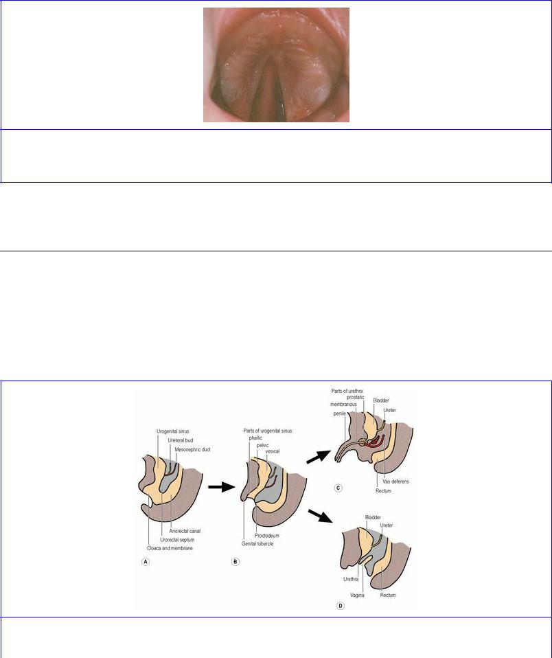
separate the two halves of the maxilla. Irregular formations of incisor and canine teeth often accompany these defects of palatal development.
Figure 1.26 Cleft palate. Palatal fusion has commenced anteriorly but a large posterior defect persists. (Provided by Mr S van Eeden, Alder Hey Hospital, Liverpool.)
A less common defect arises from the failure of fusion of the lateral nasal process with the maxillary process, producing a groove (facial cleft) on the face along the line of the nasolacrimal duct.
Development of the cloaca
At the caudal end of the embryo, the hindgut and the allantois (a diverticulum from the yolk sac) meet in a common cavity, the cloaca, bounded distally by the cloacal membrane (Fig. 1.27A). From the dorsal wall of the allantois, the urorectal septum grows downwards to meet the cloacal membrane, so dividing the cloaca and membrane into two (Fig. 1.27B): at the front are the urogenital sinus and urogenital membrane, and at the back the anorectal canal and the anal membrane, which lies in a small ectodermal depression, the proctodeum. The tip of the urorectal septum forms the perineal body.
Figure 1.27 Development of the cloaca: A the urorectal septum grows down to divide the cloaca into the urogenital sinus and the anorectal canal; B the uppermost (vesicourethral) part of the sinus

becomes the bladder and the proximal part of the prostatic urethra, with the pelvic and phallic parts distally; C in the male the pelvic part becomes the prostatic urethra distal to the opening of the ejaculatory ducts, and the phallic part becomes the dorsal part of the penile urethra; D in the female the bladder and urethra are from the vesicourethral part of the sinus.
The urogenital sinus (endoderm) has three unequally sized parts. The uppermost and largest is the vesical (vesicourethral) part, which forms most of the bladder epithelium (with surrounding mesoderm forming the muscle and connective tissue) and the female urethra (Fig. 1.27D). The lower end of the mesonephric duct (see p. 286) opens into this part of the sinus, with the ureter arising as a bud from the duct. The lower ends of the duct and ureter become incorporated into the developing bladder, so forming the trigone and in the male the part of the urethra proximal to the opening of the ejaculatory duct (Fig. 1.27C).
The middle or pelvic part of the sinus forms the rest of the prostatic urethra, the membranous urethra and the prostate (with surrounding mesoderm forming the fibromuscular stroma). In the female it contributes to the vagina (derived principally from the paramesonephric ducts; see p. 307).
The lowest or phallic part of the sinus becomes the dorsal part of the penis and penile urethra or the lower part of the vagina. At the front of the urogenital membrane (which breaks down) is a midline mesodermal swelling, the genital tubercle (Fig. 1.27B), which becomes the glans penis or clitoris. Leading back from the tubercle on either side are the urogenital folds, which in the female remain separate as the labia minora. In the male they unite at the back to form the midline raphe of the scrotum, the rest of the scrotum coming from the pair of genital (labioscrotal) swellings which develop lateral to the urogenital folds and which in the female become the labia majora. The front parts of the urogenital folds unite from the scrotum forwards as the ventral part of the penis and penile urethra; failure of such fusion results in hypospadias, where the urethra opens on the ventral aspect of a malformed penis.
Cardiac and venous development
Early development of the heart
Primitive blood vessels are laid down by angioblasts on the wall of the yolk sac. Two such vessels fuse together to make a single heart tube which develops muscle fibres in its wall and becomes pulsatile. It differentiates into four parts which in a cephalocaudal direction are the bulb, ventricle, atrium and sinus venosus. The tube grows at a greater rate than the cavity (the primitive pericardial cavity) in which it is suspended; it therefore has to bend, and it does so in such a way that the bulb and the ventricle come to lie in front of the atrium and sinus venosus. There is also a slight twisting of the bulb to the right and the larger ventricle to the left, hence the normal left-sided bulging of the definitive heart.
The upper part of the bulb is the truncus arteriosus, which divides to become the aorta and pulmonary trunk. The lower part of the bulb becomes most of the right ventricle, with the original ventricle forming most of the left ventricle. The atrium becomes divided into two, with the sinus venosus becoming mostly absorbed into the right atrium.
Development of veins

From a network of primitive veins, certain longitudinal channels develop to return blood to the sinus venosus. It receives blood from three sources: from the placenta by umbilical veins, from the yolk sac (which becomes the alimentary canal) by vitelline veins, and from the general tissues of the embryo b y cardinal veins. In each group there are right and left veins, with anastomosing cross-channels between each pair, and the whole or part of one longitudinal vein of each pair disappears: right umbilical; left vitelline; and left cardinal.
The vitelline veins, with their cross-channels, contribute to the formation of the portal vein and the upper end of the inferior vena cava.
The left umbilical vein joins the left branch of the portal vein, but its blood short-circuits the liver by passing along a venous shunt, the ductus venosus, which joins the inferior vena cava on the cranial side of the liver. After birth the left umbilical vein and its continuation, the ductus venosus, become reduced to fibrous cords, the ligamentum teres and ligamentum venosum.
On each side, a vein from the head and neck (internal jugular) and from the upper limb (subclavian) unite to form the anterior cardinal vein. Similarly, from the lower limb and pelvis, external and internal iliac veins form the posterior cardinal vein, into which drain segmental veins (intercostal and lumbar). The anterior and posterior veins unite to form the short common cardinal vein which opens into the sinus venosus. The essential features of subsequent changes are the obliteration of the major portions of the left anterior and posterior cardinals, with the persistence of a cross-channel at each end of the trunk: the left brachiocephalic vein and the left common iliac vein.
In the thorax the right anterior cardinal vein forms the right brachiocephalic vein and part of the superior vena cava, the rest being derived from the common cardinal. The azygos and hemiazygos veins develop from the right posterior cardinal vein and the supracardinal vein that replaces the left posterior cardinal vein.
In the abdomen other longitudinal channels appear, both medial (subcardinal) and dorsal (supracardinal) to the original posterior cardinal. The end result is the formation of the inferior vena cava and its tributaries from different parts of these vessels and their intercommunications. In the lower abdomen the common iliac veins are behind the corresponding arteries, but higher up the renal veins are in front of the renal arteries, due to their development from dorsal or ventral venous channels.
Fetal circulation
The fetal blood is oxygenated in the placenta not in the lungs. The economy of the fetal circulation is improved by three short-circuiting arrangements, all of which cease to function at the time of birth: the ductus venosus, the foramen ovale and the ductus arteriosus.
Ductus venosus
Oxygenated blood returns from the placenta by the (left) umbilical vein, which joins the left branch of the portal vein in the porta hepatis. This oxygenated blood short-circuits the sinusoids of the liver; it is conveyed directly to the inferior vena cava by the ductus venosus. This channel lies along the inferior surface of the liver, between the attached layers of the lesser omentum. After birth, when blood no longer flows along the thrombosed umbilical vein, the blood in the ductus venosus clots and
the ductus venosus becomes converted into a fibrous cord, the ligamentum venosum, lying deep in the cleft bounding the caudate lobe of the liver. The intra-abdominal part of the umbilical vein persists as a fibrous cord, the ligamentum teres. The two are continuous.
Foramen ovale
The interatrial septum of the fetal heart is patent, being perforated by the foramen ovale. Blood brought to the right atrium by the inferior vena cava is directed by its ‘valve’ through the foramen and so enters the left atrium. The oxygenated placental blood is thus made to bypass the right ventricle and the airless lungs, and is directed into the left ventricle and aorta and so to the carotid arteries.
After birth the foramen ovale is closed by fusion of the primary and secondary septa (see p. 206). After closure all the blood in the right atrium passes into the right ventricle and so to the lungs.
Ductus arteriosus
It has already been noted that oxygenated blood in the umbilical vein passes via the ductus venosus, inferior vena cava and right atrium through the foramen ovale to the left side of the heart and so to the head. Venous blood from the head is returned by way of the brachiocephalic veins to the superior vena cava. In the right atrium this venous bloodstream crosses the stream of oxygenated blood brought there via the inferior vena cava. The two streams of blood scarcely mix with each other. The deoxygenated blood from the superior vena cava passes through the right atrium into the right ventricle and so into the pulmonary trunk. It now short-circuits the airless lungs by the ductus arteriosus. This is a thick-walled artery joining the left branch of the pulmonary trunk to the aorta, distal to the origin of the three branches of the aortic arch. The deoxygenated blood thus passes distally along the aorta and the common and internal iliac arteries, and via the umbilical arteries, to the placenta to be reoxygenated.
After birth the ductus arteriosus is occluded by contraction of its muscular walls. It persists as a fibrous band, the ligamentum arteriosum, which connects the commencement of the left pulmonary artery to the concavity of the arch of the aorta. The umbilical arteries close off and become fibrous cords: the medial umbilical ligaments.

Part four. Anatomy of the child
The proportions of the newborn child differ markedly from the form of the adult. Some of its organs and structures are well developed and even of full adult size (e.g. the internal ear), while others have yet to develop (e.g. corticospinal tracts to become myelinated, teeth to erupt, secondary sex characters to appear).
General features of the newborn
In comparison with the adult the neonate is much more fully developed at its head end than at its caudal end. The large head and massive shoulders stand out in marked contrast to the smallish abdomen and poorly developed buttocks.
Due to the shortness of the newborn baby's neck, its lower jaw and chin touch its shoulders and thorax. Gradually the neck elongates and the chin loses contact with the chest. The head thus becomes more mobile, both in flexion–extension and in rotation.
The abdomen is not prominent at birth but becomes gradually more and more so. The ‘pot-belly’ of the young child is due mainly to the large liver and the small pelvis; the pelvic organs lie partly in the abdominal cavity. In later childhood the pelvic organs and much of the intestinal tract sink into the developing pelvic cavity and the rate of growth of the abdominal walls outpaces that of the liver. In this way, the disposition of the viscera and the contour of the abdominal wall become as in the adult, and the bulging belly flattens.
Some special features of the newborn
Skull
The most striking feature of the neonatal skull is the disproportion between the cranial vault and facial skeleton; the vault is very large in proportion to the face. In Figure 1.28 the photograph of a full-term fetal skull has been enlarged to the same vertical projection as a normal adult skull and this procedure shows in striking manner the disproportion between the two. In the fetal skull the vertical diameter of the orbit equals the vertical height of maxilla and mandible combined. In the adult skull the growth of the maxillary sinuses and the growth of alveolar bone around the permanent teeth has so elongated the face that the vertical diameter of the orbit is only one-third of the vertical height of maxilla and mandible combined.
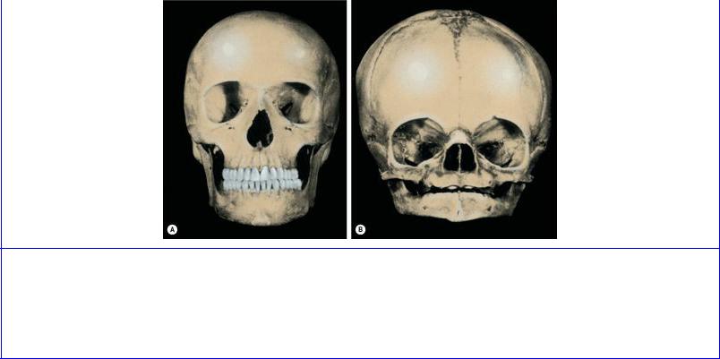
Figure 1.28 Normal adult and fetal skulls. The fetal skull (B) is projected to the same vertical height as that of the adult. Note the disproportion of the vertical extent of the face. The distance from the lower margin of the orbit to the lower border of the mandible in the adult is three times the diameter of the orbit; in the fetal skull it is equal to the diameter of the orbit.
Most of the separate skull and face bones are ossified by the time of birth but they are mobile on each other and are fairly readily disarticulated in the macerated skull. The bones of the vault do not interdigitate in sutures, as in the adult, but are separated by linear attachments of fibrous tissue and, at their corners, by larger areas, the fontanelles.
The anterior fontanelle lies between four bones. The two parietal bones bound it behind, the two halves of the frontal bone lie in front. It overlies the superior sagittal dural venous sinus. The anterior fontanelle is usually not palpable after the age of 18 months.
The posterior fontanelle lies between the apex of the squamous part of the occipital bone and the posterior edges of the two parietal bones. It is closed by the age of 3 months.
At birth the frontal bone consists of two halves separated by a median metopic suture; this is obliterated by about 8 years. The metopic suture may persist in up to about 8% of individuals, depending on ethnic origin, and must not be mistaken for a fracture line in a radiograph of the skull.
The petromastoid part of the neonatal temporal bone encloses the internal ear, middle ear and mastoid antrum, all parts of which are full adult size at birth. But the mastoid process is absent and the stylomastoid foramen is near the lateral surface of the skull, covered by the thin fibres of sternocleidomastoid—the issuing facial nerve is thus unprotected and vulnerable at birth. The mastoid process develops with the growth of the sternocleidomastoid muscle and the entry of air cells into it from the mastoid antrum. The process becomes palpable in the second year.
The tympanic part is present at birth as the C-shaped tympanic ring, applied to the undersurface of the petrous and squamous parts and enclosing the tympanic membrane, which is slotted into it. The external acoustic meatus of the newborn is wholly cartilaginous. The tympanic membrane is almost as big as in the adult, but faces more downwards and less outwards than the adult ear drum; lying more obliquely it seems somewhat smaller when viewed through the otoscope. The tympanic ring elongates

by growth from the lateral rim of its whole circumference, the tympanic plate so produced forming the bony part of the external acoustic meatus and pushing the cartilaginous part of the meatus laterally, further from the ear-drum. As the tympanic plate grows laterally from the tympanic ring the tympanic membrane tilts and comes to face rather more laterally and less downwards than in the neonate.
The mandibular fossa (which forms part of the temporomandibular joint) is shallow at birth and facing slightly laterally; with development the fossa deepens and faces directly downwards.
The maxilla, between the floor of the orbit and the gum margin, is very limited in height and is full of developing teeth. The maxillary sinus is a narrow slit excavated into its medial wall. Eruption of the deciduous teeth allows room for excavation of the sinus beneath the orbital surface, but the maxilla grows slowly until the permanent teeth begin to erupt at 6 years. At this time it ‘puts on a spurt’ of growth. The rapid increase in size of the sinus and the growth of the alveolar bone occur simultaneously with increased depth of the mandible. These factors combine to produce a rapid elongation of the face.
The hard palate grows backwards to accommodate the extra teeth; and forward growth of the base of the skull continues at the spheno-occipital synchondrosis (see p. 512) until 18 to 25 years of age.
The mandible is in two halves at birth and their cartilaginous anterior ends are separated by fibrous tissue at the symphysis menti. Ossification unites the two halves in the first year. At first the mental foramen lies near its lower border. After eruption of the permanent teeth the foramen lies higher, and is halfway between the upper and lower borders of the bone in adults. In the edentulous jaw of the elderly, absorption of the alveolar margin leaves the mental foramen nearer the upper border of the mandible (Fig. 1.29). Forward growth of the mandible changes the direction of the mental foramen. At birth the mental neurovascular bundle emerges through the foramen in a forward direction. In the adult the mental foramen is directed backwards. At birth the angle is obtuse and the coronoid process lies at a higher level than the condyle. With increase in the length and height of the mandible, to accommodate the erupting teeth, the angle diminishes. In the adult the angle approaches a right angle, and the condyle is at the same level or higher than the coronoid process. In the edentulous mouth of the elderly the angle of the mandible increases again and the neck inclines backwards, lowering the level of the condyle.

Figure 1.29 Age changes in the mandible: A birth; B adult; C old age.
Neck
The newborn baby has a very short neck. The subsequent elongation of the neck is accompanied by positional changes in the covering skin; an incision over the lower neck in an infant usually results in the scar lying over the upper sternum by later childhood.
The left brachiocephalic vein crosses the trachea so high in the superior mediastinum that it encroaches above the jugular notch into the neck, especially if it is engorged and the head extended; this should be remembered by the surgeon performing tracheotomy on the young child.
The shortness of the neck of the newborn involves a higher position of its viscera. The larynx is nearer the base of the tongue and the upper border of the epiglottis is at the level of the second cervical vertebra. From these elevated positions their descent is slow and they reach their adult levels only after the seventh year. The larynx and trachea are of small bore at birth. The vocal cords are about 5 mm long by the end of the first year. Laryngitis and tracheitis in infancy thus carry far more risk of respiratory obstruction than they do in later years. Up to the age of puberty there is no difference between the male and female larynx. At puberty the male larynx increases rapidly in size and the median angle of the thyroid cartilage moves forwards (laryngeal prominence). Consequently the vocal cords elongate from 8 to 16 mm within a year, resulting in the characteristic ‘breaking’ of the voice. Castration or failure of testicular hormone prevents this change taking place.
Thorax
The thoracic cage of the child differs from that of the adult in being more barrel-shaped. A crosssection of the infant thorax is nearly circular; that of the adult is oval, the transverse being thrice the length of the anteroposterior diameter. The large thymus extends from the lower part of the neck through the superior into the anterior mediastinum; it regresses at puberty. The ribs lie more nearly
horizontal, so the cage is set at a higher level than in the adult. The high thorax involves a higher level of the diaphragm, with consequent increase of abdominal volume.
Abdomen
At birth the liver is relatively twice as big as in the adult and its inferior border is palpable below the coastal margin. The kidneys are always highly lobulated at birth with very little perinephric fat; grooves on the surface of the adult organ frequently persist as visible signs of the original fetal lobulation. The suprarenal is enormous at birth, nearly as large as the kidney itself. The caecum is conical and the appendix arises from its apex in the fetus; this arrangement is usually still present at birth. During infancy and early childhood the lateral wall of the caecum balloons out and the base of the appendix comes to lie posteriorly on the medial wall. The appendiceal mucous membrane is packed with massed lymphoid follicles in the child. These become much more sparse in later life. The pelvic cavity is very small at birth and the fundus of the bladder lies above the pubic symphysis even when empty.
Upper limb
The upper limb is more fully developed than the lower limb at birth. The grasping reflex of the hand is very pronounced. Growth in length occurs more at the shoulder and wrist than at the elbow. Amputation through the humerus in a young child requires a very generous flap of soft tissue lest the growing bone should later protrude through the stump.
Lower limb
At birth the lower limb is not only poorly developed, but occupies the fetal position of flexion, a position which is maintained for 6 months or more. In preparation for standing and walking the limb not only becomes more robust, but undergoes extension and medial rotation that carry the flexor compartment around to the posterior aspect of the limb. The inverted foot of the newborn gradually becomes everted harmoniously with the changes in position of the knee and hip joints. Growth of the limb proceeds more rapidly at the knee than at the hip or ankle. It is not symmetrical across the lower epiphysis of the femur, and ‘knock knee’ (genu valgum) is normal in the child.
Vertebral column
Until birth the column is C-shaped, concave ventrally. This is imposed by constriction in utero. After birth the column is so flexible that it readily takes on any curvature imposed by gravity. The cervical curve opens up into a ventral convexity when the infant holds up its head, and the lumbar curve opens up into a ventral convexity when the infant walks. The extension of the hip that accompanies walking tilts the inlet of the pelvis forwards, so that the axis of the pelvic cavity is no longer in line with that of the abdominal cavity. This forward tilt of the pelvis necessitates forward curvature (lordosis) of the lumbar spine in order to keep the body vertical in the standing position.
The spinal cord extends to the third lumbar vertebra at birth and does not ‘rise’ to the L1/L2 junction until adult years.