


Part eleven. Mouth and hard palate
The mouth is for eating and talking through, and its structure is adapted accordingly. It also serves as an emergency airway in dyspnoea but its structure has nothing to do with this function; it merely provides a bigger airhole than the narrow nostrils. The tongue is for grasping food, for moving it during mastication, and for helping to swallow it. The delicate movements of the tongue turn laryngeal noise into articulate speech. In addition its mucous membrane is highly sensitive, even more than fingertips, and it also possesses the sense of taste.
The mouth extends from the lips to the palatoglossal arches (anterior pillars of the fauces). It is enclosed by the lips and cheeks; the slit-like space between lips/cheeks and teeth/gingivae (gums) is the vestibule of the mouth. The space inside the teeth and gums is the mouth (oral) cavity proper. The floor is the mylohyoid muscle, and the roof is the hard palate. Rising from the floor of the mouth, the tongue occupies much of the oral cavity.
The lips and cheeks are covered with hairy skin, except for the red margin of the lips, which is devoid of hair, and has a rich capillary blood supply, hence the colour. The red margin is highly sensitive and is represented by a large area in the sensory cortex. It is the main exploratory sensory area in babies, before they learn to use their hands for stereognosis.
The oral cavity is lined with stratified squamous epithelium, which is keratinized on the gums, hard palate and much of the dorsum of the tongue, but not elsewhere. The mucous membrane is adherent on lips and cheek to the face muscles, on tongue to the muscles thereof, and on the hard palate to the periosteum of the bone. It is therefore seldom caught between the teeth when chewing.
On the mucous membrane of the cheek the parotid duct opens opposite the second upper molar tooth (Fig. 6.17). Nearby are the tiny openings of the ducts of the molar glands which lie on the outer surface of the buccinator. There are many other mucous glands (buccal and labial) scattered in the mucous membrane of the vestibule.
Nerve supply. Much of the mucous membrane of the inside of the cheeks and lips is supplied by the buccal branch of the mandibular nerve, with contributions from the mental branch of the inferior alveolar (also mandibular) and the infraorbital branch of the maxillary nerve; the last two also supply the red margin of the lower and upper lips respectively.
The gingivae (gums) are firmly attached to the periosteum of the alveolar bone and extend to surround the necks of the teeth. They consist of dense vascular fibrous tissue covered by epithelium. At the gingival crest, the epithelium dips down to line a sulcus, at the floor of which the epithelium is attached to the surface of the tooth. The change from alveolar mucosa (continuous with that of the cheek) to gingival mucosa is marked by an abrupt change of colour, from red shiny alveolar to pink opaque gingival.
The upper gums are supplied by the superior alveolar, greater palatine and nasopalatine nerves
(maxillary), while the lower receive their innervation from the inferior alveolar, buccal and lingual nerves (mandibular). The buccal nerve does not usually innervate the upper gums.
Teeth
The bulk of a tooth consists of dentine, a hard avascular calcified tissue penetrated by minute canals, the dentinal tubules. The part of the tooth that projects into the mouth is the crown which is covered by enamel, the hardest tissue in the body, and the part held in the jaw is the root which is covered by cementum, a calcified tissue rather like bone. The junction between enamel and cementum is the cervical margin or neck. Because enamel and cementum meet, dentine is not normally exposed on the surface. Inside the dentine is the pulp cavity. The cavity is filled by dental pulp, loose connective tissue, with nerves (below), blood vessels (see p. 363) and lymphatics (see p. 331), all of which gain access to the pulp through the apical foramen. The pulp is covered with a single layer of tall columnar cells, the odontoblasts, lying in contact with the inner surface of the dentine. Throughout life they retain the power to produce dentine within the pulp cavity if the surface of the dentine is breached. The odontoblasts give off fine cytoplasmic processes that occupy the dentinal tubules.
The tooth is slung in its bony socket by the periodontal ligament, consisting of collagen fibres passing obliquely from the alveolar bone towards the apex of the tooth. It is really the modified periosteum of the alveolar bone and is radiolucent; it shows as a clear interval between tooth and bone shadows in a radiograph.
Permanent dentition
The human adult has from the midline 2 incisors, 1 canine, 2 premolars and 3 molars; that is, 8 teeth in each half-jaw, or 32 teeth in all. The shape of a tooth is adapted to its function. The incisors are for biting and cutting, the canines for holding and tearing, the premolars and molars for chewing and grinding. In clinical dentistry it is common to refer to teeth by number (1 to 8 starting from the midline) rather than by name.
The teeth can be distinguished from one another by the characteristics of their roots and crowns. The upper molars have three roots each; two are lateral and one is medial. The lower molars have two roots each, one anterior and one posterior. All the other teeth have a single root, except for the first upper premolar which usually has a bifid root.
The incisor crowns are chisel-shaped. Upper and lower incisors do not meet edge to edge, but by a sliding overlap, like the blades of a pair of scissors. The canine crowns are pyramidal or conical, The premolar (bicuspid teeth) crowns have two cusps (lingual and buccal). Upper molars have four, lower molars five, cusps on their crowns.
Nerve supply
The term nerve supply of a tooth really means the nerve supply of the pulp; some fine nerve filaments may enter some dentinal tubules, but most of the dentine and all the enamel and cementum have no innervation. The pulp and periodontal ligament share the same nerve.
The upper teeth are supplied by the superior alveolar nerves, anterior, middle and posterior, which form a plexus above the apices of the teeth. The middle nerve may be absent.
In the lower jaw the molars and premolars are supplied by the main trunk of the inferior alveolar nerve, whose terminal incisor branch supplies the canine and incisors, overlapping to the opposite central incisor.
Dental anaesthesia
The alveolar bone of the maxilla is relatively porous, so anaesthetic solution deposited in the gingivae opposite the apex of a tooth root will readily penetrate the bone to anaesthetize the tooth for dental procedures. Infiltration of the buccal aspect of the jaw will allow painless drilling of the tooth, but for extraction the palatal aspect must be infiltrated as well.
For the teeth of the lower jaw infiltration anaesthesia is usually effective only for the incisors. The other mandibular teeth are embedded in bone that is denser and does not allow sufficient penetration of the anaesthetic agent. For these teeth, inferior alveolar nerve block is required; for extraction it is necessary to include block of the nearby lingual and buccal nerves as well in order to anaesthetize the adjacent soft tissues.
For infiltration anaesthesia on the buccal (outer) aspect of the jaw, the needle is inserted opposite the appropriate tooth just below or into the buccal fold (where the mucosa is reflected between jaw and cheek), with the tip of the needle directed to the level of the apex of the tooth. On the palatal side, the point of insertion of the needle is midway between the gingival margin and the midline of the palate.
For inferior alveolar and lingual nerve block, the needle is inserted orally through the buccinator above the level of the occlusal surface of the molar teeth and in front of the pterygomandibular raphe, which raises a visible and palpable ridge in the opened mouth; the needle passes behind the (palpable) deep tendinous band of the temporalis muscle (see p. 357). The line of approach is from the premolar teeth of the opposite side, and a small injection is made 0.5 cm from the mucosal surface, when the needle is above the lingual nerve; the main injection is made another 1 cm deeper above the lingula, where the inferior alveolar nerve enters the mandibular foramen, which is situated midway between the anterior and posterior borders of the mandibular ramus. Entry of the anaesthetic agent within the parotid fascia around the deep part of the parotid, and its diffusion through the gland substance, may cause a transient facial paralysis.
Tooth position
The teeth of the upper jaw lie in a continuous curve, like a horseshoe. In the alveolar bone the outer (buccal) plate is thinner than the inner (palatal) plate. In the lower jaw the curve of the anterior teeth straightens out in the molar region. In the alveolar bone of the mandible the labial (outer) plate is thinner than the lingual (inner) plate over incisors, canines and premolars, but in the posterior molar region the lingual plate is thinner than the buccal; the lingual nerve lies here beside the third molar tooth and is at risk when the tooth is extracted.
The attachment of mylohyoid is below the apices of most of the mandibular teeth—an apical abscess thus points in the mouth. The apices of the second and third molars lie below the mylohyoid line and an apical abscess bursting through the inner plate points in the neck.
Deciduous dentition
The deciduous, or milk, teeth begin to erupt at about the sixth month and are completely erupted at the end of the second year. They consist of 5 teeth in each half-jaw, 20 in all. There are 2 incisors, 1 canine and 2 molars. They are shed as the permanent teeth erupt. The deciduous molars are replaced by the permanent premolars, not by permanent molars which have no counterpart in the deciduous
dentition.
Development and eruption of teeth
Teeth are derived by budding of the epithelium (ectoderm) lining the mouth. The buds of ectoderm produce only the enamel; they evoke a reaction in the surrounding mesoderm, which differentiates to produce the dentine and cementum under the influence of neural crest cells.
In the mouth cavity (stomodeum) of the 5-week embryo (12 mm long) an ingrowth of ectoderm occurs over the site of the future gums. A curved sheet of ectoderm grows into the adjacent mesoderm, tilting medially. This is the primary dental lamina. From its outer surface a series of buds grow into the mesoderm, one for each deciduous tooth. At a later stage a similar series of buds grow (more medially) from the depths of the primary dental lamina, one bud for each permanent tooth. When these epithelial buds are well formed the primary dental lamina becomes absorbed. Remnants of this epithelium may later grow into cysts or tumours.
The developed tooth erupts by a combination of elongation of the root and absorption of the overlying bone. The elongating root remains ensheathed in an upgrowth of alveolar bone.
The approximate normal times of eruption are:
Deciduous teeth
6 months |
|
Lower central incisors |
7 months |
|
Upper central incisors |
8–9 months |
|
Lateral incisors |
1 year |
|
First molars |
18 months |
|
Canines |
2 years |
|
Second molars. |
Permanent teeth |
|
|
|
|
|
6 years |
First permanent molars |
|
7 years |
Central incisors |
|
8 years |
Lateral incisors |
|
9 years |
First premolars |
|
10 years |
Second premolars |
|
11 years |
Canines |
|
12 years |
Second permanent molars |
|
17–21 years |
Third permanent molars (wisdom teeth). |
|
A lower tooth usually precedes its opposite number in the upper jaw. The first permanent molar (the 6-year molar) erupts before any deciduous teeth have been shed. The second permanent molar does not erupt until 12 years of age. In the intervening period the five deciduous teeth in each half-jaw are replaced. The order of replacement is first the incisors, central and lateral, then the milk molars, first and second and, last of all, the long-rooted canine.

Hard palate
The palate is the roof of the mouth. Between the teeth it lies on a basis of bone, the hard palate. Behind the teeth and hard palate the soft palate projects down.
The hard palate is made up of the palatal process of the maxilla and the horizontal plate of the palatine bone, meeting at a cruciform suture formed of intermaxillary, interpalatine and palatomaxillary sutures. In the midline at the front of the hard palate lies the incisive fossa, into which open the incisive canals, each ascending into its half of the nasal cavity. The greater palatine foramen lies medial to the last molar tooth; just behind it the lesser palatine foramina perforate the palatine bone.
The mucous membrane of the front of the hard palate is strongly united with the periosteum and the attachment of the periosteum to the bone is secured by multiple fibrous tissue pegs (Sharpey's fibres) that leave a finely pitted bone surface on the dried skull. This fixation is for mastication; the moving bolus does not displace the mucous membrane. There are transverse masticatory ridges in this part of the mucoperiosteum. From a little papilla overlying the incisive fossa a narrow low ridge, the palatine raphe, runs anteroposteriorly; the submucosa is absent here. Over the horizontal plate of the palatine bone mucous membrane and periosteum are separated by a mass of mucous gland tissue; Sharpey's fibres are few here, and the bone surface is smooth. From the hard palate the mucous membrane curves down to the undersurface of the soft palate. The stratified squamous epithelium is keratinized on the hard palate, and non-keratinized on the soft palate.
Blood supply is by the greater palatine artery (from the third part of the maxillary artery), which emerges from the greater palatine foramen and passes forwards around the palate (lateral to the nerve) to enter the incisive canal and pass up into the nose. Veins accompany the artery back to the pterygoid plexus. Other veins pass back to the supratonsillar region and join the pharyngeal plexus.
Lymph drainage is to retropharyngeal and deep cervical lymph nodes.
Nerve supply is by the greater palatine nerve (from the maxillary via the pterygopalatine ganglion) as far forward as the incisive fossa. The anterior part of the palate, behind the incisor teeth (the area of the premaxilla) is supplied by the two nasopalatine nerves, from the same source.
Tongue
The tongue is essentially a mass of skeletal muscle covered by mucous membrane, and with a midline fibrous septum separating the two muscular halves. It has a dorsum, tip, inferior surface and root. The anterior two-thirds, or oral part, of the dorsum faces upwards towards the hard palate, and the posterior one-third, or pharyngeal part, faces backwards towards the oropharynx. The stratified squamous epithelium is keratinized on the oral part and non-keratinized on the pharyngeal part. The tip is the most anterior and mobile part and merges into the inferior surface. The mucous membrane of the inferior surface is thin and smooth, similar to that of the floor of the mouth and cheek.
The oral anterior two-thirds of the dorsum of the tongue is covered by mucous membrane into which the underlying muscles are inserted. The surface is roughened by the presence of three types of papillae: filiform, fungiform and vallate. The filiform papillae are minute conical projections that give rise to the velvety appearance of the tongue. Fungiform papillae are visible as discrete pink

pinheads, more numerous towards the edges of the tongue; each bears a few taste buds (there are none on filiform papillae). The vallate papillae are about a dozen in number and are arranged in the form of a V with the apex pointing backwards, just in front of an ill-defined shallow groove, the sulcus terminalis, which marks the junction of the oral and pharyngeal parts of the tongue. Each is a cylindrical projection surrounded by a circular sulcus and a raised outer wall (Fig. 6.33). There are many taste buds and serous glands in the sulcus that surrounds each vallate papilla. There are no other glands on the dorsum of the anterior two-thirds of the tongue. The vallate papillae are far back on the oral surface and so not in contact with the food being chewed, but food juices and saliva reach them and so flavours are transmitted to them.
Figure 6.33 Dorsum of the tongue.
There are scattered mucous and serous glands under the tip and sides. On the undersurface behind the tip there is a rather large mixed gland, the anterior lingual gland, on each side of the midline. From each gland small ducts open on the undersurface of the tongue. A retention cyst of this gland is the probable cause of the clinical condition known as a ranula.
The posterior third of the dorsum of the tongue slopes downwards from the sulcus terminalis as the anterior wall of the oropharynx. At the apex of the sulcus is a small depression, the foramen caecum, the remains of the upper end of the thyroglossal duct (see p. 26). There are no papillae behind the sulcus. The smooth mucous membrane has a nodular appearance from the presence of underlying masses of mucous and serous glands and aggregations of lymphoid follicles. The latter constitute the ‘lingual tonsil’, part of Waldeyer's ring (see p. 387). Between the tongue and epiglottis there is a midline flange of mucous membrane, the median glossoepiglottic fold, on each side of which is a depression, the vallecula (see p. 387), bounded laterally by a similar mucosal fold, the lateral glossoepiglottic fold, extending from the side of the epiglottis to the wall of the pharynx; as the latter fold is not attached to the tongue it would be better named pharyngoepiglottic.
When the tip of the tongue is raised to the roof of the mouth, the inferior surface of the tongue can be inspected. A small midline septum of mucous membrane (lingual frenulum) unites it to the floor of the mouth. Lateral to this the deep lingual vein can usually be seen through the mucosa (the lingual artery and nerve that are near it are not visible), and farther laterally still is another fold of mucosa, the fimbriated fold. Foliate papillae are a series of vertical parallel folds of mucous membrane on the
sides of the posterior part of the tongue (Fig. 6.33). They are the site of numerous taste buds.
The palatoglossal arches (anterior pillars of the fauces) are ridges of mucous membrane raised up by the palatoglossus muscles. They extend from the undersurface of the front of the soft palate to the sides of the tongue in line with the vallate papillae. The whole constitutes the oropharyngeal isthmus. In front of it is the mouth, behind it is the pharynx; and it is narrower than either. It is closed by depression of the palate and elevation of the dorsum of the tongue, and narrowed by contraction of the palatoglossus muscles.
Muscles
The muscles of the tongue are divided into intrinsic and extrinsic groups; the intrinsic muscles are wholly within the tongue and not attached to bone, while the extrinsic muscles have a bony attachment. There are four muscles in each group in each half of the tongue, with a midline fibrous septum dividing the organ into two symmetrical halves. The muscles of the intrinsic group are the superior and inferior longitudinal, transverse and vertical, and the extrinsic group comprises genioglossus (which is the largest of all the muscles and makes up the bulk of the tongue), hyoglossus, styloglossus and palatoglossus.
Genioglossus arises from the superior mental spine (genial tubercle) of the mandible (see Fig. 8.5B, p. 510), whence the fibres radiate backwards in a fan-shaped manner to be inserted into the mucous membrane of the tongue, with the lowest fibres passing down to the hyoid body.
Hyoglossus arises from the length of the greater horn of the hyoid bone and from the lateral part of its body. It extends upwards as a quadrilateral sheet, its upper border interdigitating at right angles with the fibres of styloglossus, and is attached to the side of the tongue. Superficial (lateral) to the muscle from above downwards lie the lingual nerve, submandibular duct, and the hypoglossal nerve with its accompanying veins, while passing deep to its posterior border from above downwards are the glossopharyngeal nerve, stylohyoid ligament and the lingual artery with its accompanying veins lying on the anterior fibres of the middle constrictor (Fig. 6.24).
Styloglossus arises from the front of the lower part of the styloid process and the upper part of the stylohyoid ligament. It passes forwards below the superior constrictor to be inserted into the side of the tongue, interdigitating with the upper fibres of hyoglossus.
Palatoglossus descends from the undersurface of the palatine aponeurosis to the side of the tongue, forming with its fellow of the opposite side the palatoglossal arch. It is described further with the soft palate (see p. 389).
Blood supply
The tongue is supplied by the lingual artery (see p. 342), which runs above the greater horn of the hyoid bone deep to hyoglossus and passes forwards to the tip. Beneath hyoglossus it gives off dorsal lingual branches into the posterior part. At the anterior border of hyoglossus it gives a branch to the sublingual gland and the floor of the mouth. There are small contributions from the tonsillar branch of the facial artery and from the ascending pharyngeal artery. The fibrous septum dividing the two halves of the tongue prevents any significant anastomosis of blood vessels across the midline. Accordingly the septum is a less vascular site for surgical division of the tongue for hemiglossectomy or
enhancing access to the oropharynx.
Venous tributaries accompanying the lingual artery and its dorsal branches form the lingual vein. The venous return from the tip is by the deep lingual vein, visible on each side of the midline on the undersurface. It runs back superficial to hyoglossus and is joined at the anterior border of that muscle by the sublingual vein (from the sublingual gland) to form the vena comitans of the hypoglossal nerve. It continues backwards close to the nerve and has a variable ending, joining either the lingual, facial or internal jugular veins. The lingual vein usually joins the internal jugular near the greater horn of the hyoid bone.
Lymph drainage
A significant feature of the tongue's lymph drainage (Fig. 6.34), which is through the floor of the mouth or pharyngeal wall, is that lymph from one side, especially of the posterior part, may reach nodes of both sides of the neck (in contrast to the blood supply which remains unilateral). The tip may drain to submental nodes or directly to deep cervical nodes. Marginal lymphatics from the rest of the anterior part tend to drain to ipsilateral submandibular nodes and then, or sometimes directly, to deep cervical nodes. Central lymphatics from the anterior part descend between the genioglossi and drain to deep cervical nodes of either side. The posterior part drains directly and frequently bilaterally to deep cervical nodes. The deep cervical nodes usually involved are the jugulodigastric and juguloomohyoid nodes. All lymph from the tongue is believed to eventually drain through the juguloomohyoid node before reaching the thoracic duct or right lymphatic duct.
Nerve supply
All the muscles of the tongue, intrinsic and extrinsic, are supplied by the hypoglossal nerve (except palatoglossus, which, being essentially a palate muscle, is supplied by the pharyngeal plexus, see p. 389). The pathway of proprioceptive impulses from the tongue is probably via the lingual nerve.
The sensory supply of the mucous membrane of the oral part (anterior two-thirds), but not the region of the vallate papillae, is by the lingual nerve, whose trigeminal component mediates common sensibility (with cell bodies in the trigeminal ganglion) and whose chorda tympani component mediates taste (with cell bodies in the geniculate ganglion of the facial nerve). The parasympathetic secretomotor fibres to the anterior lingual gland run in the chorda tympani from the superior salivary nucleus, and relay in the submandibular ganglion.
The posterior one-third of the mucosa, together with the presulcal area that includes the vallate papillae, is mainly supplied by the glossopharyngeal nerve. This has fibres of both common sensibility and taste (with cell bodies in the glossopharyngeal ganglia in the jugular foramen). A small area of lingual mucosa forming the anterior wall of the vallecula is supplied by the nerve of the vallecular mucosa, the internal laryngeal (with cell bodies in the inferior vagal ganglion).
Movements
The intrinsic muscles alter the shape of the tongue, the extrinsic muscles stabilize the organ and by their contraction alter its position, as well as its shape. The tongue rests on the floor of the mouth (see p. 335) and this highly mobile shelf enhances the mobility of the tongue.
The position of the tongue is altered by the mylohyoid muscles, on which the tongue rests. The mobile
floor of the mouth can be elongated or shortened, raised or lowered, thus still further altering the position of an already mobile organ.
In the first (voluntary) stage of swallowing contraction of the vertical intrinsic muscle makes a longitudinal groove on the dorsum; the heaped-up tip and edges are in contact with the hard palate and teeth. The liquid or moist bolus is thus imprisoned in the groove. Contraction of mylohyoid now raises the floor of the mouth, compressing the tongue against the hard palate. The vertical intrinsic fibres relax from before backwards obliterating the groove in the same sequence, forcing the bolus backwards.
In the unconscious, the tongue muscles relax and the organ may fall backwards to obstruct the pharyngeal part of the airway. Pulling the tongue forwards is an important element in restoring a patent airway in cardiopulmonary resuscitation. This can be achieved by pulling forwards the mandible to which genioglossus is attached.
Development
Tongue muscles are derived from occipital myotomes which migrate forwards carrying their nerve supply with them (hypoglossal nerve). The migration passes ventrally medial to the internal jugular vein and around the internal and external carotid arteries. The epithelium is derived from the lining of the floor of the pharynx, and comes from parts of the first, third and fourth arches. The mucosa of the anterior two-thirds is from the ectodermal lining of the midline tuberculum impar and the pair of lateral lingual swellings of the first arch (lingual and chorda tympani nerves, see Fig. 1.19, p. 24). The mucosa of the posterior third is from the endodermal lining of the midline hypobranchial eminence of the third arch (glossopharyngeal nerve), with a small contribution from the fourth arch (internal laryngeal nerve). Tissue of the second arch is not represented because third arch tissue overgrows it in a forward direction to meet that from the first arch. The thyroglossal duct grows downwards from the junction between the tuberculum impar and the hypobranchial eminence, the site being indicated by the foramen caecum.
The sublingual gland is almond-shaped and lies in between mylohyoid and the side of the tongue (genioglossus), under the mucous membrane of the floor of the mouth. Laterally it lies against the sublingual fossa of the mandible (see Fig. 8.5B, p. 510). Its upper surface raises the sublingual fold in the floor of the mouth. At the front the two glands almost meet each other. The gland is mucussecreting and of its 15 or so ducts half open into the submandibular duct, the remainder separately on the sublingual fold.
It is supplied by the lingual artery and by branches of the submental artery which pierce mylohyoid muscle to reach it. The venous return is by corresponding veins. Postganglionic parasympathetic secretomotor fibres are supplied to the gland by the lingual nerve; they originate from the submandibular ganglion where preganglionic chorda tympani fibres synapse (see p. 338).

Part twelve. Pharynx and soft palate
Pharynx
The pharynx is a fibromuscular tube, attached above to the base of the skull and continuous below with the oesophagus. It is about 12 cm in length. Its anterior wall is largely deficient and it thereby communicates with the nose, mouth and larynx. On account of these communications it is descriptively divided into three parts: nasal, oral and laryngeal, i.e. nasopharynx, oropharynx and laryngopharynx.
Muscles and fascia
The muscular wall is surprisingly thin. It consists of three curved sheets of muscle, the superior, middle and inferior constrictors (supplemented by three smaller muscles: stylopharyngeus, palatopharyngeus and salpingopharyn-geus). They overlap posteriorly, being telescoped into each other like three stacked cups. But the muscle does not extend up to the base of the skull; here the immobile wall of the nasopharynx consists of a rigid membrane, the pharyngobasilar fascia. This is a fibrous thickening of the submucosa that fills in the gap between the skull and the upper border of the superior constrictor, making a fourth but fibrous cup stacked inside the other three. The attachment of this fascia to the base of the skull (Fig. 6.35) can be traced from the pharyngeal tubercle, a midline thickening, in front of the foramen magnum, to which the pharyngeal raphe is attached (see below). The attachment then passes laterally, convex forwards over longus capitis to the petrous part of the temporal bone just in front of the carotid canal. From here it passes forwards and medially below the cartilaginous part of the auditory tube, to the sharp posterior border of the medial pterygoid plate, along which it continues down to the hamulus. Suspended from the base of the skull, and sweeping around from one medial pterygoid plate to the other, the pharyngobasilar fascia holds the nasopharynx permanently open for breathing. As it descends inside the superior constrictor it diminishes in thickness and peters out below the level of the hard palate.
The quadrangular area at the apex of the petrous bone in front of the carotid canal lies within a lateral recess of the pharynx. The levator palati muscle arises here and is intrapharyngeal, covered medially by mucous membrane. The cartilaginous part of the auditory tube enters the nasopharynx above the pharyngobasilar fascia, which is firmly attached to its lower surface.
Superior constrictor
The superior constrictor fibres arise from the lower part of the posterior border of the medial pterygoid plate down to the tip of the hamulus, outside the pharyngobasilar fascia, and from the pterygomandibular raphe, which runs from the hamulus to the mandible just above the posterior end of the mylohyoid line. The superior constrictor passes backwards from the pterygomandibular raphe; buccinator passes forwards from it (Fig. 6.13).
From its origins the muscle sweeps around the pharynx, its fibres diverging mostly upwards to meet their opposite fellows in the midline pharyngeal raphe (Fig. 6.36) at the back. The upper end of this raphe forms a fibrous band which receives the uppermost constrictor fibres and is attached to the pharyngeal tubercle. The lowest fibres extend at the back as far down as the level of the vocal folds, lying within the middle constrictor.

Figure 6.36 Pharynx, from behind. On the right the inferior constrictor has been removed to show the extent of the middle constrictor and the attachment of stylopharyngeus to the posterior border of the thyroid lamina.
There is a gap laterally between the superior and middle constrictors, through which stylopharyngeus passes down into the pharynx, and styloglossus and the glossopharyngeal and lingual nerves pass forwards to the tongue (Fig. 6.24).
Middle constrictor
The middle constrictor arises from the lower part of the stylohyoid ligament, the lesser horn of the hyoid bone and the greater horn, deep to hyoglossus. Its fibres diverge upwards and downwards as they sweep backwards around the pharynx to enclose the superior constrictor and end in the median raphe and the lowest fibres arch down as far as the vocal folds, lying within the inferior constrictor.
The anterior gap between the middle and inferior constrictors is closed by the thyrohyoid membrane (see p. 392), which thereby contributes to the wall of the laryngeal part of the pharynx (Fig. 6.37). Passing through this gap by piercing the membrane are the internal laryngeal nerve and superior laryngeal vessels.

Figure 6.37 Pharyngeal constrictors, from the right.
Inferior constrictor
This has two parts, named from their origins. The thyropharyngeus part arises from the oblique line of the thyroid cartilage and in continuity below this from a fibrous arch that spans the cricothyroid muscle (Fig. 6.37). It encloses the middle and superior constrictors as its fibres curve backwards and upwards around them to the midline raphe. The cricopharyngeus, rounded and thicker than the flat sheets of the other constrictors, extends uninterruptedly from one side of the cricoid arch to the other around the pharynx. There is no raphe here. The muscle acts as a sphincter at the lower extent of the pharynx, and is continuous with the circular muscular coat of the oesophagus (Fig. 6.41). It is composed largely of fibres of the ‘slow twitch’ variety and is always closed, except for momentary relaxation during deglutition. It imparts some resistance to the passage of an endoscope (overcome by swallowing). The closure of the cricopharyngeus prevents air from being sucked into the upper oesophagus when intrathoracic pressure falls; air is sucked only into the permanently open trachea. Passing upwards deep to the lower border of the inferior constrictor are the recurrent laryngeal nerve and inferior laryngeal vessels.
The junction between the oblique fibres of thyropharyngeus and the horizontal fibres of cricopharyngeus near the midline is a potentially weak area at the back of the pharyngeal wall (Fig. 6.36), and through this area (Killian's dehiscence) a pouch of mucosa may become protruded (pharyngeal diverticulum) (Fig. 6.38). Inappropriate contraction of cricopharyngeus during swallowing is an aetiological factor and surgical management includes division of its horizontal fibres.
Figure 6.38 Barium meal radiograph of a subject with a pharyngeal pouch. A residual film of barium sulphate has outlined the posterior surface of the tongue, the median and lateral glossoepiglottic folds and the piriform fossae.
Palatopharyngeus
Palatopharyngeus is described with the soft palate (see p. 389). As the muscle fibres pass down from the palate they lie internal to the superior constrictor (Fig. 6.41).
Salpingopharyngeus
Salpingopharyngeus is a very slender muscle that arises from the lower part of the cartilage of the auditory tube (see p. 416) and runs downwards (Fig. 6.41) to blend with palatopharyngeus.
Stylopharyngeus
Stylopharyngeus arises from the deep aspect of the styloid process high up. It slopes down across the internal carotid artery (Fig. 6.23), in front of which it crosses the lower border of the superior constrictor and passes down inside the middle constrictor, to be inserted with palatopharyngeus into the posterior border of the thyroid lamina (Fig. 6.36). The glossopharyngeal nerve curls round the posterior border of the muscle from medial to lateral, and supplies it.
Blood supply
Branches of many arteries supply the pharynx: ascending pharyngeal, ascending palatine and tonsillar (from facial), greater palatine and pharyngeal (from maxillary), lingual and the superior and inferior laryngeal arteries. Venous blood is largely collected into the pharyngeal venous plexus which like the nerve plexus (see below) is situated mainly at the back of the middle constrictor; it drains into the internal jugular vein and has connections with the pterygoid plexus.
Lymph drainage
Lymph passes to retropharyngeal lymph nodes and via these or directly to upper and lower deep cervical groups.
Nerve supply
The main motor nerve supply of the muscles of the pharynx is from the pharyngeal plexus. However, stylopharyngeus is supplied by the glossopharyngeal nerve and cricopharyngeus is also supplied by the recurrent and external laryngeal nerves. The cell bodies that supply all six muscles on each side are in the nucleus ambiguus.
The pharyngeal plexus lies on the posterolateral wall of the pharynx, mainly over the middle constrictor, and is formed by the union of pharyngeal branches from the vagus and glossopharyngeal nerves and the cervical sympathetic. The glossopharyngeal component is afferent; the pharyngeal fibres of the vagus carry motor fibres (derived from the cranial part of the accessory nerve). The sympathetic fibres are vasoconstrictor.
The mucosa of the nasopharynx is supplied by the pharyngeal branch of the maxillary nerve through the pterygopalatine ganglion. Most of the oropharynx receives its sensory supply from the glossopharyngeal nerve, but the internal laryngeal nerve supplies the valleculae and the lesser palatine nerves (maxillary) contribute to the supply of the tonsillar mucosa. The internal laryngeal nerve is sensory to the laryngopharynx.
Nasal part
The nasopharynx extends from the base of the skull to the upper surface of the soft palate, at the level of C1 vertebra. In front it communicates with the nose through the choanae. The space between the soft palate and the posterior pharyngeal wall through which the nasopharynx joins the oral part of the pharynx is the oropharyngeal isthmus. The soft palate becomes elevated during swallowing to meet
the posterior wall, so closing the isthmus. The main features within the nasopharynx are the openings of the auditory tubes, the pharyngeal recesses and the pharyngeal tonsil (Fig. 6.39)
Figure 6.39 Sagittal section of the head.
The opening of the auditory tube lies in the lateral wall and is triangular in appearance. The opening is guarded above, behind and in front by a prominent rounded ridge, the tubal elevation, formed by the trumpet-shaped medial end of the tubal cartilage as it underlies the mucous membrane, which here contains lymphatic tissue, the tubal tonsil. The tubal elevation is in the shape of an inverted J, the long limb lying posteriorly and being continued downwards as the salpingopharyngeal fold, produced by the underlying salpingopharyngeus muscle. The lower margin of the opening has a slight bulge due to the underlying levator palati muscle.
The pharyngeal recess (fossa of Rosenmüller) is a narrow vertical gutter behind the opening of the auditory tube, resulting from the angular attachment of the pharyngobasilar fascia to the base of the skull in front of the carotid canal (Fig. 6.35). A catheter missing the tubal orifice and introduced into the recess may perforate the fascia and enter the internal carotid artery, which here lies against the wall of the pharynx.
In the mucous membrane high on the posterior wall is a collection of lymphoid nodules, prominent only in chil-dren and forming the pharyngeal tonsil. When enlarged the nodules are commonly known as the adenoids.
Oral part
The oropharynx extends from the lower surface of the soft palate to the upper border of the epiglottis (halfway down C3 vertebral body). The wall of the oropharynx is formed posteriorly by all three constrictors. It closes completely behind a swallowed bolus, but is otherwise open for breathing. Anteriorly in front of the gap between the soft palate and epiglottis there is a mobile wall, the posterior part of the tongue, above which the oropharynx communicates with the mouth. At the sides there are projecting ridges, the palatopharyngeal and palatoglossal arches (pillars of the fauces), formed by the underlying corresponding muscles, with the palatine tonsils between them. The palatoglossal arches form the boundary between the pharynx and the mouth. The palatine and lingual tonsils and the valleculae are in the oropharynx.
The palatine tonsil (the pair commonly called simply ‘the tonsils’) is a large collection of lymphoid
tissue which projects into the oropharynx from the tonsillar fossa between the palatopharyngeal fold behind and the palatoglossal fold in front (Fig. 6.33). The floor of the fossa (lateral wall) is the lower part of the superior constrictor. On its lateral aspect the glossopharyngeal nerve crosses the lower part of the bed, running obliquely downwards and forwards to reach the tongue by passing under the lower border of the constrictor.
The lymphoid tissue of the tonsil extends up to the soft palate and down to the dorsum of the tongue. The medial surface is covered by pharyngeal mucosa on which are the openings of several epithelial downgrowths, the tonsillar crypts. One large downgrowth near the upper pole is the intratonsillar cleft, which is the remains of the fetal second pharyngeal pouch (see p. 26). The lateral surface is covered by fibrous tissue which forms the tonsillar hemicapsule. (A peritonsillar abscess occurs outside the capsule.) The superior constrictor separates this surface from the facial artery and two of its branches, the ascending palatine and tonsillar. The internal carotid artery is about 2.5 cm posterolateral to the tonsil.
The palatine, lingual, pharyngeal and tubal tonsils collectively form an interrupted circle of lymphoid tissue (Waldeyer's ring) at the upper end of the respiratory and alimentary tracts.
Blood supply. The tonsillar branch of the facial artery forms the main arterial supply; it enters the tonsil by piercing the superior constrictor. There are smaller contributions from the lingual, ascending pharyngeal and ascending and greater palatine vessels.
The veins form a plexus round the capsule and pierce the superior constrictor to drain into the pharyngeal plexus. One large vein descends from the soft palate between the tonsillar hemicapsule and the superior constrictor before piercing the pharyngeal wall; this is the external palatine, or paratonsillar vein and, is the usual cause of haemorrhage after tonsillectomy.
Lymph drainage. Lymphatic channels pierce the superior constrictor to reach the deep cervical nodes, especially the jugulodigastric (or tonsillar) node below the angle of the mandible.
Nerve supply. The mucous membrane overlying the tonsil is supplied mainly by the tonsillar branch of the glossopharyngeal nerve, and to a small extent by the lesser palatine nerves. The glossopharyngeal nerve also supplies the middle ear, through its tympanic branch, and tonsillitis may cause referred pain in the ear.
The valleculae lie between the epiglottis and the posterior surface of the tongue. They are shallow fossae separated by the median glossoepiglottic fold and limited inferolaterally by the lateral glossoepiglottic folds (Fig. 6.38). The nerve supply of the mucosa of the valleculae, including that of the part of the tongue that forms the anterior vallecular wall, is by the internal laryngeal nerve. A crumb that ‘goes down the wrong way’ is one that lodges in the vallecula and sets up a reflex bout of coughing (see p. 395) to dislodge it.
Laryngeal part
The laryngopharynx extends from the upper border of the epiglottis to the level of the lower border of the cricoid cartilage (C6 vertebra) where it becomes continuous with the oesophagus. In the upper part of the anterior aspect is the opening into the larynx (aditus or laryngeal inlet) (see p. 393). The piriform recesses, broad above and narrow below, lie beside the aperture of the larynx. Below the

inlet, the lower part of the pharynx (referred to clinically as the hypopharynx) possesses an anterior wall, comprising the arytenoids and the lamina of the cricoid cartilage (see p. 391) draped over with mucous membrane. The posterior wall of the laryngopharynx is formed by the three overlapping constrictors down to the level of the vocal folds (upper border of cricoid lamina). Below this (i.e. behind the cricoid lamina) there is only the thyropharyngeus, the site of the dehiscence of Killian, and finally the cricopharyngeal sphincter.
At each side of the epiglottis the lateral glossoepiglottic fold separates the oropharynx from the laryngeal part. Below the fold is the piriform recess (piriform fossa) (Fig. 6.38). This mucosa-lined space is bounded medially by the quadrangular membrane of the larynx, (see p. 392), and laterally by the thyrohyoid membrane above and the lamina of the thyroid cartilage below (Fig. 6.40). A malignancy may grow in the space provided by the piriform fossa without producing symptoms, until the patient presents with metastatic cervical lymphadenopathy. The recesses are danger sites for perforation by an endoscope.
Figure 6.40 Laryngeal part of the pharynx from behind. On the right the mucous membrane has been removed to show the anastomoses within the pharynx of the superior and inferior laryngeal vessels and of the internal and recurrent laryngeal nerves. There is no such overlap in the larynx; the vocal folds are a complete ‘watershed’.
Soft palate
The soft palate hangs down from the back of the hard palate as a mobile flap that fuses at the sides of its anterior part with the lateral wall of the pharynx and which can be raised so that the posterior part of its superior surface makes contact with the posterior wall of the pharynx to close off the nasopharynx during swallowing. It consists of an aponeurosis that is acted upon by attached muscles to alter its shape and position, but much of its bulk is due to mucous and serous glands. There are five paired muscles: tensor palati, levator palati, palatoglossus (which also belongs to the tongue), palatopharyngeus (which also belongs to the pharynx) and the muscle of the uvula. The tensor and levator are properly called tensor veli palatini and levator veli palatini, but the older and simpler
name is retained here.
Tensor palati
This muscle arises outside the palate from the scaphoid fossa at the upper end of the medial pterygoid plate, the lateral side of the cartilaginous part of the auditory tube, and the spine of the sphenoid (Figs 6.19 and 6.35). From this origin the triangular muscle passes down between the medial and lateral pterygoid plates converging to a tendon that turns medially around the pterygoid hamulus, above the fibrous arch in the origin of the buccinator (Fig. 6.13), and so gets inside the pharynx. As to whether the tendon now is attached to the palatine aponeurosis, or flattens to become the fibrous aponeurosis, is academic. The triangular aponeurosis is attached anteriorly to the inferior surface of the hard palate behind the crest of the palatine bone (Fig. 6.35). The posterolateral borders of the aponeurosis blend with the side wall of the pharynx in front, but hang free behind, forming the edge of the soft palate and meeting at the dependent uvula in the midline. The aponeurosis is not flat, but concave towards the mouth; when tensed by contraction of the tensor muscle it is flattened and therefore depressed somewhat. The increased rigidity, however, enables the levator palati to elevate the soft palate during swallowing. The main action of the tensor palati is to tense the palatine aponeurosis so that other muscles may elevate and depress it without altering its shape. When the tensor palati contracts (e.g. in swallowing and yawning) it pulls upon the cartilage of the auditory tube, opens the tube, and permits equalization of air pressure between the middle ear and nose. This action is impaired in children with cleft palate, who hence have a higher incidence of middle ear problems.
Levator palati
This muscle arises from the quadrate area on the inferior surface of the apex of the petrous bone anterior to the carotid canal and from the adjacent medial side of the cartilaginous part of the auditory tube, it forms a rounded belly that is inserted into the nasal surface of the palatine aponeurosis between the two heads of palatopharyngeus. The two levator muscles in passing down to the palate are directed forwards and medially, together forming a V-shaped sling. Their contraction pulls the palate upwards and backwards. Contraction of the levator also opens the cartilaginous tube and equalizes air pressure between the middle ear and the nose.
Palatoglossus
The muscle arises from the undersurface of the palatine aponeurosis and passes downwards to interdigitate with styloglossus. The muscle raises the palatoglossal fold of mucous membrane in front of the tonsil (the anterior pillar of the fauces), marking the junction between mouth and pharynx. Its action is sphincteric at the oropharyngeal isthmus; it raises the tongue and reduces the transverse diameter of the isthmus.
Palatopharyngeus
The muscle arises from two heads. The anterior head arises from the posterior border of the hard palate and the anterior part of the upper surface of the palatine aponeurosis (Fig. 6.42). The posterior head arises further back on the upper surface of the aponeurosis. The two heads arch downwards over the lateral edge of the aponeurosis, join, and form a muscle that passes downwards beneath the mucous membrane and submucosa of the lateral wall of the pharynx just behind the tonsil (Figs 6.41 and 6.42). The upper part of the muscle raises the palatopharyngeal fold of mucous membrane that constitutes the posterior pillar of the fauces; the lower part (blending with stylopharyngeus and

salpingopharyngeus) is inserted chiefly into the posterior border of the thyroid lamina and its horns; the muscle ought rightly to be named palatolaryngeus. Some of the anterior fibres are inserted into the upper border of the thyroid lamina just in front of the superior horn. Some of the posterior ones merge with the surrounding fibres of the inferior constrictor.
Figure 6.42 Muscles of the left half of the soft palate, viewed from within the pharynx. Part of the levator palati muscle has been removed.
The muscle is an elevator of the larynx and pharynx. It arches the palate, making it more concave on its oral surface.
Palatopharyngeal sphincter
The uppermost fibres of palatopharyngeus, arising with the anterior head, run horizontally at the level of C1 vertebra, from the anterior part of the lateral edge of the aponeurosis on one side to that on the other side. These fibres form the palatopharyngeal sphincter (Fig. 6.42); when they contract they draw the posterior wall of the pharynx forwards as a ridge (Passavant's ridge), against which the upper surface of the elevated soft palate comes into contact during swallowing. The palatopharyngeus is hypertrophied in children with cleft palate.
The musculus uvulae consists of two strips of muscle on the upper surface of the aponeurosis on either side of the midline, running from the posterior nasal spine of the palatine bone to the mucosa of the uvula. They aid palatopharyngeal closure.
Blood supply
Lesser palatine branches of the maxillary artery, the ascending palatine branch of the facial artery, and palatine branches of the ascending pharyngeal artery supply the soft palate. The venous drainage passes through the pharyngeal wall into the pharyngeal venous plexus and the pterygoid plexus.
Lymph drainage
Lymphatics from the soft palate empty into retropharyngeal and upper deep cervical lymph nodes.
Nerve supply
All the muscles of the soft palate are supplied by the pharyngeal plexus except for tensor palati, which is supplied by a branch from the nerve to the medial pterygoid (from the mandibular branch of the trigeminal nerve). The plexus fibres to the palate are from the nucleus ambiguus via the cranial part of the accessory nerve and the pharyngeal branch of the vagus. Postganglionic secretomotor fibres to the palatal glands from the pterygopalatine ganglion run with the lesser palatine nerves. They are activated from the superior salivary nucleus in the pons by way of the nervus intermedius and the greater petrosal nerve (see p. 450); these nerves also carry taste fibres (cell bodies in geniculate ganglion) from the few taste buds on the oral surface of the soft palate. Common sensation from the mucous membrane of the soft palate is transmitted by the lesser palatine nerves to the maxillary division of the trigeminal nerve, through the pterygopalatine ganglion without relay. On the oral surface there is slight overlap of glossopharyngeal sensory fibres from the lateral wall of the pharynx.
Elevation of the palate and contraction of pharyngeal muscles—the gag reflex—occur when the palate, tonsil, posterior part of the tongue or posterior pharyngeal wall are touched by an unfamiliar object, as when testing with a swab (but the passage of food over the same areas does not cause the reflex, due to conditioned familiarity). The afferent side of the reflex is glossopharyngeal and the efferent is vagal.
Structure
The soft palate is covered with non-keratinized stratified squamous epithelium on its oral surface, and on the posterior part of its nasal surface up to where it comes into contact with Passavant's ridge. The anterior part of its nasal surface is covered with respiratory mucous membrane. In the submucosa on both surfaces are mucous glands, which are most plentiful around the uvula and on the oral aspect of the soft palate. On this surface there are also scattered taste buds and lymphoid follicles.
Part thirteen. Larynx
The larynx is a respiratory organ, set in the respiratory tract between the pharynx and trachea. Although phonation is important in man, the main function of the larynx is to provide a protective sphincter for the air passages. The larynx lies below the hyoid bone in the midline of the neck at the level of C4–6 vertebrae.
Skeleton of the larynx
The framework of the larynx consists of cartilages, ligaments and membranes. There are three single cartilages (thyroid, cricoid and epiglottic) and three pairs of cartilages (arytenoid, corniculate and cuneiform). The ligaments and membranes are extrinsic (thyrohyoid membrane and cricotracheal, hyoepiglottic and thyroepiglottic ligaments) and intrinsic (quadrangular membrane and cricothyroid ligament). The vocal cords are the upper part of the cricothyroid ligament (cricovocal membrane).
Cartilages
The thyroid, cricoid and arytenoid cartilages are composed of hyaline cartilage and with age parts of them may calcify or ossify; the epiglottic, corniculate and cuneiform cartilages are elastic fibrocartilage.
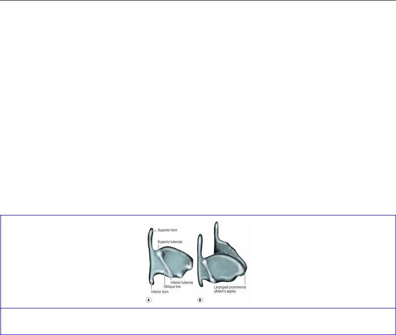
The thyroid cartilage consists of two laminae whose anterior borders are fused at a median angle, o r laryngeal prominence. A thyroid notch marks the upper end of the prominence. The posterior borders are free and projected upwards and downwards as the superior and inferior horns (Fig. 6.43). Each inferior horn articulates with the cricoid cartilage to form the cricothyroid joint. The outer surface of each lamina possesses an oblique ridge running downwards and forwards, and bounded above and below by a tubercle.
Figure 6.43 Thyroid cartilage: A from the right; B from the right and above and slightly in front.
The cricoid cartilage is the foundation of the larynx; to this signet-ring structure (Fig. 6.44) the thyroid and arytenoid cartilages are articulated by synovial joints. It is the only complete cartilaginous ring in the whole of the air passages. The anterior part of the ring is the arch; posteriorly it is projected upwards as a quadrangular flat lamina. Near the junction of the arch and lamina is an articular facet for the inferior horn of the thyroid cartilage. The upper part of the lamina has sloping shoulders, which carry articular facets for the arytenoids. A vertical ridge in the midline of the lamina produces a shallow concavity on each side for the attachment of the posterior
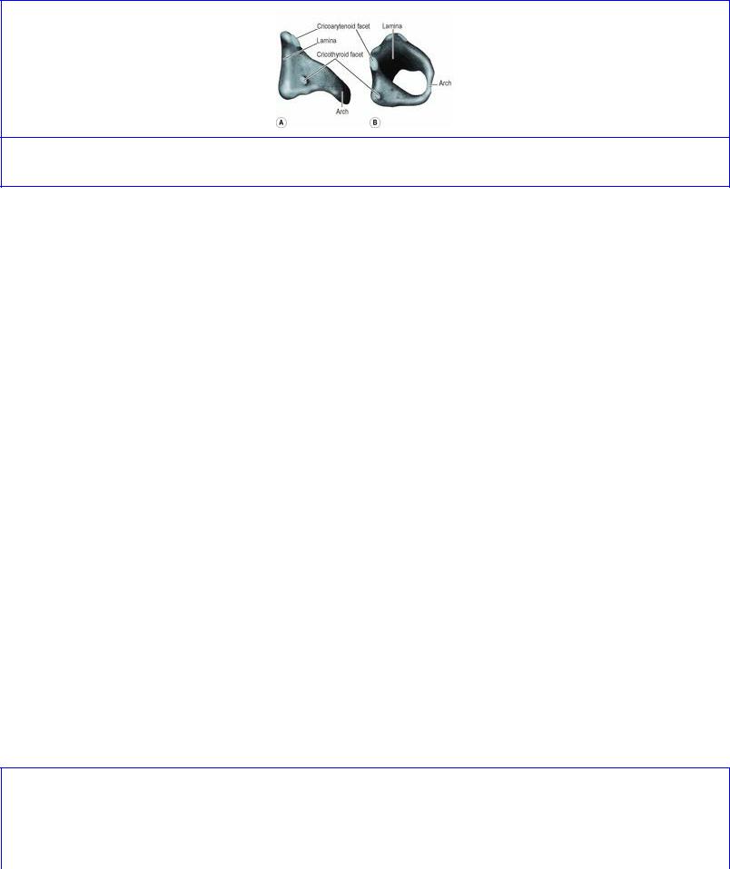
cricoarytenoid muscle; the ridge gives attachment to longitudinal muscle fibres of the oesophagus (see p. 209).
Figure 6.44 Cricoid cartilage: A from the right; B from the right, above and in front.
The epiglottic cartilage is a slightly curled, leaf-shaped structure, prolonged below into a slender process (the stalk of the leaf) attached in the midline to the back of the laryngeal prominence, below the thyroid notch. The epiglottic cartilage leans back from its attached stalk to overhang the vestibule of the larynx. Fibrous tissue attaching the front and sides of the epiglottis to the body and greater horns of the hyoid bone (hyoepiglottic ligaments) form the framework of the glossoepiglottic folds that bound the valleculae (see p. 387). The posterior surface below the apex is pitted by mucous glands. A bulge on the lower part of this surface is the tubercle of the epiglottis.
Each of the pair of arytenoid cartilages is a three-sided pyramid with anterolateral, medial and posterior surfaces. The inferior base has a forward projection, the vocal process, and a lateral projection, the muscular process. The base articulates with the sloping shoulder on the upper border of the cricoid lamina. A very small corniculate cartilage articulates with the apex of each arytenoid cartilage and a tiny cuneiform cartilage lies nearby in the aryepiglottic fold; they are unimportant.
Joints
The cricothyroid joint, between the inferior horn of the thyroid cartilage and the facet on the side of the arch of the cricoid, is synovial. Movement between the cricoid and thyroid occurs round an axis that passes transversely between the two joints, so that one cartilage can rock backwards and forwards on the other. The recurrent laryngeal nerve lies immediately behind this joint.
The cricoarytenoid joint is also synovial. The capsule here is lax, allowing both rotary and lateral gliding movements. When the arytenoids are pulled laterally and downwards they slide apart from one another along the sloping shoulders of the cricoid lamina. This gliding of the arytenoids opens the gap between the vocal folds (the rima of the glottis) in the shape of a V; rotation opens the glottis in the shape of a diamond. In man there is a greater range of gliding than of rotary movement, and the open human glottis resembles a V and not a diamond (Fig. 6.45).

Figure 6.45 Movements of the arytenoid cartilages. In A, the vocal folds are adducted. In B, rotation of the arytenoids, as in animals, produces a diamond-shaped opening. In C, lateral excursion of the arytenoids produces the human V-shaped opening.
Ligaments and membranes
Of the extrinsic membranes, the thyrohyoid membrane connects the whole length of the upper border of the thyroid laminae and the superior horns to the body and greater horns of the hyoid bone (Fig. 6.37). The thyrohyoid membrane passes up behind the body of the hyoid bone to be attached to its upper border; a bursa lies between the membrane and the back of the bone. It is here that remnants of the thyroglossal duct (see p. 26) may persist, necessitating resection of the central part of the bone to give adequate removal.
Figure 6.46 Skeleton of the larynx: A interior, viewed from the right with the right quadrangular membrane and the right halves of the thyroid cartilage and hyoid bone removed; B similar view showing muscles attached to the right arytenoid cartilage.
The thyrohyoid membrane forms the lateral wall of the piriform recess and is perforated by the internal laryngeal nerve and the superior laryngeal vessels. It is not part of the larynx, but anchors the skeleton of the larynx to the hyoid bone.
The epiglottis is attached to the hyoid bone and thyroid cartilage by the hyoepiglottic and thyroepiglottic ligaments. The former are described above; the latter is a strong band attaching the stalk of the cartilage to the angle between the thyroid laminae just below the thyroid notch.
T he cricotracheal membrane connects the lower border of the cricoid cartilage to the first cartilaginous ring of the trachea.
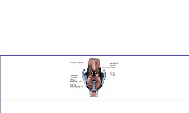
Of the intrinsic membranes, the quadrangular membrane is a thin fibroelastic membrane that extends between the arytenoid cartilage and the epiglottis (Fig. 6.46). Its anterior border is attached to the side of the lower half of the epiglottis. Its posterior border, much shorter, is attached to the anterolateral surface of the arytenoid. Its lower border is free, constituting the vestibular ligament (‘false vocal cord’). The mucous membrane covering its much longer upper border constitutes the aryepiglottic fold. The two aryepiglottic folds form the margins of the oval inlet of the larynx.
The other intrinsic ligament is the cricothyroid ligament. This is composed of mainly elastic tissue and has distinct anterior and lateral parts, continuous with each other. The anterior part is a thick band in the midline connecting the upper border of the cricoid to the lower border of the thyroid, the anterior (median) cricothyroid ligament. The paired, thinner lateral cricothyroid ligaments (also termed cricothyroid or cricovocal membranes) are attached below to the upper border of the cricoid, but as they ascend they converge and pass up deep to the lamina of the thyroid on each side (Fig. 6.46). Each lateral ligament has a free, thickened superior edge, attached in front to the back of the angle of the thyroid cartilage, midway between the notch and the lower border, and at the back to the vocal process of the arytenoid cartilage. This free edge constitutes the vocal ligament or vocal cord (hence the term cricovocal membrane). The free edge of the quadrangular membrane, the vestibular ligament, lies above the vocal ligament and there is a gap between the two ligaments. The quadrangular and cricovocal membranes are lined on their inner aspects by mucous membrane, and that part of it which covers the vestibular and vocal ligaments forms the vestibular and vocal folds respectively.
Cavity of the larynx
The inlet (aditus) of the larynx, through which it communicates with the pharynx, faces backwards and upwards and is bounded in front by the upper edge of the epiglottis, at the sides and back by the aryepiglottic folds, and in the posterior midline by the transverse mucosal fold between the arytenoids (Figs 6.40 and 6.47). The space below the level of the inlet down as far as the vestibular folds is the vestibule. In the gap between the vestibular and vocal ligaments, the mucous membrane of the larynx bulges outwards, forming a deep horizontal groove, the ventricle or laryngeal sinus (Fig. 6.48). Opening from its anterior end is a small pouch of mucous membrane, the laryngeal saccule, which extends upwards between the vestibular fold and the thyroid lamina.
Figure 6.47 Larynx from behind after removal of the mucous membrane of the laryngopharynx.
Figure 6.48 Anterior half of the larynx and upper trachea which have been sectioned in the coronal plane.
The gap between the vocal folds is the rima of the glottis (or simply ‘the glottis’), the anteroposterior slit through which air passes (Fig. 6.45). The anterior 60% of the glottis (intermembranous part) is bounded on each side by the vocal fold itself. The posterior 40% (intercartilaginous part) lies between the vocal processes of the arytenoid cartilages and the medial margins of their bases (covered of course by mucous membrane). In the resting state during quiet respiration, the glottis is triangular and about 8 mm wide at the back; the sagittal length is about 23 mm in the male and 17 mm in the female. On looking down into the larynx from above, as with a laryngoscope, the vestibular folds appear as bulges of mucosa above and lateral to the vocal folds, which are lower and closer together and move with respiration and phonation.
Below the glottis the infraglottic part of the larynx extends down to the level of the lower border of the cricoid cartilage where it becomes continuous with the trachea.
Mucous membrane
As part of the respiratory tract, the larynx in general is lined by pseudostratified columnar ciliated epithelium. The anterior surface of the epiglottis (not in the larynx) faces the tongue. Its mucosa is covered by stratified squamous epithelium, which ‘climbs over’ from the front of the epiglottis on to the aryepiglottic folds and the upper part of the posterior epiglottic surface, before being replaced by the ciliated variety. However, over the vocal folds the epithelium is always stratified squamous. The folds are a whitish colour since blood vessels do not show through here, due to the firm attachment of the mucosa to the vocal ligaments. The saccules contain many mucous glands whose secretion flows down to lubricate the vocal folds, where mucous glands are absent. Taste buds are present on the posterior epiglottic surface and the aryepiglottic folds.
The lamina propria is loose in all parts except over the vocal folds where the mucous membrane is very firmly attached. It therefore allows great swelling except at the glottis; in ‘oedema of the glottis’ the swelling accumulates above the rima, but may still cause dangerous obstruction to the airflow.
Muscles
The muscles of the larynx alter the size and shape of the inlet, or affect the vocal ligaments causing

their movements or changing their tension. The muscles that act on the inlet are the aryepiglottic and oblique arytenoid muscles, assisted by the transverse arytenoid and thyroepiglottic muscles. Those that affect the vocal ligaments are the posterior and lateral cricoarytenoids, oblique and transverse arytenoids, thyroarytenoids and vocalis, and the cricothyroids. Apart from transverse arytenoid, all the muscles are paired.
The transverse arytenoid is attached to the posterior surfaces of the arytenoid cartilages (Fig. 6.47). It draws the arytenoids (and their vocal processes) nearer to each other and adducts the vocal folds, helping to close the glottis. The oblique arytenoids pass from the back of the muscular process of one arytenoid to the apex of the opposite one, crossing each other on the posterior surface of the transverse arytenoid, and have the same action. Some fibres continue from the arytenoid apex into the aryepiglottic fold and reach the edge of the epiglottis, so forming the aryepiglottic muscle (Fig. 6.40); they approximate the aryepiglottic folds and close the laryngeal inlet.
The posterior cricoarytenoid arises from the concavity on the back of the lamina of the cricoid whence its fibres converge on the back of the muscular process of the ipsilateral arytenoids (Fig. 6.47). Its upper fibres are almost horizontal, its lower lateral fibres almost vertical. Their combined action is to move the arytenoid laterally and rotate its vocal process outwards. It is the most important muscle of the larynx as it is the only muscle that abducts the vocal folds and opens the glottis.
The lateral cricoarytenoid arises from the upper border of the cricoid arch and passes upwards and backwards to be attached to the front of the muscular process of the arytenoid (Fig. 6.46B). By drawing the muscular process forwards it rotates the vocal process inwards and closes the glottis.
The thyroarytenoid muscle extends backwards and laterally from the angle of the thyroid to the anterolateral surface of the arytenoid and the lateral surface of its vocal process (Fig. 6.46B). It shortens and relaxes the vocal ligament, thereby altering the pitch of the voice. A part of this muscle runs parallel and lateral to the vocal ligament, and some of its fibres arise from the ligament rather than the thyroid; these form the vocalis muscle and act on the posterior part of the vocal ligament.
Many thyroarytenoid fibres ascend up to the aryepiglottic fold and some even reach the side of the epiglottis. They constitute the thyroepiglottic muscle and they open the laryngeal inlet by abducting the aryepiglottic fold. Some of these fibres pass lateral to the saccule, which they compress.
The cricothyroid muscle is a fan-shaped muscle on the outer surface of the larynx; it arises from the lateral aspect of the cricoid arch and is attached to the inferior horn and adjacent lower border of the thyroid lamina (Fig. 6.49). Its contraction makes the thyroid tilt slightly downwards and forwards, thereby lengthening and tensing the vocal ligament.

Figure 6.49 Cricothyroid muscle: A from the right; B from the front.
Swallowing. Protection of the inlet during swallowing is provided by the sphincteric action of the aryepiglottic muscles. A second sphincter is provided by closure of the glottis, but swallowed material very rarely enters the vestibule. Elevation of the larynx beneath the posteriorly bulging tongue displaces the epiglottis backwards, assisting closure of the larynx. A large passing bolus may fold the epiglottis over the closed inlet, but the epiglottis is not essential for the protection of the airway. Indeed the epiglottis often stays upright during swallowing, food passing beside it on either side into the piriform fossa (the lateral food channel; see Fig. 6.38).
Phonation. Phonation or voice production involves the making of sounds that can be varied in pitch, intensity and quality (timbre). The stream of air emitted during phonation emerges as a series of discrete jets, as from a siren. This is not only a more effective means of sound production, but is very economical of expired air. At rest the vocal folds are separated. During phonation they are held together. The apposed vocal folds are blown apart by the pressure of the air below them, and elastic recoil returns them to their original position; the rapid repetition of these movements results in vibration of the folds, so giving rise to sound waves with a certain pitch. The frequency of emission of the jets depends on the length and tension of the folds, and it is these features that are adjusted by the intrinsic muscles to vary the pitch. The intensity of the sound varies with the pressure of the air forced through the glottis. The quality or timbre of the voice depends on the resonating chambers above the glottis; these include the vestibule of the larynx, pharynx, mouth, nose and paranasal sinuses, and their overall shape and volume can be altered by the soft palate, tongue and other muscles. Depression of the larynx (see p. 337) increases the volume of the resonating chambers. Articulation depends on breaking up the sound into recognizable consonants and vowels by the use of tongue, teeth and lips.
I n whispering the vocal folds are separated, and vibrations are imparted to a constant stream of expired air. This is inefficient as a means of sound production, and is very wasteful of air.
Various muscular efforts such as heavy lifting, coughing and abdominal straining are accompanied by closure of the glottis, and also by some medial movement of the vestibular folds and compression of the laryngeal ventricle.
A cough or sneeze is an explosion of compressed air. The vocal folds are powerfully adducted, a strong expira-tory contraction is made to build up the intrathoracic pressure (see p. 188), the folds are then suddenly abducted and the blast of compressed air explodes through the larynx (its expulsive force increased by the simultaneous ‘choke-barrel’ narrowing of the trachea). In the cough reflex, afferents from the mucous membrane supplied by the glossopharyngeal and vagus nerves pass to the nucleus of the tractus solitarius (see p. 480). There are widespread connections in the brainstem and spinal cord for the efferent side of the reflex, which involves muscles of the larynx, pharynx, palate, tongue, diaphragm and other thoracic and abdominal muscles.
Abdominal straining is made more effective by adduc-tion of the vocal folds. The diaphragm is weaker than the muscles of the anterior abdominal wall. To prevent loss of intra-abdominal pressure by upward displacement of the diaphragm the folds are closed after a deep breath and the diaphragm is held down by a cushion of compressed air. This manoeuvre is used for evacuation of pelvic effluents and also for the straining of heavy lifting. Escape of a jet of compressed air causes the
characteristic accompanying grunt.
Blood supply
Above the vocal folds blood is brought to the larynx by the superior laryngeal branch of the superior thyroid artery. This enters the piriform recess below the internal laryngeal nerve by piercing the thyrohyoid membrane (Fig. 6.40). The superior laryngeal veins accompany the artery and empty into the superior thyroid veins.
The lower half of the larynx is supplied from the inferior laryngeal branch of the inferior thyroid artery; it accompanies the recurrent laryngeal nerve beneath the inferior constrictor of the pharynx. Venous return is by the inferior laryngeal veins to the inferior thyroid veins.
Lymph drainage
From the supraand infraglottic parts of the larynx, lymphatics accompany the superior or inferior thyroid vessels and drain to the upper or lower groups of deep cervical nodes respectively. A few infraglottic lymphatics pass through the cricothyroid membrane and drain initially to prelaryngeal and to pretracheal nodes.
Nerve supply
All the muscles of the larynx are supplied by the recurrent larnygeal nerve except cricothyroid which is innervated by the external laryngeal nerve. All the motor fibres in both nerves are from cell bodies in the nucleus ambiguus derived mainly via the cranial part of the accessory nerve.
The recurrent laryngeal nerve enters the pharynx by passing upwards under the lower border of the inferior constrictor behind the cricothyroid joint. By this stage it has often divided into an anterior (motor) and a posterior (sensory) branch, at the level of the upper border of the isthmus of the thyroid gland. The nerve reaches the lower part of the piriform recess and then penetrates the laryngeal wall.
With complete recurrent laryngeal nerve paralysis the vocal fold takes up a variable position. Respiratory problems are rare but there may be stridor (noisy respira-tion) if the airflow is substantially increased for any reason. The voice is initially hoarse but with compensatory movement of the other vocal fold disability is reduced. With acute bilateral complete palsies there is significant inspiratory stridor and an immediate tracheostomy may be needed. The position of the vocal folds following partial lesions of the recurrent laryngeal nerves is contentious. The traditional view, that the only abductor muscle (posterior cricoarytenoid) is more vulnerable, is disputed and the vocal folds may assume a paramedian or intermediate (half abducted) position with differing effects on phonation and respiration.
Paralysis of the external laryngeal nerve affecting cricothyroid may pass unnoticed, or perhaps cause some hoarseness of the voice which appears to recover (due to hypertrophy of the opposite cricothyroid) but with a residual inability to produce higher frequencies, as in the high notes in singing. Examination reveals that the vocal fold on the damaged side is slightly bowed and at a lower level than the normal, due to loss of the tension normally provided by cricothyroid.
The mucous membrane of the larynx above the level of the vocal folds is supplied by the internal laryngeal nerve; that of the folds and the larynx below them is supplied by the recurrent laryngeal
nerve.
The sympathetic supply (vasoconstrictor) comes in with the superior and inferior laryngeal arteries from the middle and inferior cervical sympathetic ganglia.
Development
The larynx develops from the laryngotracheal groove at the caudal end of the floor of the primitive pharynx, with the laryngeal cartilages being derived from the fourth and sixth arches (see pp. 25 and 26).
Laryngotomy
In very acute airway obstruction at the level of the glottis or above, when an endotracheal tube cannot be successfully introduced through the oral or nasal cavities, an emergency laryngotomy is preferred to tracheostomy (see p. 341). The laryngeal prominence and cricoid cartilage are palpated and entry is made through the cricothyroid ligament between the cricoid and the lower border of the thyroid cartilage. There are no large midline vessels here; an anastomosis between the small cricothyroid branches of the superior thyroid arteries, high up on the cricothyroid ligament, does not usually cause problems. The proximity of the vocal cords, at the level of the middle of the thyroid angle, must be borne in mind both during the procedure and thereafter; an airway introduced through a laryngotomy is usually not maintained for more than 48 hours, lest it leads to subglottic stenosis. This site is also used for the insertion of a minitracheal tube (for suction rather than as an airway).

Part fourteen. Orbit and eye
The eye (eyeball) is the organ of vision and the principal component of the visual apparatus. This is lodged in the orbit, together with the extraocular muscles which move the eye, nerves, vessels, the lacrimal gland, fascia and fat.
Orbit
The orbit is a bony cavity shaped like a four-sided pyramid lying on its side, with the apex at the back and the base forming the orbital margin on the front of the facial skeleton (Fig. 6.50). The orbital fascia is the periosteum of the orbit which, at the back, becomes continuous with the dura mater and the sheath of the optic nerve, which enters the orbit through the optic canal at the apex. The relations of the orbit are important. Above is the anterior cranial fossa, with the meninges and the frontal lobe of the cerebral hemisphere. Medially are the nasal cavity and ethmoid sinuses. Below lies the maxillary sinus. Posterolaterally are the infratemporal fossa and the middle cranial fossa.
Figure 6.50 Bones of the left orbit, viewed along the orbital axis which is at 25° to the sagittal plane.
The roof of the orbit is the orbital part of the frontal bone, with the lesser wing of the sphenoid at the most posterior part. The frontal sinus frequently extends into its anteromedial part.
The medial wall, 5 cm long, extends in front from the anterior lacrimal crest on the frontal process of the maxilla, backwards across the lacrimal bone and the paper-thin orbital plate of the ethmoid, to the body of the sphenoid. The posterior lacrimal crest is a vertical ridge on the lacrimal bone. Between the two crests is the fossa for the lacrimal sac, which leads down into the nasolacrimal canal. At the junction of medial wall and roof lie the anterior and posterior ethmoidal foramina, between the ethmoid and frontal bones; the anterior foramen is about 24 mm behind the anterior lacrimal crest, the posterior foramen about 12 mm behind this and the optic nerve emerges through the optic canal about 6 mm further back. A knowledge of these mean measurements is of particular value during endoscopic surgical procedures on the ethmoid sinuses. The medial walls lie anteroposterior, parallel with each other. The very thin bone of the orbital plate separates the orbits from the ethmoidal air cells.
The lateral wall, 5 cm long, is composed of the zygomatic bone (the thickest and strongest part of the orbit walls) and the greater wing of the sphenoid. Posteriorly there is a gap, the superior orbital fissure, between lateral wall and roof (greater and lesser wings of the sphenoid), leading into the middle cranial fossa (Fig. 6.50). Another gap, the inferior orbital fissure, diverges from the medial

end of this fissure between lateral wall and floor (greater wing and maxilla); it leads into the pterygopalatine and infratemporal fossae. The lateral wall slopes at 45° to the sagittal plane; the two lateral walls are at right angles to each other, and, if prolonged backwards, would meet at a right angle in the pituitary fossa.
T he floor, formed mainly by the orbital surface of the maxilla (grooved and canalized by the infraorbital nerve), is completed laterally by the zygomatic bone and posteriorly by the tiny orbital process of the palatine bone. Blunt force applied to the face may cause a ‘blow-out’ fracture of the thin bone of the orbital floor or medial wall. Fracture of the floor may lead to herniation of orbital fat into the maxillary sinus, entrapment of an extraocular muscle (causing diplopia) or injury to the infraorbital nerve.
The orbital margin has four curved sides. The supraorbital margin (frontal bone) is notched or canalized a third of the way from its medial end for the passage of the supraorbital nerve and artery. The lateral margin is formed by corresponding processes of the frontal and zygomatic bones, which meet at a palpable suture line. The infraorbital margin is formed by the zygomatic bone and maxilla. The infraorbital foramen lies about 1 cm below the middle of this margin. The medial margin of the orbit is formed by the anterior lacrimal crest (maxilla) and the frontal bone.
Eyelids
The eyelids protect the eye from injury and assist in the distribution of tears over the anterior surface of the eyeball. They are covered in front with loose skin and behind with adherent conjunctiva. Their fibrous framework is the orbital septum, thickened at the margins of the lids to form the tarsal plates. The orbicularis oculi muscle lies in front of the septum. The eyelids meet at the medial and lateral angles (or canthi). The lateral canthus is in direct contact with the eyeball. The medial canthus is separated by a small triangular space, the lacus lacrimalis, in the centre of which is a small pink elevation, the caruncle. A semilunar conjunctival fold lies on the lateral side of the caruncle.
The orbital septum is attached to the margins of the orbit (Fig. 6.51). It has a wide ‘buttonhole’ in it: the palpebral fissure between the lids. It is greatly thickened above and below the buttonhole to form the crescent-shaped superior and inferior tarsal plates. The plates are formed of dense fibrous tissue, not cartilage as might be imagined from their stiffness. From the medial end of the buttonhole a thick medial palpebral ligament anchors the tarsal plates to the anterior lacrimal crest. The corresponding, much thinner, lateral palpebral ligament is attached to the marginal tubercle (of Whitnall) on the zygomatic bone just inside the orbital margin (Fig. 6.50). The levator palpebrae superioris is attached to the superior tarsal plate (see p. 401). The tarsal (Meibomian) glands are modified sebaceous glands embedded within the substance of the tarsal plates. Their ducts discharge an oily secretion at the eyelid margin; this delays evaporation of tears and discourages spilling of excess tears.

Figure 6.51 Left orbital septum and tarsal plates.
The thin skin of the eyelids is adherent to the margins of the palpebral fissure where it becomes continuous with the conjunctiva. The eyelashes are located here; they do not possess arrector pili muscles. Sebaceous glands open into each hair follicle. Ciliary glands (modified sweat glands) between the follicles open on to the eyelid margin. A hordeolum, or stye, is an infection of these glands.
Movements. The lower lid possesses very little mobility, the upper lid a great deal. The lids are closed gently by the palpebral fibres and forcibly when the orbital fibres of the orbicularis oculi join in (see p. 350). The lids are opened in the ordinary way by levator palpebrae superioris (see p. 401). The levators hold the lids open while the orbital fibres of orbicularis are contracting to lower the eyebrows as a pair of sun visors. The sun visors can be brought together by corrugator supercilii, causing (unwanted) vertical wrinkles between them. This small muscle arises medially from the frontal bone and passes laterally through orbicularis to skin above the middle of the supraorbital margin.
The conjunctiva is a transparent membrane attached to the sclera at the margins of the cornea, with which it blends. It is loosely attached elsewhere over the anterior part of the sclera and thence reflected to the inner surfaces of the eyelids. It is firmly attached to the tarsal plates and blends with the skin at the margins of the lids. The subtarsal sulcus is a shallow groove on the back of the lids, about 2 mm from the margin, where foreign bodies tend to lodge. The conjunctival epithelium is stratified columnar, except near the eyelid margin and close to the corneoscleral junction, where it changes to non-keratinized stratified squamous. Mucus-secreting goblet cells are scattered in the conjunctival epithelium and small accessory lacrimal glands are scattered in the subconjunctival connective tissue.
The blood supply of the eyelids is from the medial palpebral branches of the ophthalmic artery and the lateral palpebral branches of the lacrimal artery (branch of ophthalmic), which form a pair of arcades in each lid. The palpebral (eyelid) conjunctiva is very vascular (hence the pink colour); the bulbar (ocular) is only slightly vascular and is transparent. The venous drainage of the lids is to ophthalmic and angular veins. Lymphatic drainage from the lateral two-thirds of the lids (and lacrimal gland) is to preauricular nodes, and from the medial third to submandibular nodes.
The skin of the upper lid receives nerve supply from the lacrimal, supraorbital, supratrochlear and infratrochlear nerves, and that of the lower lid from the infraorbital nerve. The same nerves supply the corresponding palpebral and bulbar conjunctiva. The cornea has a separate supply from the long and short ciliary nerves (see p. 403).

Lacrimal apparatus
The production of tears and the removal of excess tears is the function of the lacrimal apparatus, which consists of the lacrimal gland, lacrimal canaliculi, lacrimal sac and the nasolacrimal duct.
Lacrimal gland
This is a serous gland with a large orbital and a small palpebral part. The orbital part lies in the lacrimal fossa on the lateral part of the roof of the orbit, above the lateral part of the aponeurotic tendon of levator palpebrae superioris. The gland curls round the lateral margin of the tendon and the palpebral part is visible through the superior fornix of the conjunctiva. The gland drains by a dozen ducts that run from the palpebral part into the lateral extent of the superior fornix. Closure of the eyelids begins at the lateral side of the upper lid and moves medially, so spreading tears across the eye. Under normal conditions the lacrimal gland secretes just enough tears to replace those lost by evaporation. Secretomotor fibres from the superior salivary nucleus travel in the greater petrosal nerve and relay in the pterygopalatine ganglion. The postganglionic fibres run with the zygomatic branch of the maxillary nerve, and reach the gland via an anastomotic connection between its zygomaticotemporal branch and the lacrimal nerve.
At the medial end of each lid margin is a low elevation, the lacrimal papilla, surmounted by a minute lacrimal punctum. This opens into a lacrimal canaliculus, a tiny canal which conveys excessive tears to the lacrimal sac (Fig. 6.52).
Figure 6.52 Magnified view of a left dacrocystogram (lacrimal canaliculogram).
The lacrimal sac lies in the lacrimal groove formed by the maxilla and lacrimal bone, crossed in front by the medial palpebral ligament, and some deeper fibres of orbicularis oculi are inserted into the walls of the sac (see p. 350). When the palpebral and lacrimal parts of orbicularis oculi contract, the lids are closed and the puncta turned inwards to dip into the lacus lacrimalis. Simultaneously the sac is drawn widely open, so that tears are sucked in through the canaliculi.
The nasolacrimal duct, 2 cm long, slopes downwards, backwards and laterally, in conformity with the pear-shaped nasal cavity, to open high up in the anterior part of the inferior meatus 2 cm behind the nostril. The mucous membrane is raised into several variable folds which act as valves to prevent air being blown up the duct into the lacrimal sac. The duct and sac are lined by ciliated columnar epithelium.

Muscles of the orbit
The eyeball is moved by extrinsic or extraocular muscles: four rectus (superior and inferior, medial and lateral) and two oblique (superior and inferior). The orbit also contains the levator palpebrae superioris for moving the upper lid.
The two eyes face forwards and the long axes of the eyes lie in the sagittal plane, parallel with each other and with the medial walls of the orbits, but the lateral walls slope backwards and medially, making a right angle with each other. The optic nerve and ocular muscles come from the apex of the orbit, at the back of the medial wall, and pass forwards and laterally to their ocular attachments. The actions of the superior and inferior recti are therefore not ‘straight’, despite their names. Hence the need for two oblique muscles, to act in concert with these two recti and produce direct upward and downward movements of the eyes.
The superior orbital fissure is retort-shaped, with the broad end medially (Fig. 6.50). A common tendinous ring surrounds the ‘bulb of the retort’ and the optic canal ( Fig. 6.53). It is attached to a small bony projection on the inferior margin of the fissure, and is adherent to the dural sheath of the optic nerve. From the ring the four recti arise; from the bone above the ring the levator palpebrae superioris and the superior oblique take origin. As these muscles pass forwards from the apex of the orbit they broaden out, to form a cone of muscles around the eye. Many nerves pass through the superior orbital fissure (Fig. 6.29). Three pass through the lateral part, outside the fibrous ring, and they remain outside the cone of muscles. They are the lacrimal, frontal and trochlear nerves. The rest of the lateral part of the fissure is closed by the fibrous layer of the dura mater of the middle cranial fossa. The nerves that pass through the tendinous ring and enter the cone of muscles are the oculomotor, abducens and nasociliary. Only the posterior and anterior ethmoidal and infratrochlear branches of the nasociliary nerve come out of the cone.
Figure 6.53 Diagram of the tendinous ring muscle attachments, and of structures passing through the superior orbital fissure and optic canal.
Levator palpebrae superioris arises from the undersurface of the lesser wing of the sphenoid at the apex of the orbit. It is a flat muscle that broadens as it passes forwards (Fig. 6.54). The thick frontal nerve lies on its upper surface, dividing towards the front of the orbit into its supraorbital and supratrochlear branches. At the anterior end the muscle forms an aponeurotic tendon that is widened
on each side up to a crescentic margin. This broad tendon penetrates the orbital septum and is inserted into the front of the superior tarsal plate (Fig. 6.55). Some fibres are attached to the skin of the upper lid. A thin sheet of smooth muscle lies beneath the main tendon and is inserted into the upper margin of the tarsal plate.
Figure 6.54 Right orbit. Dissection from above after removal of the roof (part of the anterior cranial fossa).
Figure 6.55 Sagittal section of the right orbit.
The superior division of the oculomotor nerve supplies the muscle. The branch either pierces the superior rectus or passes on its medial side to enter the lower surface of the levator. The nerve also carries postganglionic sympathetic fibres (from the superior cervical ganglion, via the internal carotid plexus) for the smooth muscle part.
The muscle opens the eye by elevating the upper lid. Complete oculomotor palsy causes complete ptosis (drooping of the upper lid); division of the cervical sympathetic chain causes partial ptosis.
The superior, medial, inferior and lateral rectus muscles arise from the common tendinous ring. The

superior and medial recti also have origin from the dural sheath of the optic nerve. The superior, inferior and lateral muscles pass forwards and laterally, the medial directly forwards. They all pierce the fascial sheath of the eyeball and are inserted into the sclera anterior to the coronal equator of the eye.
The superior oblique arises from the body of the sphenoid, passes forward above the medial rectus (Fig. 6.54) and gives way to a slender tendon (Fig. 6.104), which passes through the trochlea (where it is lubricated by a synovial sheath). It then turns backwards and laterally to pierce the fascial sheath and pass under the superior rectus to be inserted into the posterosuperior lateral quadrant of the sclera (i.e. behind the coronal equator of the eye). The trochlea (pulley) is a loop of fibrocartilage attached to the trochlear fossa of the frontal bone just behind the orbital margin.
The inferior oblique arises from the maxilla on the floor of the orbit, near the anterior margin. The muscle passes obliquely backwards and laterally below inferior rectus (Fig. 6.56) and then curves upwards deep to lateral rectus to be attached to the posteroinferior lateral quadrant of the sclera (i.e. behind the coronal equator of the eye).
Figure 6.56 Nerves of the right orbit and the ciliary ganglion: lateral aspect.
A mock ‘chemical formula’ is an aid to memorizing the nerve supplies of the eye muscles: LR 6SO4. This signifies that the Lateral Rectus is supplied by the sixth (abducens) nerve, the Superior Oblique by the fourth (trochlear) nerve. All the other muscles (superior, medial and inferior rectus and inferior oblique) are supplied by the third (oculomotor) nerve. The nerves all enter the ocular surfaces of the respective muscles, except the trochlear which enters the superior surface of superior oblique. Because of the decussation of fibres in the midbrain (see p. 477), the trochlear nerve nucleus of one side supplies the superior oblique of the eye of the opposite side.
The actions of the muscles that move the eyes are considered below.
Fascial sheath of the eye
A thin fascial sheath of the eye (fascia bulbi or Tenon's capsule) closely surrounds the eyeball and separates it from orbital fat. Anteriorly the sheath is attached to the sclera just behind the corneoscleral junction. Posteriorly the sheath is pierced by ciliary vessels and nerves, and fuses with the sclera and the dura around the optic nerve at its attachment to the eye. The sheath is pierced by the

tendons of the four recti and two obliques and is reflected as a sleeve proximally (i.e. away from the eye) around each tendon. Triangular expansions from the sleeves of the medial and lateral recti form the medial and lateral check ligaments, which are attached respectively to the lacrimal and zygomatic bones. The sleeve of the inferior rectus is thickened on its underside and blends with the sleeve of the inferior oblique as well as with the check ligaments, forming a hammock-like support for the eye, the suspensory ligament (of Lockwood). If the suspensory ligament remains intact when the floor of the orbit is fractured, or the maxilla removed surgically, the eye does not sag. Double vision (diplopia) following a blow-out fracture of the orbital floor is usually due to entrapment of the inferior rectus.
Nerves of the orbit
The optic nerve enters the orbit through the optic canal, accompanied by the ophthalmic artery below and lateral to the nerve. The nerve is really an extension of the white matter of the brain; it is covered by pia, arachnoid and dura mater as far as the back of the eye. Its length in the orbit is 25 mm. It curves laterally and downwards as it passes forwards to meet the sclera 3 mm medial to the posterior pole. The central artery and vein of the retina pierce the nerve about halfway along its course to the eye. After passing within the fibrous ring, the optic nerve is usually crossed above from lateral to medial by the ophthalmic artery, with the nasociliary nerve and the superior ophthalmic vein behind the artery. The ciliary ganglion (see below) lies on the lateral side of the optic nerve one-third of the way from optic canal to eye, and the anterior part of the nerve is closely surrounded by the short ciliary nerves and vessels.
The continuation of the subarachnoid space around the optic nerve accounts for the appearance of papilloedema in increased intracranial pressure.
T he infraorbital nerve enters the orbit through the inferior orbital fissure accompanied by the zygomatic nerve and infraorbital artery. The infraorbital nerve and artery occupy the groove in the posterior part of the orbital floor. Both enter the infraorbital canal and proceed to the face, also supplying the maxillary sinus and some upper teeth (see p. 376).
The zygomatic nerve passes along the lateral wall and divides into its zygomaticotemporal and zygomaticofacial branches. The former gives a communicating branch to the lacrimal nerve, so providing the secretomotor fibres for the lacrimal gland, and traverses a canal in the zygomatic bone to enter the infratemporal fossa; the latter traverses a separate canal to emerge on the face.
The lacrimal, frontal and trochlear nerves enter the orbit through the superior orbital fissure, outside the tendinous ring (Figs 6.53 and 6.54).
The lacrimal nerve, the smallest of the three main branches of the ophthalmic, runs forward on the lateral wall of the orbit along the upper border of the lateral rectus muscle. It picks up a secretomotor branch from the zygomaticotemporal nerve which it gives off to the lacrimal gland. It pierces the orbital septum to supply both surfaces of the conjunctiva in the upper fornix and the skin of the outer part of the upper lid.
The frontal nerve is the large main branch of the ophthalmic nerve which runs straight forward above the levator muscle in contact with the periosteum of the orbital roof. It divides into the small
supratrochlear and (laterally) the large supraorbital nerves which pass to the forehead.
The trochlear nerve (fourth cranial), lying medial to the frontal nerve, passes forward and sinks into the superior oblique muscle (Fig. 6.54).
The oculomotor nerve enters the tendinous ring in two divisions, superior and inferior, with the nasociliary nerve between them and the abducent nerve below all three (Figs 6.53 and 6.29).
The superior division of the oculomotor nerve (third cranial) runs forwards above the optic nerve and supplies the overlying superior rectus and levator palpebrae muscles. It carries sympathetic fibres from the internal carotid cavernous plexus to the smooth muscle part of the levator.
The inferior division of the oculomotor nerve is larger; it gives off the nerve to the inferior rectus and the nerve to the medial rectus, which passes below the optic nerve to reach that muscle. The rest of the inferior division continues as the nerve to the inferior oblique; it gives off the parasympathetic root to the ciliary ganglion (Fig. 6.56).
The abducens nerve (sixth cranial) simply passes forward, diverging away from the optic nerve, and enters the lateral rectus muscle (Fig. 6.29).
The nasociliary nerve, the third branch of the ophthalmic, runs forward and then crosses from lateral to medial above the optic nerve (Fig. 6.56) and behind the ophthalmic artery to approach the medial wall of the orbit. Here it becomes the anterior ethmoidal by entering the anterior ethmoidal foramen (Fig. 6.104), under the frontal bone and above the ethmoidal labyrinth; it then runs forward in a groove on the cribriform plate and enters the nose through a slit at the side of the crista galli. Before entering the foramen, the nasociliary gives off the infratrochlear nerve which passes forward on the medial wall of the orbit just below the trochlea, supplies the lacrimal sac and conjunctiva, and continues above the medial palpebral ligament to skin of the upper lid and bridge of the nose.
The nasociliary nerve gives off the posterior ethmoidal nerve, which leaves the orbit through the posterior ethmoidal foramen to supply the posterior ethmoidal and sphenoidal air sinuses.
The nasociliary nerve also gives off a sensory branch to the ciliary ganglion, and a pair of long ciliary nerves which pierce the sclera medial to the short ciliary nerves. The long ciliary nerves supply the ciliary body, iris and cornea, and carry postganglionic sympathetic fibres from the superior cervical ganglion (derived from the internal carotid plexus) for the dilator pupillae.
The ciliary ganglion is a minute body (2 mm diameter) lying on the lateral side of the optic nerve (Fig. 6.56), between the nerve and the lateral rectus. Three roots enter its posterior end (see Fig. 1.15, p. 21). The sensory root is a branch of the nasociliary nerve and passes through the ganglion without relay to supply the eye, but not the conjunctiva. The sympathetic root is a branch from the internal carotid plexus, which enters through the fibrous ring and passes through the ganglion without relay, carrying vasoconstrictor fibres to the vessels of the eye. The parasympathetic root is from the nerve to the inferior oblique, the cell bodies being in the Edinger–Westphal nucleus (see p. 477); the fibres relay in the ganglion and supply the ciliary body (for accommodation) and the sphincter pupillae. The branches of the ganglion are the 8–10 short ciliary nerves, which contain fibres from all three roots of the ganglion. The nerves pierce the back of the sclera around the attachment of the

optic nerve.
Vessels of the orbit
The ophthalmic artery is a branch of the internal carotid given off as the vessel emerges from the roof of the cavernous sinus. It passes through the optic canal, inferolateral to the optic nerve and within its dural sheath. In the orbit it pierces the dura and spirals around the lateral side of the optic nerve to pass forwards above the nerve, anterior to the nasociliary nerve. Its many branches accompany all the branches of the nasociliary, the frontal and the lacrimal nerves. Thus it supplies the ethmoidal air cells, nasal cavity, external nose, eyelids and forehead, in all of which places its branches anastomose with branches of the external carotid, establishing connections between internal and external carotid systems.
Within the orbit the ophthalmic artery supplies all the extraocular muscles, the lacrimal gland and the eye. This last is by two sets of vessels. The central artery supplies the optic nerve and retina, while the posterior ciliary arteries pierce the sclera to enter the choroid coat of the eye. Choroidal capillaries supply the outer layers of the retinae, but there is no anastomosis between the two sets of vessels; the central artery is an end artery. Anterior ciliary arteries, from the muscular branches to the recti, pierce the anterior part of the eye.
Two ophthalmic veins drain the orbit and receive tributaries that correspond in the main with the branches of the ophthalmic artery. The superior ophthalmic vein commences above the medial palpebral ligament and passes back above the optic nerve to drain into the cavernous sinus through the superior orbital fissure. It communicates at its commencement with the angular vein. The inferior ophthalmic vein commences at the front of the orbital floor and runs back to drain through the inferior orbital fissure into the pterygoid plexus and either directly into the cavernous sinus through the superior orbital fissure or by joining the superior ophthalmic vein.
There are no lymphatics in the eyeball. The lymphatics of the eyelids drain with those of the face.
Movement of the eyes
Normal binocular vision depends on the properly coordinated activity of the 12 muscles that move the two eyes. Three of the rectus muscles—medial, superior and inferior—are concerned with turning the eye in, and the lateral rectus and the two obliques turn it out.
The actions of the medial and lateral recti are simple. Each lies in a horizontal plane and turns the eye in or out respectively. There are no secondary movements.
The actions of the superior and inferior recti and obliques are more complex. Each is inserted in front of the coronal equator, and the line of pull passes medial to the axis of rotation of the eye. Thus the superior rectus turns the eye up and in, and the inferior oblique turns it up and out; combined they produce a vertical upward movement, for their medial and lateral components cancel each other out. Similarly the inferior rectus turns the eye down and in, the superior oblique down and out; combined they turn it vertically down. Pure up and down movement is thus produced by one rectus acting with its oppositely named oblique (superior rectus and inferior oblique, inferior rectus and superior oblique).
On account of the concurrent action of the superior and inferior recti, the elevating action of the inferior oblique and the depressing action of the superior oblique only become independently demonstrable when the eye is turned in; the more the eye is turned out, the less is their contribution to the up or down movement. Hence the function of the trochlear nerve (action of superior oblique) is tested by asking the subject to look downwards and inwards at the tip of the nose. Similarly the elevating and depressing actions of the superior and inferior recti are best demonstrated when the eye is turned out.
The obliquity of pull of the superior and inferior recti and obliques produces a certain amount of torsion or wheel rotation of the eye around an anteroposterior axis. Thus, as viewed by an observer from the front, 12 o'clock on the right cornea rotates to 1 o'clock (intorsion) by the action of superior rectus and oblique, or to 11 o'clock (extorsion) by the action of inferior rectus and oblique.
Control of conjugate gaze
Moving the eyes from side to side implies turning one eye in and the other out; this is conjugate horizontal gaze and, if looking to the left for example, depends essentially on the coordinated activity of the left lateral rectus and right medial rectus. The neural pathways (Fig. 6.57) involve fibres from the visual cortex to the frontal eye field in the middle frontal gyrus, from which fibres pass to a region of the reticular formation of the opposite side adjacent to the abducens nucleus in the pons, the pontine paramedian reticular formation. From here some fibres pass to the abducens nucleus of the same side, so activating the lateral rectus of that eye; other fibres cross the midline to join the medial longitudinal fasciculus and run to the part of the oculomotor nucleus that controls the medial rectus of the other eye.
Figure 6.57 Pathway for conjugate gaze. The para-abducent nucleus (pontine paramedian reticular formation) projects to the abducens nucleus of the same side and the oculomotor nucleus of the opposite side.
Similar pathways for control of conjugate vertical gaze involve the oculomotor and trochlear nuclei and a rostral interstitial nucleus of the medial longitudinal fasciculus in the midbrain.

Ocular nerve paralyses
Each complete nerve lesion produces a characteristic pattern of strabismus (squint) and diplopia (Fig. 6.58). The simplest nerve lesion is that of the sixth nerve (abducens). The eye cannot look outwards due to paralysis of the lateral rectus (Fig. 6.58A) and, when trying to look straight ahead, it is turned in by the unopposed action of the medial, superior and inferior recti (oculomotor nerve).
Figure 6.58 Position of the right eye in right ocular nerve paralyses, when attempting to look in the direction of the arrow in A and B, and looking forward in C. A Abducens nerve; B trochlear nerve; C oculomotor nerve.
With a fourth nerve paralysis (trochlear), the eye cannot look downwards as far as it should when the eye is turned in (Fig. 6.58B) because of the superior oblique paralysis, so that the patient complains of diplopia when reading or difficulty going down stairs. There is also some degree of extorsion, because the superior oblique which normally produces intorsion is not available to counteract the extorting effect of the inferior oblique. To compensate for the extorsion, the patient characteristically tilts the head towards the opposite shoulder, in order to bring (for example in a right fourth nerve palsy) the 11 o'clock position on the cornea up to 12 o'clock; at the same time the good eye (the left in this example) uses its intact control mechanisms to produce intorsion.
With a third nerve lesion (oculomotor), the most obvious feature is ptosis of the upper lid due to paralysis of the levator. When the lid is manually lifted up, the eye is seen to be looking down and out (Fig. 6.58C) due to the unopposed actions of the lateral rectus (sixth nerve) and superior oblique (fourth nerve). The eye cannot be turned up, in or further down due to paralysis of the superior, medial and inferior recti, but on looking outwards the diplopia will disappear because the lateral rectus (sixth nerve) is acting normally. The pupil is dilated and does not react to light or on accommodation, due to interruption of the parasympathetic fibres that run in the oculomotor nerve (see p. 407) though the consensual reflex in the opposite eye is preserved.
Structure of the eye
The eye contains the light-sensitive retina and, like a camera, it is provided with a lens system for focusing images (the cornea, lens and refractive media) and with means of controlling the amount of light admitted (the iris diaphragm). Like a camera, its inside is black to prevent internal reflections.

The relatively large area behind the lens is occupied by the vitreous body. In front of the lens is the small area filled by aqueous humour and incompletely divided into anterior and posterior chambers by the iris. The space bounded by the inner margin of the iris is the pupil.
The wall of the eye, enclosing the refractive media, is made up of three coats. The outer coat is fibrous and consists of the sclera and cornea; a vascular coat (the choroid, ciliary body and iris) intervenes between this and the innermost nervous coat (the retina). The sclera can be regarded as a cup-like expansion of the dural sheath of the optic nerve. The choroid, similarly, is an expansion of the arachnoid and pia, the retina being an expansion of the white matter of the brain (optic nerve).
Fibrous coat
The sclera is the posterior five-sixths of the outer coat of the eyeball. The sclera is opaque (the ‘white’ of the eye) and consists of dense collagen fibres, interspersed with elastic fibres. It is thinnest at the coronal equator and where it is pierced by the recti. It is thickest at the back but weakest at the entrance of the optic nerve, whose perforating fibres give it a sieve-like appearance, the lamina cribrosa. If a sustained increase of intraocular pressure occurs (chronic glaucoma) the lamina cribrosa yields and bulges posteriorly (‘cupping’ of the disc).
The sheath of dura mater around the optic nerve blends with the sclera. The sclera receives the insertions of the ocular muscles. It is pierced by the ciliary nerves and arteries around the entrance of the optic nerve, and by the venae vorticosae (the choroid veins) just behind the coronal equator. The anterior ciliary arteries (from muscular branches to the recti) perforate the sclera near the corneoscleral junction.
The fascial sheath of the eye and the bulbar conjunctiva are connected to the sclera by loose connective tissue, which is vascular under the conjunctiva; engorgement of these vessels produces a circumcorneal injection indicative of inflammation within the eye. The rest of the sclera is almost avascular.
Just behind the corneoscleral junction, within the sclera is a circularly running canal, the sinus venosus sclerae (canal of Schlemm) (Fig. 6.59). Posterior to the canal is a triangular projection, the scleral spur, pointing forwards and inwards, to which the ciliary muscle is attached. The canal is lined with endothelium. Aqueous humour from the anterior chamber filters into the canal through trabecular tissue on its anterior wall. The canal is connected with anterior scleral veins, into which it drains.

Figure 6.59 Section through the eye in the region of the corneoscleral junction.
Anteriorly the cornea is continuous with the sclera (Fig. 6.59), with the difference that its laminae of fibrous tissue are transparent instead of opaque white. It bulges forward from the sclera at the corneoscleral junction or limbus, being the segment of a smaller sphere. It occupies the anterior onesixth of the eye and is completely avascular. At the limbus the conjunctival epithelium becomes continuous with the corneal epithelium, which is a very regular stratified squamous type of about five layers of cells. It is separated from the corneal stroma by the anterior limiting layer (Bowman's membrane), a homogeneous layer with scattered collagen fibrils and much ground substance.
The corneal stroma or substantia propria consists of over 200 lamellae of collagen fibrils, with scattered fibroblasts. The transparency is due to the precise lattice arrangement of its lamellae embedded in a ground substance. The normal lack of vascularity (and of lymph vessels) accounts for the success of corneal grafts, which are thus not invaded by T lymphocytes. The inner surface of the stroma lies against the posterior limiting layer (Descemet's membrane), which is the basement membrane of the innermost single layer of corneal endothelium.
The cornea is supplied by the short (mainly) and long ciliary nerves. The corneal reflex is elicited clinically by the gentlest touching of the cornea (not the conjunctiva) with a wisp of cotton wool; both eyes should shut. The pathway is via the trigeminal ganglion to the main sensory nucleus, whence impulses pass by way of the reticular formation to reach both facial nerve nuclei and so stimulate both orbicularis oculi to close the lids on both sides.
Vascular coat
The intermediate coat of the eye, frequently known as the uveal tract, consists of a continuum of vascular tissue which is made up of the choroid, the ciliary body and iris.
The choroid is a thin, pigmented layer lining the inner surface of the sclera, with the delicate connective tissue of the suprachoroid lamina intervening. Anteriorly it merges into the ciliary body (Fig. 6.59). Posteriorly it is perforated by the optic nerve, to which it is firmly attached. Its inner surface is firmly attached to the pigment layer of the retina, and the choroid capillaries provide nutrition for the rods and cones of the retina. The veins collect into four or five large venae vorticosae, which pass through the sclera just behind the coronal equator.
The ciliary body is continuous with the choroid behind, and the iris in front. It lies as a flat ring applied to the inner surface of the sclera. Being thicker in front and thinner behind, the ciliary body appears triangular in section. The two long sides of the triangle are in contact with the sclera externally and the vitreous body internally. The periphery of the iris is attached halfway along the short anterior base of the triangle. The scleral surface of the ciliary body contains the ciliary muscle. The vitreous surface of the ciliary body is lined by two layers of epithelium. The outer layer is pigmented and the inner layer non-pigmented; they respectively represent the pigmented layer and the nervous part of the retina. This surface appears smooth where it is continuous with the choroid at the ora serrata, but further forward this surface is projected into 70–80 small ciliary processes which lie in reciprocal grooves on the anterior surface of the vitreous body.
The ciliary muscle consists of smooth muscle; its function is to focus the lens for near vision. Its outermost fibres are longitudinal and pass back into the stroma of the choroid; the innermost are circular and run circumferentially near the periphery of the lens. Between the two are radial fibres which radiate in from the scleral spur. Contraction of the ciliary muscle relaxes the suspensory ligament, allowing the lens to bulge and focus near objects on the retina. The muscle is supplied from the Edinger–Westphal part of the oculomotor nucleus in the midbrain, by fibres which relay in the ciliary ganglion and enter the eye in the short ciliary nerves.
The iris is attached at its periphery to the middle of the anterior surface of the ciliary body; peripheral to this attachment the ciliary body itself and a narrow rim of sclera form the iridocorneal angle of the anterior chamber (Fig. 6.59). From its peripheral attachment the iris is pushed slightly forwards, in the form of a very low cone, by contact with the anterior convexity of the lens. The iris is perforated centrally by the pupil, the varying size of which controls the amount of light entering the eye.
The main bulk of the iris is made up of vascular connective tissue in which melanocytes are present. Behind the stroma are two epithelial layers. The cells of the anterior layer contain few melanin granules, while those of the posterior layer are packed with melanin granules. The colour of the iris is determined by the amount of pigment in the iris. When pigment is lacking, as at birth, the iris is blue. As the amount of pigment cells increase the iris colour becomes darker. The colour of the pigment, too, varies in different individuals.
The sphincter pupillae is a circular band of smooth muscle lying in the stroma of the iris at the margin of the pupil. It is supplied, like the ciliary muscle, from the Edinger–Westphal part of the oculomotor nucleus. The dilator pupillae is a thin sheet of radial fibres of smooth muscle at the back of the stroma of the iris, extending from the ciliary body to the sphincter pupillae. It is supplied by the cervical sympathetic. The preganglionic cells lie in T1 segment of the spinal cord. The stroma of the iris and the sphincter and dilator muscles are derived from the neural crest (see p. 23).
Control of the pupil and reflexes
The size of the pupil depends on the interplay between the sphincter innervated by parasympathetic fibres and the dilator which receives a sympathetic supply. When a light is shone into one eye, the pupil of that eye constricts; this is the direct pupillary light reflex. The pupil of the other eye also constricts; this is the indirect or consensual light reflex. The neural pathway is as follows (Fig. 6.60). Some fibres of the optic tract bypass the lateral geniculate body by running in the superior brachium to reach the midbrain at the level of the superior colliculus and enter the pretectal nucleus.
From there the cell bodies send their axons to the Edinger–Westphal nucleus, from which fibres reach the ciliary ganglion via the oculomotor nerve and its branch to the inferior oblique. The short ciliary nerves from the ganglion supply the sphincter pupillae. Because the pretectal nucleus sends fibres to the Edinger–Westphal nucleus of both sides, both pupils will constrict. The partial crossing of the fibres of one optic nerve in the optic chiasma also ensures that both pretectal nuclei are stimulated.
Figure 6.60 Pathway for pupillary light reflexes.
Pathological studies indicate that in the main oculomotor nerve trunk the pupillary fibres lie on the surface of the nerve and have a blood supply from vessels of the nerve sheath, not from those of the nerve trunk. Thus these fibres may be affected by pressure (e.g. from an aneurysm of the posterior communicating artery) but not by infarction of the nerve trunk (as in diabetes).
The eye automatically focuses and converges for near vision, and this change in lens curvature is accompanied by pupillary constriction to sharpen the focus. These three components— accommodation, convergence and pupillary constriction—constitute the accommodation– convergence reflex, or near reflex. The changes accompany conscious vision: they involve cortical as well as subcortical pathways. From the visual cortex efferent fibres pass to the pretectal area and thence to the oculomotor nucleus including the Edinger–Westphal part, which by ciliary ganglion relay activates the ciliary muscle and the sphincter pupillae. Contraction of the ciliary muscle releases tension on the suspensory ligament of the lens, so allowing the lens by its own elasticity to thicken and focus for near vision. Accompanying the accommodation changes, the medial rectus muscle of each eye contracts to provide the necessary convergence for near vision.
The sympathetic path to the pupil is very long (Fig. 6.61). From cells in the hypothalamus (whose cortical control is uncertain), fibres run down through the brainstem and spinal cord to lateral horn cells in T1 segment of the cord. Preganglionic fibres enter the sympathetic trunk via the white ramus communicans of T1 nerve and pass up to the superior cervical ganglion. From there postganglionic fibres accompany the internal carotid artery into the skull and cavernous sinus, leaving the artery to join the ophthalmic nerve and become distributed to the eye by the nasociliary and its long ciliary branches.

Figure 6.61 Sympathetic pathway to the pupil.
Damage to any of the above fibres can interrupt this pathway. Thus such conditions as vascular or degenerative lesions of the brainstem or spinal cord, pressure on the T1 nerve root by a cervical rib, involvement of the sympathetic trunk by carcinoma of the lung, thyroid or oesophagus, or metastatic lymph nodes, may give rise to Horner's syndrome. The characteristic features include slight constriction of the pupil (due to unopposed parasympathetic activity and really a failure of dilatation) but which still reacts to light and accommodation, partial ptosis (due to paralysis of the smooth muscle part of levator palpebrae) and reduction of sweating on the forehead or a larger area of the head (unless the lesion is above the superior cervical ganglion, when there is no loss of sweating).
Nervous coat
The retina is the delicate innermost membrane of the eye. Its outer surface is attached to the choroid, and its inner surface is in contact with the vitreous body. The light-sensitive area ends abruptly, halfway between equator and corneoscleral junction, at a dentate line, the ora serrata. Forward of this a thin insensitive layer passes on in continuity as the epithelial layers of the ciliary body and iris. At the entrance of the optic nerve is a circular pale area, 1.5 mm in diameter; this is the optic disc. It overlies the lamina cribrosa of the sclera. The optic disc is excavated to a variable degree, producing the physiological cup. There are no rods or cones in the optic disc, hence it is insensitive to light: the ‘blind spot’. The disc and whole surrounding area of the back of the eye as seen with the ophthalmoscope constitute the fundus of the eye.
At the posterior pole of the eye (3 mm lateral to the optic disc) is a shallow depression; it is completely free of blood vessels and is yellowish, hence called the macula lutea. In the centre of the macula is a shallow pit, the fovea centralis, of comparable size to the disc. This is the thinnest part of the retina. There are no blood vessels and no rods here, but there is a high concentration of cones. It is the area of the most acute vision.
The outer layer of the retina consists of a single layer of pigmented epithelial cells firmly attached to the choroid. Next to this layer lie the light receptors (the rods and cones), forming a layer less firmly attached to the pigment cells, so that in detachment of the retina the pigment cells remain in position while the rods and cones, with the other layers of the retina, become displaced inwards from them.
The physiological arrangement of the nervous elements is very similar to that in any other sensory pathway (e.g. in the spinal cord). From the sense receptor, the first neuron has its cell body
peripherally placed (in the retina this is the bipolar cell). It leads by synapse to the second neuron (in the retina this is the ganglion cell) whose axon passes to the thalamus (in this case the lateral geniculate body) whence, after relay, the third neuron leads through the retrolentiform part of the internal capsule to the visual cortex (see Fig. 7.9, p. 465).
The light receptors are of two kinds, rods and cones. Only the rods contain the photoreceptor protein rhodopsin, or visual purple. (Cones contain related photo-sensitive pigments with different absorption properties.) Rods do not register colour, but are sensitive to dim light (scotopic vision). The periphery of the retina contains rods only. Several rods by their bipolar cells share one ganglion cell (from which one axon passes to the lateral geniculate body). They are of low threshold; together they ‘whisper up the line’ to the thalamus and cortex. The cones have a higher threshold (photopicvision) and they register colour. Cones alone occupy the fovea centralis. Beyond this they share equally with the rods, but they fall short of the periphery of the retina. Each cone is connected to a separate ganglion cell; alone it ‘shouts up the line’ to the thalamus and cortex.
Customarily 10 retinal layers are distinguished from outside inwards; they are as follows:
1.Pigment epithelium
2.Rods and cones (processes)
3.External limiting lamina
4.Outer nuclear layer (rod and cone cell bodies with their nuclei)
5.Outer plexiform layer
6.Inner nuclear layer (bipolar cells)
7.Inner plexiform layer
8.Ganglion cell layer
9.Nerve fibre layer (axons of ganglion cells which pass into the optic nerve at the disc)
10.Inner limiting lamina.
The central artery of the retina passes through the lamina cribrosa within the optic nerve and in the optic disc divides into an upper and lower branch. Each gives off nasal and temporal branches. The upper and lower temporal branches curve up and down respectively to clear the macula lutea. The branches of the central artery are end arteries. They supply the neurons (bipolar and ganglion cells) of the retina. The light receptors (rods and cones and their nuclei in the outer nuclear layer) are supplied by diffusion from the capillaries of the choroid. The retinal veins run with the branches of the central artery. The central vein leaves via the optic disc and emerges from the optic nerve and its coverings to join the superior ophthalmic vein.
Development. The retina is developed from a hollow outgrowth, the optic vesicle, which protrudes from the cerebral vesicle. The optic vesicle becomes invaginated to form the optic cup, consisting of two layers of cells. The outer layer differentiates to form the pigment cell layer. The inner layer forms the remaining layers of the retina with the rods and cones outermost (next to the pigment cells). The ganglion cells and their axons are innermost; light has therefore to pass through them to activate the receptors.

Refracting media
Most of the refraction of light takes place at the junction of air and corneal epithelium. Beyond the cornea light passes through the aqueous humour, the lens and vitreous body to reach the retina.
The aqueous humour is a clear fluid that lies between the back of the cornea and the front of the lens. The space is divided by the iris into anterior and posterior chambers, which communicate with each other through the pupil (Fig. 6.62). The anterior chamber is 3 mm deep centrally. Aqueous humour is produced by the ciliary processes by diffusion from the capillaries and transported by the ciliary epithelium into the posterior chamber; it passes through the pupil into the anterior chamber. At the margin of the anterior chamber is the iridocorneal angle (see p. 407) and here aqueous humour filters through trabecular tissue into the canal of Schlemm (see p. 405). Obliteration of the angle therefore prevents absorption of aqueous humour, with consequent rise of intraocular tension, leading to the condition of glaucoma. Aqueous humour is an avenue for nutrients and metabolic exchange for the avascular cornea and lens.
The posterior chamber is bounded in front by the iris and behind by the lens and its suspensory ligament. It is triangular in cross-section (pupil and lens in contact with each other forming the apex of the triangle). The base of this triangle is formed by the ciliary processes. Aqueous humour lies between the fibres of the suspensory ligament as far back as the anterior surface of the vitreous.
The lens is a transparent biconvex body enclosed in a transparent elastic capsule. It is 10 mm in diameter and 4 mm thick. Its posterior surface, resting on the vitreous, is more highly convex than the anterior. The latter surface is in contact with the pupillary margin of the iris. The lens capsule is an elastic membrane that envelops the whole lens. The capsular epithelium lies anteriorly, deep to the capsule. Centrally it is a single layer of cubical cells, but more peripherally the cells elongate to produce fibres which, by their accumulation, make up the lens substance.
The suspensory ligament of the lens, or zonule, is a series of delicate fibrils attached to the ciliary processes and, through the furrows between them, further back on the ciliary body. The fibres pass centrally to attach themselves to the lens, mostly in front of, but a few behind, the circumference. In the rest position they hold the lens flattened under tension; when relaxed by contraction of the ciliary muscle, elasticity of the lens causes its anterior surface to bulge, so thickening it (as in accommodation, see p. 407).
The vitreous body is a colourless, jelly-like mass which occupies the posterior four-fifths of the eyeball. It comprises about 99% water and a sparse cellular and fibrous content. It is indented in front by the posterior convexity of the lens and, beyond this, has radial furrows reciprocal with the ciliary processes. It is traversed from the front towards the optic disc by the tiny hyaloid canal, the site of the embryonic hyaloid artery. The vitreous body is attached to the optic disc and just in front of the ora serrata; elsewhere it lies free, in contact with the retina.