

Part eleven. Kidneys, ureters and suprarenal glands
Kidneys
The kidneys lie high up on the posterior abdominal wall (Fig. 5.43) behind the peritoneum, largely under cover of the costal margin. At best only their lower poles can be palpated in the normal individual. Each kidney lies obliquely, with its long axis parallel with the lateral border of psoas major. It lies well back in the paravertebral gutter, so that the hilum, a vertical slit-like depression at the medial border transmitting the renal vessels and nerves and the renal pelvis (the beginning of the ureter), faces somewhat forwards as well as medially (Fig. 5.44). As a result of this slight ‘rotation’ of the kidney an anteroposterior radiograph gives a somewhat foreshortened picture of the width of the kidney. The normal kidney measures about 12 × 6 × 3 cm and weighs 130–150 g. The hilum of the right kidney lies just below, and of the left just above, the transpyloric plane 5 cm from the midline. The bulk of the right lobe of the liver accounts for the lower position of the right kidney. The upper pole of the left kidney overlies the eleventh rib, that of the right kidney the twelfth rib. Each kidney moves in a vertical range of 2 cm during the full respiratory excursion of the diaphragm.
The surfaces of the kidney, covered by its capsule, are usually smooth and convex though traces of lobulation, normal in the fetus, are often seen. The pelvis emerges from the hilum, behind the vessels, to pass down as the ureter.
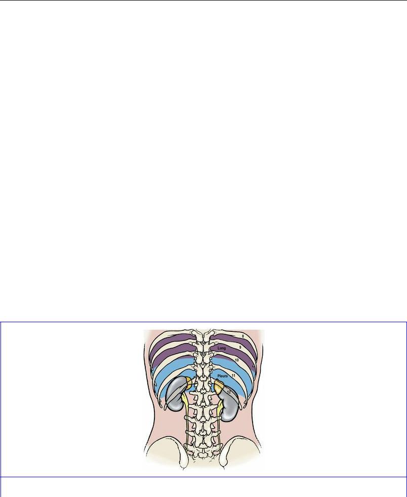
Posteriorly the relations of both kidneys are similar, comprising mostly the diaphragm and quadratus lumborum muscles, with overlap medially on to psoas and laterally on to transversus abdominis. The upper pole lies on those fibres of the diaphragm which arise from the lateral and medial arcuate ligaments. A small triangular part of the costodiaphragmatic recess of the pleura lies behind the diaphragm and is an important posterior relation (Fig. 5.48), which is at risk in the lumbar approach to the kidney (see p. 286). The subcostal vein, artery and nerve, on emerging from beneath the lateral arcuate ligament, lie behind the kidney, as do the iliohypogastric and ilioinguinal nerves as they emerge from the lateral border of psoas. The hilum of the kidney lies over psoas and the convexity of the lateral border lies on the aponeurosis of origin of transversus abdominis.
Figure 5.48 Relationship of the pleural sacs to the upper poles of the kidneys, from behind. The

ureters lie medial to the tips of the lumbar transverse processes.
The suprarenal glands surmount the superior poles of both kidneys and overlap a small part of their anterior surfaces. The rest of the upper halves of each kidney lie in contact with peritoneum, which on the right kidney is the peritoneum of the hepatorenal pouch (part of the greater sac), and on the left is the peritoneum of the lesser sac (part of the stomach bed) medially, and the peritoneum of the greater sac laterally (between the kidney and the spleen), with the splenorenal ligament passing forwards between these areas (Fig. 5.49). The hilum is separated from the peritoneum, on the right side by the second part of the duodenum and on the left side by the body of the pancreas and splenic vessels (Fig. 5.26). The lateral part of the lower pole is separated from peritoneum by the hepatic and splenic flexures of the colon on the right and left sides respectively. The medial part of the lower pole, on each side, lies in contact with peritoneum which separates it from coils of jejunum; between peritoneum and kidney are ascending branches of the right and left colic arteries.
The perinephric fat lies outside the renal capsule (Fig. 5.49) and plays a part in retaining the kidney in position. Nephroptosis (‘floating kidney’) may develop after severe loss of weight. The renal fascia (of Gerota) surrounds the perinephric fat. It is not a very obvious membrane in the living, but appears more convincingly in the embalmed cadavre. It is a condensation of the areolar tissue between the parietal peritoneum and the posterior abdominal wall and restrains the extension of a perinephric abscess. It ascends as a dome over the upper pole of the kidney and the suprarenal. However, a fascial septum separates the two organs, which explains why in nephrectomy the latter gland is not usually displaced (or even seen). At the lateral renal border the anterior and posterior layers fuse, while at the hilum the fascia is attached to the renal vessels and the ureter. When traced downwards, the fascia fades into the extraperitoneal tissue around the ureter. Pus in the perinephric space and injections into it do not usually track downwards, but increasing pressure may force the fascia to rupture and allow such contents to flow downwards retroperitoneally towards the pelvis.
The renal pelvis is the funnel-shaped commencement of the ureter, and is normally the most posterior of the three main structures in the hilum (though an arterial branch or venous tributary may lie behind it). The capacity of the average pelvis is less than 5 mL.
Blood supply and segments
The wide-bored renal arteries have a blood flow in excess of 1 litre per minute. They leave the abdominal aorta at right angles and lie behind the pancreas and renal veins.
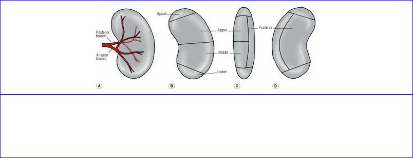
Based on its blood supply, each kidney possesses five segments (Fig. 5.50). In the region of the hilum the artery typically gives rise to an anterior and a posterior division. The posterior division supplies the posterior segment, while the anterior division gives branches that supply the apical, upper, middle and lower segments. The pattern of branching of the vessels may vary, but there are always five segments with no collateral circulation between them. Abnormal or aberrant renal arteries, such as a vessel running from the aorta to the lower pole are, in fact, segmental vessels with an unusual origin (persistence of a fetal vessel, see below). They are not usually accompanied by veins.
Figure 5.50 Arterial segments of the left kidney. A shows branches of the renal artery; B, C and D indicate the segments as seen from the front, the lateral side and the back respectively. The posterior division of the artery supplies the posterior segment and the anterior division supplies the other four. There may be variations in the pattern of division but the segments are constant.
Veins from the renal segments communicate with one another (unlike the arteries) and eventually form five or six vessels that unite at the hilum to form the single renal vein (see p. 277). The usual order of structures in the hilum of each kidney is vein, artery, ureter from front to back.
Lymph drainage
The lymphatics of the kidney drain to para-aortic nodes at the level of origin of the renal arteries (L2).
Nerve supply
Renal nerves are derived from both parts of the autonomic system. The sympathetic preganglionic cells lie in the spinal cord from T12 to L1 segments and they send preganglionic fibres to the thoracic and lumbar splanchnic nerves. These fibres synapse in the coeliac and renal ganglia. They are vasomotor in function. Afferent fibres, including those subserving pain, accompany the sympathetic nerves, as for most other viscera. The pathway for the pain of renal colic from a stone in the calyces or renal pelvis passes to the coeliac plexus and thence by the splanchnic nerves to the sympathetic trunk and via white rami communicantes to T12–L1 spinal nerves and so into the spinal cord by the posterior nerve roots. The pain may thus be referred to the back and lumbar region, and radiate to the anterior abdominal wall and down to the external genitalia. It is possible that some afferents run with
the vagal fibres, and this could explain the nausea and vomiting that may accompany renal pain.
Structure
The internal structure of the kidney is displayed when the organ is split open longitudinally. A dark reddish cortex lies beneath the capsule and extends towards the pelvis as the renal columns, lying between a number of darker and triangular striated areas, the pyramids of the medulla. The apices of several pyramids open together into a renal papilla, each of which projects into a minor calyx. The minor calyces unite to form two or three major calyces which open into the renal pelvis.
The histological and functional unit of the kidney is the nephron, and there are about 1 million in each kidney. Each nephron consists of a glomerulus and a tubule system. The glomerulus is a tuft of capillaries surrounded by very thin epithelial cells (podocytes), the whole forming a mass which projects into a rounded capsule (of Bowman). The epithelium covering the capillaries is continuous with that forming the boundary of Bowman's capsule, which in turn continues into the epithelium of the tubule system. The part of the tubule adjacent to Bowman's capsule is the proximal convoluted tubule, and this leads into the thin-walled loop of Henle and so to the distal convoluted tubule and finally to the collecting tubule and collecting duct. The glomeruli and convoluted tubules are in the cortex, and the loops of Henle and collecting tubules and ducts in the medulla. The collecting ducts unite with one another, and the largest open at the tip of a renal papilla in a minor calyx. The glomerular capillaries are supplied by an afferent arteriole, and leaving them is an efferent arteriole which breaks up into peritubular capillaries surrounding the proximal and distal convoluted tubules. Urine is a glomerular filtrate (deproteinized plasma) which passes into the space of Bowman's capsule and so into the tubule system where it is modified by selective absorption and secretion. Certain arteriolar cells and distal convoluted tubule cells constitute the juxtaglomerular apparatus which secretes renin.
The pelvis, like the ureter, is lined by transitional epithelium and there is smooth muscle in its wall. Specialized muscle cells in the walls of the minor calyces act as ‘pacemakers’ that initiate contractile waves which pass down into the ureter.
Development
Three separate excretory organs appear in vertebrate evolution: the pronephros; mesonephros; and metanephros (see p. 23). The first two consist of excretory tubules arranged segmentally and they empty into the same duct. The third consists of a mass of tubules having no segmental arrangement and it drains into a new duct that develops specifically for the purpose (the ureter).
The pronephros is very evanescent, but its duct persists. Mesonephric tubules then develop and open into the pronephric duct which is henceforth called the mesonephric (Wolffian) duct (see p. 231). Caudal to the mesonephros the intermediate cell mass gives rise to about a million new tubules, forming the metanephros. The latter induces a bud, the ureter, to grow from the caudal end of the mesonephric duct. The ureteric bud separates from the mesonephric duct, leaving the latter to form part of the bladder (see p. 298) and in the male the vas deferens and associated structures (see p. 301). The bud grows up and divides into the calyces of the pelvis (major and minor) and the collecting tubules of the medullary pyramids, into which the distal convoluted tubules of the metanephros come to drain. The fetal and neonatal kidney has a lobulated appearance, reflecting the way metanephric tissue overlies tubular budding from the calyces.
The definitive kidney (metanephros) develops in the pelvis and is supplied from the internal iliac artery. It subsequently migrates to its adult position, gaining successively new arteries of supply from the common iliac and then from the aorta. The older vessels degenerate as the new ones appear, until the (usually) single definitive artery forms. The hilum is at first anterior but the kidney rotates 90° medially.
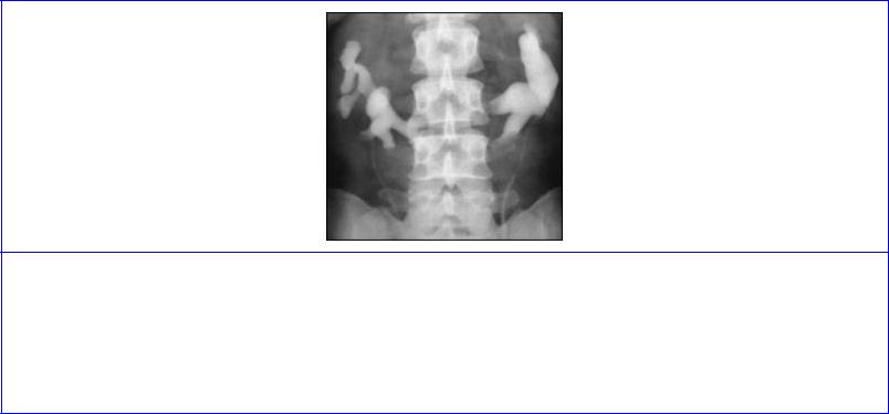
Anomalies. Persistence of fetal lobulation is of no significance. Persistence of one of the fetal arteries is common (30% of individuals), especially a vessel from the aorta to the lower pole. Whether such vessels should be called accessory, abnormal, aberrant, supernumerary or whatever is debated. Fusion of the lower poles of the kidneys gives rise to horseshoe kidney (1 in 800); the ureters pass anterior to the isthmus of kidney substance, as does the inferior mesenteric artery which limits ascent of the horseshoe (Fig. 5.51) . Polycystic disease (1 in 500) in which both kidneys are riddled with cysts is a hereditary disorder and may be associated with cysts in the liver, pancreas and lungs. One person in 500 has only one kidney (renal agenesis), a situation which must be excluded before considering nephrectomy.
Figure 5.51 Intravenous pyelogram showing a horseshoe kidney. The isthmus, which is not clearly seen, lies in front of the upper part of L4 vertebra, its further ascent being prevented by the origin of the inferior mesenteric artery from the front of the aorta. The calyces and pelvis on each side are generally rotated anteriorly causing the characteristic radiological appearance of calyces pointing medially.
Surgical approach
For many operations on the kidney including removal (nephrectomy) and removal of stones (nephrolithotomy) a lumbar approach is used (see p. 233). The renal fascia and perirenal fat are incised to expose the kidney, whose upper pole is freed leaving the suprarenal gland within its own compartment of the fascia. The overlying peritoneum is pushed away forwards and medially. The renal vessels can then be exposed, ligated and divided (the artery before the vein) to mobilize the organ further and transect the ureter. On the right a diseased kidney may adhere to the colon, duodenum, inferior vena cava or suprarenal gland, and on the left to the colon, spleen, pancreas or suprarenal. The right renal vein is only 2.5 cm long, so the inferior vena cava is very near the operation area. Operations on the kidney are also conducted through minimal access utilising laparoscopic techniques.

For percutaneous renal biopsy, the lower pole of the kidney is entered by an approach 2.5 cm below the twelfth rib and at a distance from the midline determined radiologically. Damage to a renal vessel or calyx is a potential hazard, and the needle is only advanced while the patient is holding the breath so that the kidney is not torn by respiratory movement.
For transplantation, the donor kidney is placed retroperitoneally in the iliac fossa with the hilum parallel to the external iliac vessels. The renal artery is anastomosed to the internal or external iliac artery and the renal vein to the external iliac vein. The ureter is implanted into the bladder.
Ureters
The ureter is 25 cm long. Its points of narrowest calibre are at the pelviureteric junction, where it crosses the pelvic brim, and as it passes through the bladder wall.
The ureter passes down on psoas major under cover of the peritoneum and crosses in front of the genitofemoral nerve, being itself crossed anteriorly by the gonadal vessels. On the right the upper part is behind the third part of the duodenum, while lower down it is crossed anteriorly by the right colic and ileocolic vessels and by the root of the mesentery. On the left it is lateral to the inferior mesenteric vessels and is crossed anteriorly by the left colic vessels and, at the pelvic brim, by the apex of the sigmoid mesocolon. It leaves the psoas muscle at the bifurcation of the common iliac artery, over the sacroiliac joint, and passes into the pelvis (see p. 298). It adheres to the peritoneum of the posterior abdominal wall when that membrane is stripped off the posterior abdominal wall. It can be distinguished from vessels and nerves in the living body in that it is a whitish, non-pulsatile cord which shows peristaltic activity when gently pinched with forceps.
Its surface markings are of use in palpating it for tenderness and in identifying radiographic shadows. On the anterior abdominal wall it can be marked from the tip of the ninth costal cartilage (see p. 234) to the bifurcation of the common iliac artery (see p. 276).
More important is the line of projection of the ureter on a radiograph. It lies medial to the tips of the transverse processes of the lumbar vertebrae (Fig. 5.24 and 5.48) and crosses the pelvic brim at the sacroiliac joint. From here its pelvic shadow passes to the ischial spine and thence, foreshortened, to the pubic tubercle.
Blood supply
The upper end is supplied by the ureteric branch of the renal artery and the lower end by branches from the inferior and superior vesical and uterine arteries. The middle stretch of the ureter is supplied by branches from the abdominal aorta, the gonadal, common iliac and internal iliac arteries. All these vessels make a fairly good anastomosis with each other in the adventitia of the ureter, forming longitudinal channels. The blood supply is endangered if the ureter is stripped clean of its surrounding tissue.
Lymph drainage
The lymphatics run back alongside the arteries; the abdominal portion of the ureter drains into paraaortic nodes, the pelvic portion into common iliac and internal iliac nodes.
Nerve supply

Although sympathetic fibres from T10–L1 segments of the cord reach the ureter via the coeliac and hypogastric plexuses, together with parasympathetic fibres from the pelvic splanchnic nerves, their functional significance is not clear. Intact innervation of the renal pelvis or ureter is not necessary for the initiation or propagation of peristalsis from the calyceal pacemakers. There are no ganglion cells in or on the ureter. Pain fibres accompany sympathetic nerves, as from the kidney (see p. 285).
Structure
The ureter is a tube of smooth muscle lined internally by mucous membrane. The muscle often appears histologically to be arranged as a middle circular layer with inner and outer longitudinal layers. However, it is more accurate to consider the muscle as a single coat with fibres running in many different directions because they are parts of intertwining helices. The lax mucous membrane is lined by transitional epithelium; there is no muscularis mucosae.
Development
The ureter is of mesodermal origin; it is derived by a process of budding from the caudal end of the mesonephric duct (see p. 286). Its upper end divides into two or three (the major calyces of the renal pelvis) and further subdivisions produce the minor calyces and collecting tubules. Low division of the ureteric bud produces double ureter.
Suprarenal glands
These glands lie anterosuperior to the upper part of each kidney (Fig. 5.43). They are somewhat asymmetrical, yellowish in colour, and lie within their own compartment of the renal fascia. Adrenal glands is an alternative name.
The right suprarenal gland is pyramidal in shape and surmounts the upper pole of the right kidney. It lies on the diaphragm and encroaches on to the front of the right kidney. The anterior surface is overlapped medially by the inferior vena cava. The rest of the anterior surface is in contact above with the bare area of the liver, and is covered below by the peritoneum of the posterior wall of the hepatorenal pouch.
The left suprarenal gland is crescentic in shape and drapes over the medial border of the left kidney above the hilum. It lies on the left crus of the diaphragm and overlaps the front of the left kidney. The upper part of the anterior surface is covered by the peritoneum of the posterior wall of the lesser sac, forming part of the stomach bed; the lower part is in contact with the body of the pancreas and the splenic vessels.
Blood supply
Both glands receive blood from three sources: directly from the aorta and from the renal and inferior phrenic arteries, the last providing two or three small branches. In contrast there is usually a single vein. The right vein is only a few millimetres long and enters the vena cava; the left vein is longer and enters the left renal vein.
Lymph drainage
To para-aortic nodes.
Nerve supply
The main supply is by myelinated preganglionic sympathetic fibres from the splanchnic nerves via the coeliac plexus; the fibres synapse directly with medullary cells (see p. 18). Blood vessels receive the usual postganglionic sympathetic supply. Cortical control is not neural but by ACTH from the anterior pituitary.
Structure
The suprarenal gland has an outer yellow cortex completely enclosing a much thinner grey medulla. The cortex, whose principal products are cortisol, aldosterone, androgens and related hormones, consists of three layers or zones. They are, from the surface inwards, the zona glomerulosa (with small rounded cells), the zona fasciculata (parallel rows of pale-staining vacuolated cells) and the zona reticularis (a network of smaller and darker-staining cells). The rather small central medulla has larger cells secreting the catecholamines adrenaline (epinephrine) (80%) and noradrenaline (norepinephrine) (20%) and some dopamine. Many of the medullary cells exhibit the chromaffin reaction: they contain fine cytoplasmic granules (the catecholamine precursors) which are coloured brown by chromium salts. Dilated capillaries are usually prominent in the medulla but not in the cortex.
Development
The medulla is derived by migration of cells from the neural crest and is ectodermal in origin while the cortex is derived in situ from the mesoderm of the intermediate cell mass (see p. 23).
Surgical approach
For bilateral adrenalectomy the glands are usually approached from the front. A bilateral subcostal ‘rooftop’ incision provides appropriate transperitoneal access. For exposure of the right gland, after retraction of the right lobe of the liver, a Kocher manoeuvre (see p. 269) is employed to mobilize the second part of the duodenum and head of the pancreas from the upper pole of the kidney and inferior vena cava. For the left gland, after division of the phrenicocolic ligament to mobilize the splenic flexure of the colon, the posterior layer of the splenorenal ligament is incised and the spleen turned medially with the tail of the pancreas. On each side the suprarenal vein is ligated before the numerous small arteries; the right vein is particularly short and the vena cava is easily torn. The glands must be handled as little as possible before venous ligation to prevent surges of hormone release. The anterior, transperitoneal approach is also used for laparoscopic bilateral adrenalectomy.
A posterolateral extraperitoneal approach through the bed of the twelfth rib provides access for unilateral adrenalectomy. The removal of a large adrenal tumour may require a higher approach, through the bed of the eleventh or tenth ribs, and division of the diaphragm.
Part twelve. Pelvic cavity
Bony pelvis
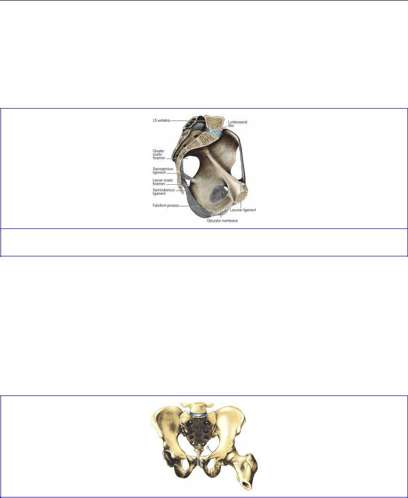
The individual features of the hip bone (see p. 164), sacrum (see p. 436) and coccyx (see p. 438) are considered separately. When articulated the bones enclose a cavity; from the brim of the cavity the ala of each ilium projects up to form the iliac fossa, part of the posterior abdominal wall. The pelvic brim is formed in continuity by the pubic crest, pectineal line of the pubis, arcuate line of the ilium, and the ala and promontory of the sacrum. The plane of the brim is oblique, lying at 60° with the horizontal (Fig. 5.53); the vagina lies in the same plane. From the brim the pelvic cavity projects back to the buttocks.
Figure 5.53 Ligaments of the left half of the pelvis.
The pelvic joints and ligaments are described on pages 323 onwards.
Sex differences are due to the two facts that the female pelvis is broader than that of the male for easier passage of the fetal head and that the female bones, including the head of the femur, are more slender than those of the male. As viewed from the front, in the male pelvis the sturdy bones make an acute subpubic angle (Fig. 5.52), pointed like a Gothic arch, while in the female the slender bones make a wide subpubic angle, rounded like a Roman arch. The outline of the pelvic brim differs. In the male the sacral promontory indents the outline, and the brim is widest towards the back (a ‘heartshaped’ outline) while in the female there is less indentation of the outline by the sacral promontory and the brim is widest further forwards (a ‘transversely oval’ outline).

Figure 5.52 Male pelvis, from the front. An imaginary horizontal plane through the top of the pubic symphysis traverses the tip of the coccyx, the ischial spine (S), the centre of the acetabulum and femoral head, and the tip of the greater trochanter.
Position of the pelvis
In the erect individual the anterior superior iliac spines and the upper margin of the symphysis pubic lie in the same vertical plane. The upper border of the symphysis pubis, the spine of the ischium, the tip of the coccyx, the head of the femur and the apex of the greater trochanter lie in the same horizontal plane (Fig. 5.52). This plane passes through the pelvic cavity at a level with the tip of the finger of the clinician during rectal or vaginal examination. The lower poles of the ovaries in the female and the seminal vesicles in the male lie in this plane.
Pelvic walls
The word pelvis is Latin for a basin and, when tilted forwards into the anatomical position, the bony pelvis does bear some resemblance to a pudding basin but with much of the front wall missing. The deficiency is made good by the lower part of the anterior abdominal wall where the aponeuroses of all three anterolateral muscles lie in front of rectus abdominis.
The pelvic brim divides the ‘false pelvis’ (above the brim, and part of the general abdominal cavity) from the ‘true pelvis’ or pelvic cavity (below the brim).
The muscles of the true pelvis are obturator internus and piriformis (which are also classified as lower limb muscles), and levator ani and coccygeus, (which, with their fellows of the opposite side, constitute the pelvic floor or pelvic diaphragm).
The side wall of the pelvis is formed by the hip bone, clad with obturator internus and its fascia. The curved posterior wall is formed by the sacrum with piriformis passing laterally into the greater sciatic foramen.
Piriformis
Piriformis arises from the front of the middle three pieces of its own half of the sacrum, the muscle taking origin from the lateral mass and extending medially between the anterior sacral foramina (see Fig. 6.97, p. 436); thus the emerging sacral nerves and sacral plexus lie on the muscle (Fig. 5.56). It runs transversely to the greater sciatic foramen. The pelvic surface of the muscle and the sacral plexus are covered by pelvic fascia attached to the sacral periosteum at the margin of the muscle. The course of the muscle in the gluteal region, its nerve supply and action are described on page 125.
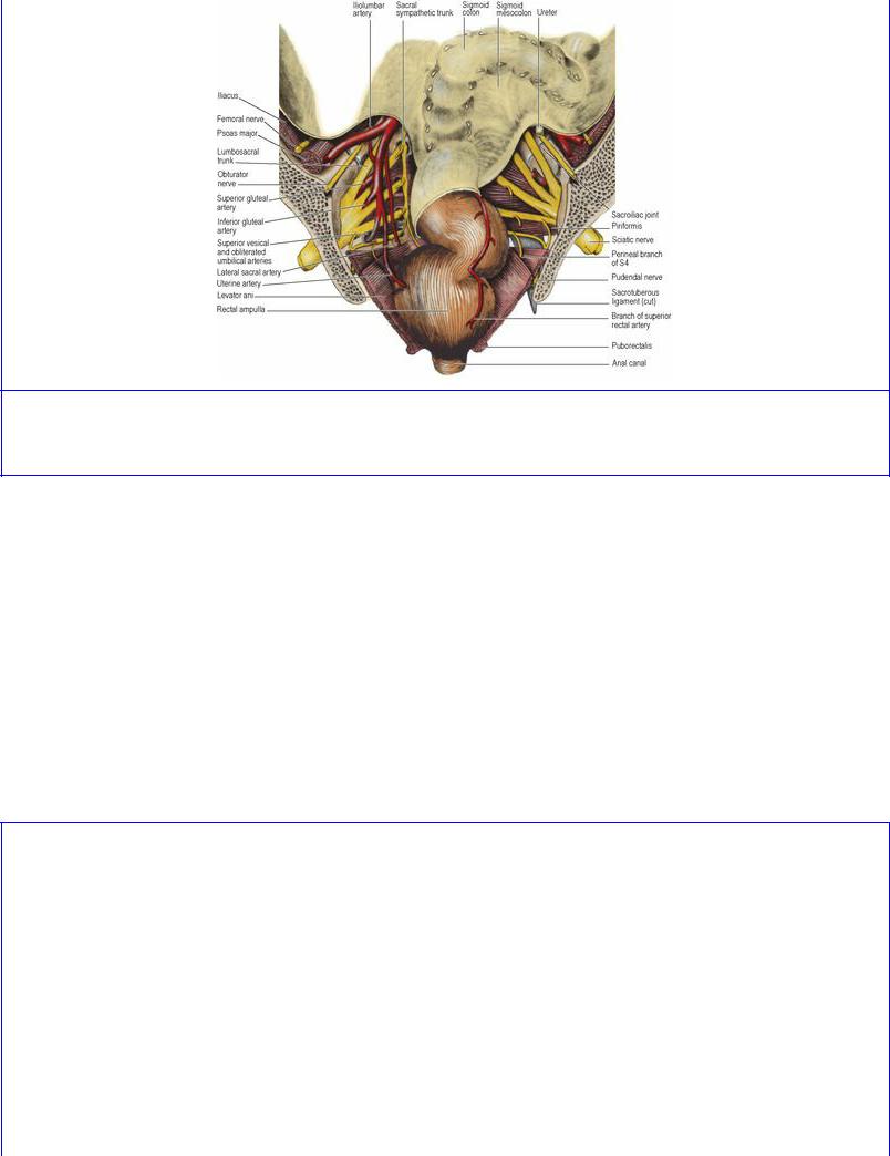
Figure 5.56 Posterior half of a coronal section of the pelvis. (The pelvic veins are not depicted. The rectum is shown in a distended state.)
Obturator internus
The large obturator foramen contains in life a felted mass of fibrous tissue, the obturator membrane (Fig. 5.53), with a gap above that converts the obturator notch into a canal for the obturator nerve and vessels. The muscle arises from the whole membrane and from the bony margins of the foramen. The origin extends posteriorly as high as the pelvic brim and across the flat surface of the ischium to the margin of the greater sciatic notch (see Fig. 3.49, p. 167). On the ischial tuberosity the origin extends down to the falciform ridge. From this wide origin the muscle fibres converge fan-wise towards the lesser sciatic notch (Fig. 5.65). Tendinous fibres develop on the muscle surface where it bears on the lesser sciatic notch and the bone often shows low ridges and grooves where the tendon takes a rightangled turn to pass into the buttock. The bone here is lined by hyaline cartilage and is separated from the tendon by a bursa. The muscle is described further on pages 125–126.
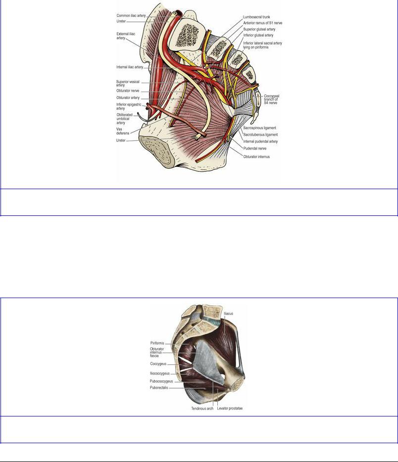
Figure 5.65 Vessels and nerves of the right half of the pelvis, in a median sagittal section.
The muscle is covered with a strong membrane, the obturator fascia (Fig. 5.54). This is attached to bone at the margins of the muscle and fuses below with the falciform process of the sacrotuberous ligament on the ischial tuberosity (Fig. 5.53). The tendinous arch of origin of levator ani slopes across the obturator internus fascia (the pelvic cavity is above this line, the ischioanal fossa below it).
Figure 5.54 Muscles of the left half of the pelvis.
Pelvic floor
The pelvic floor consists of a gutter-shaped sheet of muscle, the pelvic diaphragm, slung around the midline viscera (urethra and anal canal and, in the female, the vagina).
The muscles of the pelvic floor are the levator ani and the coccygeus. They arise in continuity from
the body of the pubis, from the tendinous arch over the obturator fascia, and from the spine of the ischium, and are inserted into the coccyx and the postanal plate (see below). From their origin the muscle fibres slope downwards and backwards to the midline; the pelvic floor so produced is a gutter that slopes downwards and faces forwards.
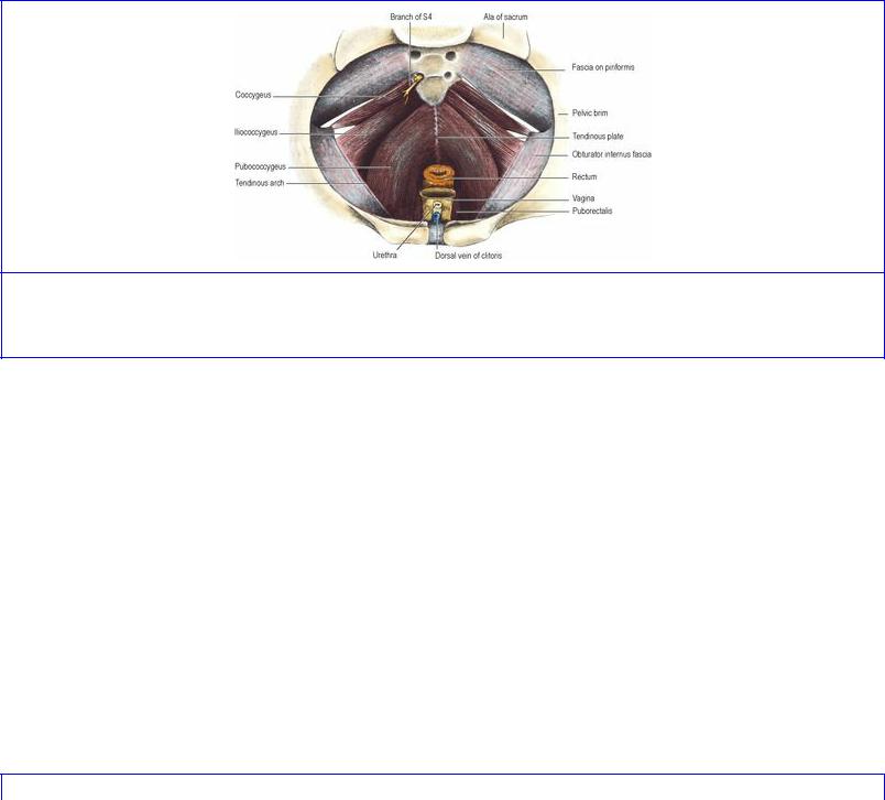
Levator ani
Levator ani consists of two main parts, pubococcygeus and iliococcygeus (Fig. 5.55). Their fibres arise in continuity from the body of the pubis to the ischial spine across the obturator fascia, along a condensation of the fascia, the tendinous arch (Fig. 5.54). The levator ani originally arose from the pelvic brim (its present origin in most mammals) and in man has migrated down the side wall of the pelvis, bringing the tendinous arch with it. Residual aponeurotic fibres of levator ani contribute to the strength of the obturator fascia above the tendinous arch.
Figure 5.55 Female pelvic floor from above. The pubococcygeus part of levator ani lies internal to the iliococcygeus part.
The pubococcygeus part is that part of levator ani which arises from the anterior half of the tendinous arch and from the posterior surface of the body of the pubis. The pubococcygeus fibres are in different functional sets. The bulk of its posterior fibres sweep backwards in a flat sheet on the pelvic surface of the iliococcygeus and form a tendinous plate in the midline, attached posteriorly to the front of the coccyx (Fig. 5.55). These constitute the pubococcygeus muscle proper. Fibres arising more anteriorly, from the body of the pubis, swing more medially and more inferiorly around the anorectal junction and join with fibres of the opposite side and the top of the external anal sphincter. This part of the muscle is called puborectalis and forms a U-shaped sling which holds the anorectal junction angled forwards (Fig. 5.68). Some of the fibres blend with the longitudinal muscle of the rectum and the conjoint longitudinal coat of the anal canal (see p. 315) and are termed puboanalis. The most medial fibres of pubococcygeus pass backwards alongside the prostate and the sphincter urethrae in the male and decussate across the midline behind the urethra; they are referred to as puboprostaticus or pubourethralis. In the female, these fibres sling around the posterior wall of the vagina and are referred to as pubovaginalis. In both sexes, fibres also attach to the perineal body.
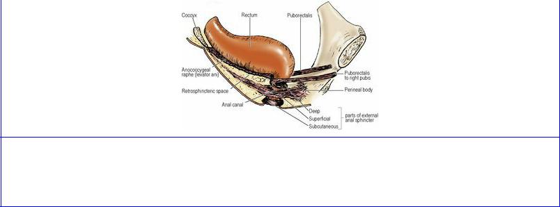
Figure 5.68 Puborectalis and the external anal sphincter from the right. The three traditional parts of the sphincter are shown as though separate, but they merge with one another and the deep part is continuous with the puborectalis part of levator ani.
The iliococcygeus part arises from the posterior half of the tendinous arch and the pelvic surface of the ischial spine and, overlapping the pelvic surface of coccygeus, its fibres are inserted into the side of the coccyx and the anococcygeal raphe (Fig. 5.68), which extends from the tip of the coccyx to the junction of rectum and anal canal. Although the iliococcygeus does not arise from the ilium, its name derives from its former origin on the iliac bone at the pelvic brim.
The postanal plate, also referred to as the anococcygeal ligament, is a layered musculotendinous structure between the anal canal and the caudal part of the vertebral column, on which the terminal rectum sits. From above downwards it consists of the superior fascia of the pelvic diaphragm (see below), the tendinous plate of pubococcygeus, the muscular raphe of iliococcygeus, and the posterior parts of puborectalis and the external anal sphincter (see p. 313).
Coccygeus
The coccygeus is best thought of as ischiococcygeus. It arises from the tip of the ischial spine and its fibres fan out to be inserted into the side of the coccyx and the lowest piece of the sacrum; it lies edge to edge with the lower border of piriformis and is overlapped anteriorly by iliococcygeus (Fig. 5.55). Its gluteal surface is fibrous tissue, and is indeed the sacrospinous ligament (Figs 5.53 and 5.54).
Nerve supply. Levator ani is mainly supplied from the sacral plexus by branches of S3 and S4 which enter the upper (pelvic) surface of the muscle. Some of these somatic fibres may travel in or very close to the pelvic splanchnic nerves. Puborectalis, pubourethralis and pubovaginalis are supplied from below by the perineal branch of S4 and the inferior rectal branch of the pudendal nerve, in common with the external anal sphincter. Levator ani, like the external anal and urethral sphincter muscles, has a high proportion of slow twitch fibres. Coccygeus is supplied by branches of S3 and S4.
Actions. The pelvic floor helps to support the pelvic viscera and retain them in their normal positions. The floor contracts to counteract increased intra-abdominal pressure, which may be momentary, as in coughing and sneezing, or more prolonged as in muscular efforts like lifting. If an expulsive effort is required, the floor relaxes. Thus in defecation (see p. 316) when the abdominal wall and diaphragm contract, puborectalis relaxes to straighten out the anorectal junction and the floor descends to become more funnel-shaped, rising again as the process comes to an end. The
pubovaginalis fibres of levator ani may be important in assisting the urethral sphincter at the end of micturition in the female. In parturition the floor initially directs the fetal head to the pelvic outlet, but the degree of stretching to which the muscular and fibrous parts of the floor are subjected may render it liable to damage by tearing.
Pelvic fascia
The parietal pelvic fascia on the pelvic surface of obturator internus is a strong membrane that fuses with the periosteum at the upper margin of the muscle. Below the tendinous arch that gives origin to levator ani, the fascia is thin where it covers obturator internus on the lateral wall of the ischioanal fossa (see p. 316). The fascia on the pelvic surface of piriformis fuses with the periosteum at the medial margins of the anterior sacral foramina. The sacral anterior primary rami emerging from these foramina thus lie behind this fascia. The internal iliac vessels are, however, in front of the fascia over piriformis; but the large (presacral) lateral sacral veins lie initially behind this fascia as they emerge from the anterior sacral foramina. From the front of the lower sacrum a condensation of connective tissue, the rectosacral fascia, which varies in its thickness, passes downwards and forwards to fuse with the mesorectal fascia (see below) 3–5 cm proximal to the anorectal junction. The large (presacral) lateral sacral veins lie behind this fascia on the front of the sacrum. The fascia on the pelvic surface of levator ani and coccygeus is the superior fascia of the pelvic diaphragm. It is attached in front to the posterior surface of the body of the pubis and at the back to the ischial spine. In between these attachments it blends with the obturator fascia and a thickening of these two fused fasciae forms the tendinous arch of origin of levator ani. The inferior fascia of the pelvic diaphragm is the thin fascia that covers the undersurface of levator ani on the sloping medial wall of the ischioanal fossa; it blends with the obturator fascia laterally and with the fascia on the external anal and urethral sphincters medially.

Part thirteen. Rectum
The Latin word ‘rectus’ means straight, and the rectum is straight in monkeys, but the human rectum follows the posterior concavity of the sacrum, and also shows three lateral curves or flexures that are most prominent when the viscus is distended: upper and lower curves convex to the right and a middle curve convex to the left, the result being that the middle part appears to bulge to the left (Fig. 5.56). The lowest part is slightly dilated as the rectal ampulla. Corresponding to the three curves seen externally, there are three sickle-shaped transverse rectal folds, formerly called rectal valves (of Houston), that project into the lumen from the wall on the concave side of these folds. They incorporate the circular muscle of the wall and are not confined merely to the mucous membrane, as is the case with the circular folds of the duodenum and jejunum. The middle fold, the largest, projects into the lumen from the right wall of the rectum just above the ampulla, at the level at which the peritoneum is reflected forwards off the rectum to form the floor of the rectovesical or rectouterine pouch (see below); it is about 8 cm from the anal orifice and is a useful visual landmark during sigmoidoscopy.
The rectum, which is about 12 cm long, is continuous with the sigmoid colon at the level of the third piece of the sacrum. The transition between the rectum and the sigmoid colon is a gradual one. At this junctional region the sigmoid mesocolon ends and the rectum has no mesentery. The taeniae of the sigmoid colon gradually broaden to form wide anterior and posterior muscular bands, which meet laterally to give the rectum a complete outer layer of longitudinal muscle; so the rectum has no sacculations. There are also no appendices epiploicae in the rectum.
The rectum turns downwards and backwards as the anal canal 2–3 cm in front of the tip of the coccyx. The anorectal junction is slung forwards by the U-loop of the puborectalis, which merges with the top of the external sphincter of the anal canal, forming a palpable ledge (the anorectal ring) on rectal examination. Hence the posterior wall of the rectum appears to make a right-angled bend at the anorectal junction. This angle widens as the puborectalis muscle sling relaxes during defecation to allow faeces to enter the anal canal.
Although the rectum has no mesentery, the connective tissue and fat around the rectum is referred to by surgeons as the mesorectum. The visceral fascia surrounding it is the mesorectal fascia (Fig. 5.57). The mesorectum is bulkier posteriorly, where it tends to be grooved in the midline. It contains the superior rectal artery and its branches, the superior rectal vein and its tributaries, lymphatic vessels and nodes. A relatively avascular areolar tissue plane lies between the mesorectal fascia and the parietal pelvic fascia; this is the plane of surgical dissection in total mesorectal excision of the rectum for carcinoma. The plane is most evident posteriorly and is minimal laterally where the inferior hypogastric plexus (see p. 311) lies tangentially on the surface of the mesorectal fascia. Crossing this interface are autonomic nerve fibres from the plexus to the rectum and occasional small middle rectal vessels. Surgical definition of surrounding connective tissue from the mesorectum comprises the iatrogenic ‘lateral ligament’ of the rectum; this is not seen on MRI or CT scanning.
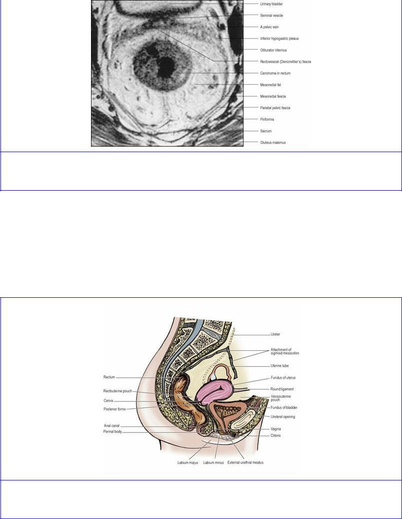
Figure 5.57 Oblique axial MRI of the rectum. There is an annular carcinoma in the rectum. (Provided by Dr G. Brown, The Royal Marsden Hospital, Sutton, Surrey.)
Peritoneum covers the upper third of the rectum at the front and sides, and the middle third only at the front; the lower third is below the level of the peritoneum which is reflected forwards on to the upper part of the bladder (in the male) or upper vagina to form the rectovesical pouch or rectouterine pouch (of Douglas) (Fig. 5.64). These pouches form the lowest parts of the peritoneal cavity, and being 7.5 and 5.5 cm from the anal margins in the male and female respectively are within reach of the fingertip on rectal examination. They are normally occupied by coils of small intestine or sigmoid colon.
Figure 5.64 Left half of the female pelvis with the attachment of the sigmoid mesocolon and the ureter entering the pelvis beneath its apex.
In front of the rectovesical pouch is the uppermost part of the base of the bladder and the tops of the seminal vesicles. Below the level of the pouch are the rest of the bladder base and seminal vesicles, the prostate, and the ends of each ureter and vas deferens. Between these structures and the rectum, a condensation of fascia forms a rectogenital septum—the rectovesical fascia of Denonvilliers (Fig. 5.58). It is connected to the floor of the rectovesical pouch above and to the apex of the prostate below. In the fetus the rectovesical pouch extends to a lower level than in the adult. Fusion of the anterior and posterior walls of the pouch as it becomes more shallow may account for the origin of this septum. The rectovesical fascia of Denonvilliers has a distinct whitish appearance in the living, is closer to the rectum than to the seminal vesicles and prostate and is usually removed in rectal excision for carcinoma (see p. 295).
Figure 5.58 Right half of a sagittal section of a male pelvis. The prostate is enlarged.
In front of the rectouterine pouch is the uppermost part of the vagina (the fornix, with the cervix of the uterus projecting into it), while below the peritoneal reflexion is more of the vagina, with the rectogenital septum intervening. This thin rectovaginal fascia fuses with the perineal body below.
Some slips from the longitudinal muscle of the rectal ampulla, the rectourethralis muscle, pass forwards to the perineal body and sphincter urethrae in the male; they must be cut at operations for excising the rectum and anal canal.
Blood supply
This is derived principally from the superior rectal artery, with contributions from the middle and inferior rectal and median sacral vessels. The lower end of the inferior mesenteric artery enters the sigmoid mesocolon and changes its name to superior rectal on crossing the pelvic brim. It crosses the left common iliac vessels medial to the ureter and descends in the base of the medial limb of the mesocolon. At the level of S3 vertebra (where the rectum begins) it divides into two branches which descend on each side of the rectum and subdivide into smaller branches. These vessels pierce the muscular wall and supply the whole thickness of the rectal wall including the mucous membrane. They continue submucosally into the anal canal, where they anastomose with branches of the inferior rectal artery. The middle rectal arteries are present in only one in five people; they are small and supply only the muscle of the mid and lower rectum. Experience in rectal surgery has shown that the inferior rectal arteries are capable of supplying the rectum from below to a level at least as high as the peritoneal reflexion from its anterior surface. The median sacral artery may make an unimportant contribution to the posterior wall in the region of the anorectal junction, but its main interest is that it
may cause bleeding at operations in this region.
Veins correspond to the arteries, but anastomose freely with one another, forming an internal rectal plexus in the submucosa and an external rectal plexus outside the muscular wall. The lower end of the internal plexus is continuous with the vascular cushions of the anal canal (see p. 315). The main route of rectal venous drainage is via the superior rectal vein to the inferior mesenteric vein, which crosses the pelvic brim between the inferior mesenteric artery and the ureter. The inferior rectal veins drain to the internal pudendal veins.
Lymph drainage
Lymphatic drainage from the rectum is mainly upwards. Lymphoid follicles in the mucous membrane drain to epicolic nodes on the surface of the rectum and to pararectal nodes in the mesorectum. The upward drainage is via nodes along the inferior mesenteric artery to preaortic nodes. Lymphatic drainage from the lower rectum to internal iliac nodes along middle rectal and inferior rectal arteries, and along the median sacral artery to nodes in the hollow of the sacrum, is minimal and unlikely to be a route for the metastatic spread of cancer that has not breached the mesorectal fascia.
Nerve supply
The sympathetic supply is by fibres that accompany the inferior mesenteric and superior rectal arteries from the inferior meseneteric plexus. The parasympathetic supply is from S2, 3 and 4 by the pelvic splanchnic nerves via the inferior hypogastric plexus; they are motor to rectal muscle. As from the bladder (see p. 297) pain fibres appear to accompany both sympathetic and parasympathetic supplies. The sensation of distension is conveyed by parasympathetic afferents.
Rectal examination
The structures that can be palpated through the anal canal in either sex include the coccyx and sacrum behind, with the ischial spines at the sides. The anorectal ring (see p. 313) can be felt posteriorly at the anorectal junction as a shelf-like projection over which the tip of the finger can be hooked when the patient bears down. In the male at the front the prostate can be felt (but normal seminal vesicles are not usually palpable). In the female the cervix is felt through the vaginal wall, with the uterosacral ligaments laterally and sometimes the ovaries (compare with vaginal examination, p. 307).
Development
The rectum and the anal canal are derived from the anorectal canal (the dorsal part of the cloaca) and the proctodeum (see p. 29). The anal membrane breaks down, at a site probably represented by the pectinate line in the anal canal (see p. 315); the anal valves are said to indicate the remains of the membrane. The part of the anal canal continuous with the rectum above the pectinate line is endodermal, and the part below which is derived from the proctodeum is ectodermal, hence the difference in the blood and nerve supplies and lymph drainage of the upper and lower parts of the canal (see p. 315).
Surgical approach
In the surgical management of carcinoma of the rectum, depending on the site of the tumour, with access through the anterior abdominal wall most or all of the rectum and its surrounding mesorectum is removed, usually with the sigmoid colon and mesocolon (Fig. 5.59), leaving the lower part and/or
anal canal to which the mobilized descending colon is anastomosed. The procedure is termed anterior resection or total mesorectal excision (TME) of the rectum. The rectovesical (rectovaginal) fascia is usually removed with the rectum after division of its attachment to the prostate (perineal body). The rectosacral fascia is divided. The ureter and the main neurovascular structures on the lateral wall of the pelvis are preserved. The superior hypogastric plexus and both inferior hypogastric plexuses are kept intact to safeguard sexual function and urinary voiding. The inferior mesenteric artery is divided close to its aortic origin to ensure removal of all lymph nodes along its courses, preserving the inferior mesenteric plexus on the aortic surface.
Figure 5.59 Specimen resulting from total mesorectal excision of the rectum in a male. The rectum has been removed with the surrounding mesorectum and the rectovesical fascia of Denonvilliers. The lower part of the excised sigmoid colon is seen at the top of the rectum. (Provided by Prof P Quirke, University of Leeds.)
For complete excision of the rectum and anal canal, the freeing of the rectum as above is supplemented by a perineal approach (abdominoperineal resection), which includes dividing the pelvic floor (levator ani). Elliptical incisions either side of the anus allow the ischioanal fossae to be entered. The coccyx is dislocated or excised and the rectosacral fascia is divided from below. Anteriorly, the dissection extends up to the transverse perineal muscles and perineal body (see p. 317). The rectourethralis muscle and rectogenital septum are divided and the plane between the septum and the prostate or vagina entered from below. A cylinder of tissue, including the freed bowel and a collar of levator ani and ischioanal fat, is removed through the perineum. The pelvic floor may be repaired by transferring a flap of muscle such as from gluteus maximus or rectus abdominis. A terminal colostomy is usually made in the left iliac fossa.

Part fourteen. Urinary bladder and ureters in the pelvis
Urinary bladder
The empty bladder is situated entirely within the pelvic cavity. As the bladder distends it domes up into the abdominal cavity. The empty bladder is a flattened three-sided pyramid, with the sharp apex pointing forwards to the top of the pubic symphysis and a triangular base facing backwards in front of the rectum or vagina. There are two inferolateral surfaces cradled by the anterior parts of levator ani, a neck where the urethra opens, and a superior surface on which the small intestine and sigmoid colon or uterus lie.
The apex has the remains of the urachus attached to it, the latter forming the median umbilical ligament which runs up the midline of the anterior abdominal wall in the median umbilical fold of peritoneum (see p. 234).
Most of the base, or posterior surface, lies below the level of the rectovesical pouch and only the uppermost portion is covered by peritoneum between the vas deferens on each side (Fig. 5.62). In addition to the latter the seminal vesicles are applied to this surface, and the ureters enter at the upper outer corner. In the female the base has a firm connective tissue union with the anterior vaginal wall and upper part of the uterine cervix with no peritoneum intervening (Fig. 5.64).
Each inferolateral surface slopes downwards and medially to meet its fellow, lying against the front part of the pelvic diaphragm and obturator internus. Where the surfaces meet below the apex there is a (retroperitoneal) space behind the pubic bones and symphysis, the retropubic space (of Retzius), containing loose fatty tissue and the fibromuscular pubovesical ligaments that extend from the bladder neck to the inferior aspect of the pubic bones.
The lowest part of the bladder is its neck, where the base and inferolateral surfaces meet and which is pierced by the urethra at the internal urethral orifice. In the male it lies against the upper surface or base of the prostate. In the female the neck is above the urethra in the connective tissue of the anterior vaginal wall.
The superior surface is covered by peritoneum which sweeps upwards on to the anterior abdominal wall. The distending bladder strips peritoneum from behind rectus abdominis, leaving the transversalis fascia on the back of the muscle; the distended bladder may thus be approached by cannula or scalpel in the midline above the pubic symphysis without entering the peritoneal cavity. At the posterior margin of this surface in the male the peritoneum continues on to the uppermost part of the base and is then continued backwards as the floor of the the rectovesical pouch, but in the female it is reflected from a little in front of the posterior margin of this surface on to the undersurface of the uterus.
The appearance of the interior of the bladder depends upon the state of distension of the organ. When collapsed the mucous membrane is thick and thrown into folds, when distended it is thin and smooth. The trabeculae of the muscle fibres can be seen through the mucous membrane. These remarks do not apply to the trigone, which varies but little with the state of distension of the organ.
The trigone is a triangular area at the base of the bladder lying between the two ureteral orifices (above and laterally) and the internal uretheral orifice (centrally and below) (Fig. 5.60). In the
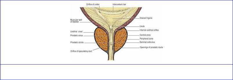
empty bladder these three openings are 2.5 cm apart from each other but when distended (as during cystoscopy) the ureteral orifices may be 5 cm apart. Being fixed on top of the prostate by the urethra, the trigone is the least mobile part of the bladder. In the female it is stabilized by the connective tissue surrounding the upper urethra at the front of the vagina. The trigone is smooth-walled and the mucous membrane is rather firmly adherent to the underlying muscle. The ureteric orifices are connected by a transverse ridge, the interureteric bar, prominent when viewed through the cystoscope; this is produced by continuity of the longitudinal muscle of the two ureters across the bladder wall. The orifices of the ureters lie at the ends of the bar; they are usually in the shape of an oblique slit, but considerable variations exist. The ureters pierce the muscle and mucosal walls very obliquely, an important factor in preventing reflux of urine when intravesical pressure rises. The ureteric orifices are closed by this pressure, except to open rhythmically in response to ureteric peristalsis each time a jet of urine is injected into the bladder (four or five times a minute normally).
Figure 5.60 Trigone of bladder, prostate and prostatic urethra: coronal section.
In the male the trigone overlies the median part of the central zone of the prostate which, after middle age, may project above the internal urethral orifice as a rounded elevation, the uvula of the bladder.
Blood supply
The superior and inferior vesical arteries provide most of the arterial blood but there are small contributions to the lower part of the bladder from the obturator, inferior gluteal, uterine and vaginal arteries.
The veins of the bladder do not follow the arteries. They form a plexus that converges on the vesicoprostatic plexus in the groove between bladder and prostate and which drains backwards across the pelvic floor to the internal iliac veins. There is a similar plexus in the female, communicating with veins in the base of the broad ligament.
Lymph drainage
The lymphatics of the bladder drain mainly to external iliac nodes. Some lymph drains to internal iliac nodes including nodes in the obturator fossa.
Nerve supply
Parasympathetic fibres which provide the main motor innervation of the bladder reach it via the
pelvic splanchnic nerves (see pp. 20 and 311). Sympathetic fibres come from L1 and 2 segments of the cord via the superior and inferior hypogastric plexuses. For most of the bladder the sympathetic fibres are vasomotor and probably inhibitory to the detrusor muscle, but they are motor to the superficial trigonal muscle and (in the male) the muscle of the bladder neck (see below). The sensation of normal bladder distension travels with parasympathetic fibres and in the spinal cord is conveyed in the gracile tract, but it appears that bladder pain (e.g. from a stone) reaches the spinal cord (lateral spinothalamic tract) by both parasympathetic and sympathetic pathways.
Control of micturition
Normal emptying of the bladder occurs by contraction of the detrusor muscle and reciprocal relaxation of the external sphincter and pelvic floor (levator ani). The accumulation of urine distends the bladder wall with adjustment of tone (accommodation) so that tension does not at first increase. Later increased tension stimulates stretch receptors from which afferent impulses pass along the pelvic splanchnic nerves to sacral segments of the cord. Here the parasympathetic cell bodies are in turn stimulated and efferent impulses travel down the pelvic splanchnic nerves to synapse with the postganglionic cells within the bladder wall and so cause contraction. This autonomic stretch reflex giving bladder control at the spinal level is typical of the infant; with training, control by higher centres becomes superimposed on the spinal activity, and bladder evacuation is assisted by voluntary contraction of abdominal muscles. There is a cortical inhibitory centre in the inferior frontal gyrus (on the medial surface of the cerebral hemisphere, some distance in front of the motor ‘perineal’ area) with fibres passing to a detrusor motor centre in the medial part of the pontine reticular formation. From there reticulospinal fibres run down the cord mixed with those of the lateral corticospinal tract to the sacral segments.
The skeletal muscle of the sphincter urethrae (external urethral sphincter; p. 307 female, p. 317 male) is controlled by the perineal branch of the pudendal nerve (see p. 321), carrying fibres predominantly from anterior horn cells of S2 segment (Onuf's nucleus). A storage centre in the lateral part of the pontine reticular formation exerts central control on this nucleus. During micturition the sphincter relaxes as the detrusor contracts. In the female the pubovaginalis part of levator ani (see p. 291) assists the external sphincter at the end of micturition.
In spinal cord transection above the level of S2 segment, afferent impulses indicating distension cannot reach consciousness, cortical control of the sacral reflex is lost, and relaxation of the sphincter urethrae cannot be prevented. Because the ‘sacral centre’ itself is intact, the bladder automatically empties when distended, as in the infant (or as in the senile where cortical control has been lost through cerebral vascular disease). If the sacral segments themselves are destroyed, the detrusor muscle is paralysed and the bladder becomes abnormally distended until overflow incontinence occurs.
Structure
The smooth muscle of the bladder wall (detrusor muscle) is composed of an interlacing network of fibres running in various directions. Both externally and internally (beneath the mucous membrane) they produce a trabeculated appearance, which is exaggerated when muscular hypertrophy occurs as a result of progressive chronic obstruction to micturition, for example by prostatic enlargement or urethral stricture. They are well supplied by parasympathetic (cholinergic) nerve fibres. However,

the trigone possesses a superficial triangular layer of muscle (superficial trigonal muscle) that is histologically and histochemically different from the rest of the bladder musculature (including the deep part of the trigone) and extends into the proximal urethra in both sexes. In further contrast to the detrusor muscle, the superficial trigonal muscle receives predominantly sympathetic (adrenergic) fibres. Contraction of this muscle may help to close the ureteral orifices.
At the bladder neck in the male, circular smooth muscle fibres form a collar around the internal urethral orifice, and extend distally to surround the proximal part of the prostatic urethra (the preprostatic part). This muscle, the preprostatic sphincter, too is profusely supplied with sympathetic (adrenergic) fibres. In the female the muscle in this region is arranged longitudinally and extends into the urethral wall. The muscle of the bladder neck has nothing to do with urinary continence; in the male it acts to prevent seminal regurgitation into the bladder during ejaculation.
The mucous membrane is thick and lax and lined by transitional epithelium. Glands are usually absent; mucus in shed urine has come from urethral glands. There is no muscularis mucosae.
Development
The endoderm of the vesicourethral part of the urogenital sinus (see p. 29) becomes the bladder epithelium, and the surrounding mesenchyme forms the muscle and connective tissue. Continued growth leads to the incorporation of the lower ends of the mesodermal mesonephric ducts into the posterior part of the bladder, so forming the trigone. These developmental differences may account for the structural differences in this region. The mesonephric ducts in the male end up at a lower level as the ejaculatory ducts entering the urethra (see p. 300). The allantois regresses to form a fibrous cord, the urachus (median umbilical ligament).
Ureters in the pelvis
The pelvic part of the ureter forms about half of its 25 cm length (see p. 286). It crosses the pelvic brim in the region of the bifurcation of the common iliac artery. On the left it underlies the apex of the sigmoid mesocolon (Figs 5.56 and 5.64). It usually runs over the external iliac artery and vein and then down the side wall of the pelvis in front of the internal iliac artery (and behind the ovary). In order from above downwards, it crosses the obturator nerve, obliterated umbilical (superior vesical) artery, obturator artery and obturator vein (Fig. 5.65). On the right the appendix, if in a pelvic position, may lie adjacent. Reaching the level of the ischial spine, it turns forwards and medially above the pelvic floor to enter the base of the bladder at its upper lateral angle. Here in the male the vas deferens crosses above the ureter and then runs down medial to the ureter. The upper end of the seminal vesicle usually lies just below the point where the ureter enters the bladder wall.
On the pelvic floor in the female, the ureter lies in the base of the broad ligament (see p. 304), where it is crossed above by the uterine artery (Fig. 5.63). Under the broad ligament the ureter penetrates the condensed tissue that forms the lateral cervical ligament (see p. 304), crossing the lateral vaginal fornix 1–2 cm from the cervix before entering the bladder in front of the fornix. The ureters are major hazards during hysterectomy, when ligating vessels and transecting ligaments.
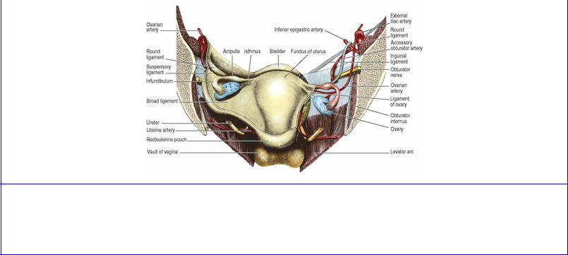
Figure 5.63 Anterior half of a coronal section of the female pelvis, from behind. The broad ligament and parietal peritoneum have been removed on the right side. The ovaries are displaced from their normal position in the parous female.
In both sexes the ureters run obliquely through the bladder wall for 1–2 cm before reaching their orifices at the upper lateral angles of the trigone.

Part fifteen. Male internal genital organs
Prostate
The prostate is a partly glandular, partly fibromuscular organ which lies beneath the bladder and above the urogenital diaphragm, and is penetrated by the proximal part of the urethra. It is normally broader than it is long, approximately 4 × 3 × 2 cm. Its female homologue is the small group of paraurethral glands (of Skene; see p. 307). The prostate provides about 30% of the volume of seminal fluid (most comes from the seminal vesicle).
The prostate has a base and an apex, and anterior, posterior and inferolateral surfaces. The base is the upper surface, fused with the neck of the bladder and perforated by the urethra which traverses the whole length of the gland (Fig. 5.60). The blunt apex is the lowest part, and the prostatic urethra emerges from the front of the apex to become the membranous urethra which is surrounded by the sphincter urethrae (see p. 317). The anterior surface is at the back of the retropubic space and is connected to the bodies of the pubic bones by the puboprostatic ligaments. The inferolateral surfaces are clasped by the pubourethralis parts of levator ani. The posterior surface is in front of the lower rectum but separated from it by the rectovesical fascia (see p. 294). The ejaculatory ducts pierce the posterior surface just below the bladder and pass obliquely through the gland for about 2 cm to open into the prostatic urethra about halfway down. The prostate's own ducts also open into this part of the urethra (see below).
A thin strong layer of connective tissue at the periphery of the gland forms the ‘true capsule’ of the prostate, and outside this there is a condensation of pelvic fascia forming the ‘false capsule’. Between these two capsules lies the prostatic plexus of veins. The gland consists of acini of varying shapes and sizes embedded in a fibromuscular stroma—a mixture of connective tissue and smooth muscle; this is the characteristic histological feature.
The prostatic urethra, 3–4 cm in length, passes through the substance of the prostate closer to the anterior than the posterior surface of the gland. It runs downwards and backwards from the internal meatus, then bends at the middle of its length and continues downwards and forwards to emerge from the anterior aspect of the apex. A midline ridge, the urethral crest , projects into the lumen from the posterior wall throughout most of the length of the prostatic urethra (Fig. 5.60). The shallow depression on either side of the crest is termed the prostatic sinus. At about the midlength of the crest the seminal colliculus, or verumontanum, forms a midline rounded eminence. The prostatic utricle, a small recess representing the fused ends of the paramesonephric (Müllerian) ducts, opens on to the middle of the verumontanum and the ejaculatory ducts open on either side of the utricle. The proximal part of the prostatic urethra, also termed the preprostatic part, is surrounded by a cylinder of smooth muscle, an extension of the circular muscle at the bladder neck; as has been noted above, this muscle contracts to prevent seminal regurgitation into the bladder during ejaculation.
The prostate is now considered to consist of a peripheral zone, a central zone and a transition zone, accounting for approximately 70%, 20% and 5% of the glandular substance, respectively, rather than being made up of lobes as previously described. The central zone is wedge-shaped and forms the base of the gland with its apex at the verumontanum (Fig. 5.60); it surrounds the ejaculatory ducts as they course through the gland. The peripheral zone surrounds the central zone from behind and below, but does not reach up to the base; it extends downwards to form the lower part of the gland.
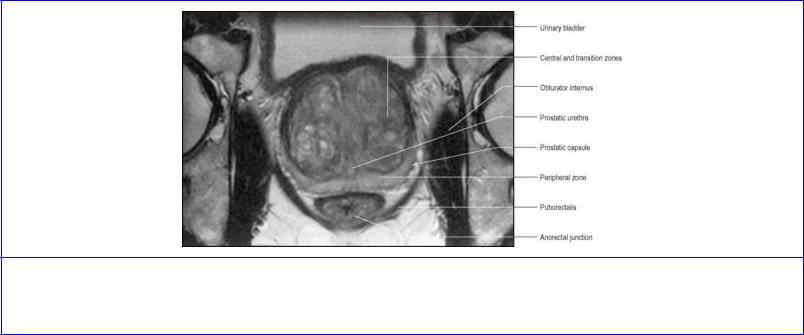
The transition zone lies around the distal part of the preprostatic urethra, just proximal to the apex of the central zone. The ducts of the transition zone open on the verumontanum, just above where the ducts of the peripheral zone open into the prostatic sinuses. Benign prostatic hyperplasia affects the transition zone which may increase markedly in size, compressing the peripheral zone (Fig. 5.61). The peripheral zone is almost exclusively the site of origin for carcinoma of the prostate. The central zone is rarely involved in any disease process.
Figure 5.61 Oblique axial MRI of the prostate. The transition zone is markedly enlarged by benign prostatic hyperplasia. (Provided by Dr G. Brown, The Royal Marsden Hospital, Sutton, Surrey.)
There is very little glandular tissue anterior to the prostatic urethra, the anterior part of the prostate being mainly fibromuscular; it is overlapped from above by the detrusor muscle of the bladder and from below by the striated muscle of the urethral sphincter.
Blood supply
The main arterial supply is from the prostatic branch of the inferior vesical artery, with some small branches from the middle rectal and internal pudendal vessels. The veins run into a plexus between the true and false capsules and this joins the vesicoprostatic plexus situated at the groove between bladder and prostate. This plexus receives the deep dorsal vein of the penis, and drains backwards into the internal iliac veins.
Lymph drainage
The lymphatics of the prostate pass across the pelvic floor mainly to internal iliac nodes; a few may reach external iliac nodes.
Nerve supply
The acini receive parasympathetic (cholinergic) inner-vation from the pelvic splanchnic nerves (see p. 311) via the inferior hypogastric plexus. The muscle fibres of the stroma, which contract to empty the glands during ejaculation (see p. 322), are under sympathetic (adrenergic) control from the inferior hypogastric plexus (see p. 311).
Development
The pelvic part of the endodermal urogenital sinus (see p. 29) gives rise to lateral epithelial buds which become the prostatic acini of the peripheral and transition zones. Dorsal outgrowths from above the level of entry of the mesonephric ducts form the acini of the central zone. The fibromuscular stroma develops from the surrounding mesenchyme.
Surgical approach
Most operations for benign prostatic hyperplasia are now carried out by the transurethral route, with the resectoscope, the area of resection being restricted to above the verumontanum so that the external urethral sphincter, which is distal to it, is not damaged during the procedure. An approach through an abdominal suprapubic incision into the retropubic space gives exposure for a total removal of the organ for prostatic carcinoma, which can also be achieved laparoscopically, or through a perineal approach. The bladder neck is anastomosed to the membranous urethra.
Vas deferens and seminal vesicle
The origin of the vas deferens as the continuation of the epididymis has been considered on page 231. It enters the abdomen at the deep inguinal ring and passes along the side wall and floor of the pelvis to reach the back of the bladder. In its course no other structure intervenes between it and the peritoneum.
After hooking around the interfoveolar ligament and inferior epigastric artery at the deep inguinal ring, it crosses the external iliac artery and vein, obliterated umbilical artery and the obturator nerve, artery and vein, lying on the obturator fascia (Fig. 5.65). It curves medially and forwards, crosses above the ureter and approaches its opposite fellow. The two ducts now turn downwards side by side (Fig. 5.62) and each dilates in fusiform manner. This dilatation is the ampulla, the storehouse of spermatozoa. The proximal part of the vas absorbs fluid produced by the seminiferous tubules of the testis, and the ductus itself makes only a small contribution to the volume of seminal fluid. The ampullae lie parallel and medial to the seminal vesicles; at their lower ends each loses its thick muscle wall and joins with the outlet of the seminal vesicle to form the ejaculatory duct. Each ejaculatory duct passes obliquely through the prostate to open on the verumontanum (Fig. 5.60).
The seminal vesicle is a thin-walled, elongated sac, like a lobulated, blind-ending tube much folded on itself. The pair produce about 60% of the seminal fluid, and are applied to the base of the bladder above the prostate (Fig. 5.62). The rectovesical fascia lies behind them and their tops are just covered by the peritoneum of the rectovesical pouch. Each lies lateral to the ampulla of the vas deferens of its own side, and at the lower end of the ampulla behind the prostate the duct of the seminal vesicle joins the vas to form the ejaculatory duct.
Blood supplies. The artery to the vas deferens is a branch of the superior vesical (or sometimes the inferior vesical) artery. It accompanies the ductus to the lower pole of the epididymis and anastomoses with the testicular artery (see p. 229). The seminal vesicles are supplied by branches from the inferior vesicle and middle rectal arteries.
Lymph drainage. Lymphatics accompany the blood vessels to the nearest iliac nodes.
Nerve supplies. The smooth muscle of the vas and seminal vesicles receives fibres from the inferior hypogastric plexus. They are mainly sympathetic motor fibres from the first lumbar ganglion via the
hypogastric plexuses; their division produces sterility, for the paralysed muscle cannot contract to expel the stored secretion and spermatozoa, i.e. there is no emission or ejaculation (see p. 322).
Structure
The striking histological feature of the vas deferens is the thickness of the muscular wall compared with the small size of the lumen. The smooth muscle of the vas is arranged as inner and outer longitudinal and a middle circular layers. The mucous membrane is columnar with stereocilia (elongated microvilli).
The muscle coat of the seminal vesicle is thinner than that of the vas. Although a single tube it is much convoluted and so appears in sections as a number of tubules, with mucosa that is very folded giving a glandular appearance. The epithelium is columnar.
Development
The vas deferens is a main derivative of the mesonephric duct (see pp. 231 and 286), and at the back of the prostate a diverticulum from the duct forms the seminal vesicle.

Part sixteen. Female internal genital organs and urethra
Uterus
The uterus is a muscular organ whose function is to provide a nidus for the developing embryo. In the virginal state it is the shape of a flattened pear. Its size is about 8 × 5 × 3 cm. It possesses a fundus, body and cervix. It receives the uterine tubes, and the cervix protrudes into the vault of the vagina where it opens.
The fundus is the part above the entrance of the tubes (Fig. 5.63). It is convex and possesses a serous coat of pelvic peritoneum which continues downwards over the front and back of the body (Fig. 5.64).
The body of the uterus tapers downwards from the fundus and is flattened anteroposteriorly. Each upper angle (cornu), at the junction of fundus and body, receives the uterine tubes. The body is enclosed by peritoneum which laterally becomes the broad ligament. The intestinal surface of the body faces upwards with coils of intestine lying upon it, while the vesical surface faces downwards resting on the bladder with the peritoneum of the vesicouterine pouch intervening (Fig. 5.64). The cavity of the uterus occupies the body. A narrow slit in the virgin, it enlarges during pregnancy by growth of the uterine walls to accommodate the fetus.
The cervix of the uterus tapers below the body and its lower end is clasped by the vault of the vagina, into which it protrudes (Fig. 5.64). It thus has vaginal (lower) and supravaginal (upper) parts. The deep sulcus which surrounds the protruding cervix is the fornix of the vagina, and is deepest posteriorly. The posterior surface of the cervix is covered by peritoneum that continues from the body on to the upper part of the fornix, forming the anterior wall of the rectouterine pouch (of Douglas). The anterior surface has no peritoneal covering, being deep to the vesicouterine pouch and attached to the bladder above the trigone by rather dense connective tissue. The ureter is about 2 cm from the cervix as it passes first lateral to and then in front of the fornix (Fig. 5.63). The body of the uterus is rarely exactly in the midline; when deviated to one side the cervix becomes deflected to the opposite side, so one ureter may be closer to the cervix than the other.
The canal of the cervix is continuous with the cavity of the body at what is commonly called the internal os. The lower opening into the vagina is the external os; this is circular in the nulliparous but usually a transverse slit after childbirth, with anterior and posterior lips, the anterior lying at a lower level than the posterior. The external os is normally on a level with the ischial spines.
Uterine tubes
Each tube is 10 cm long. The medial 1 cm (intramural part) is embedded within the uterine wall. Emerging from the cornu, the tube then lies in the upper edge of the broad ligament (Fig. 5.63), the peritoneal fold embracing it being the mesosalpinx. The part adjacent to the uterus (the isthmus of the tube) is straight and narrow. Next to it is the wider ampulla, forming more than half the length of the tube. The lateral end of the tube has a trumpet-shaped expansion, the infundibulum or fimbriated end, with a number of finger-like processes, the fimbriae, one of which is longer and typically applied to the ovary. This open end lies behind the broad ligament adjacent to the lateral pelvic wall.
The tube, formed of two layers of smooth muscle (inner circular and outer longitudinal, like the gut),
is lined by a mucous membrane thrown into folds. The surface epithelium is a mixture of ciliated and non-ciliated columnar cells. The cilia are most abundant at the fimbriated end, which is least muscular. The cilia beat towards the uterus.
Blood supply of uterus and uterine tubes
The uterus is supplied by the uterine artery, a branch of the internal iliac. It passes medially across the pelvic floor in the base of the broad ligament, above the ureter (Fig. 5.63), to reach the side of the supravaginal part of the cervix. Giving a branch to the cervix and vagina, the vessel turns upwards between the layers of the broad ligament to run in a tortuous manner alongside the uterus as far as the cornu, giving off branches which penetrate the uterine walls and anastomose across the midline with corresponding branches of the opposite uterine artery. At the junction of uterus and uterine tube the artery turns laterally and ends by anastomosing with the tubal branch of the ovarian artery, which supplies the uterine tube.
The veins of the uterus course below the artery at the lower edge of the broad ligament where they form a wide plexus across the pelvic floor. This communicates with the vesical and rectal plexuses and drains to the internal iliac veins. The tubal veins join the ovarian veins (see p. 305).
Lymph drainage
Lymph from the cervix drains to external and internal iliac nodes, and also to sacral nodes along the uterosacral ligaments. The lower part of the uterine body drains to external iliac nodes. Lymphatics from the upper part of the body, the fundus and the uterine tube accompany those from the ovaries to para-aortic nodes; a few pass to external iliac nodes, and a few from the region of the uterine cornua accompany the round ligaments to reach the superficial inguinal nodes.
Nerve supply
The nerves of the uterus are branches from the inferior hypogastric plexus (see p. 311). The smooth muscle of the uterus is sensitive to hormonal influences. The sympathetic supply is vasoconstrictor, and also has a facilitating function in relation to uterine muscle, but division of all uterine nerves or high transection of the spinal cord does not affect uterine contractility, even in labour. Pain from the cervix is usually considered to be carried by the pelvic splanchnic nerves, although from the upper cervix it appears to run with sympathetic nerves as does pain from the body of the uterus (including labour pains). The cord segments concerned are T10–L1, and pain can be referred to the corresponding dermatomes. However, presacral neurectomy (cutting the hypogastric nerves from the superior hypogastric plexus) does not abolish labour pain, although it may improve dysmenorrhoea. The abolition of uterine sensation requires the division of all nerves, or transection of the cord, above T10 level. As with most hollow viscera, distension causes pain, but both the cervix and body are relatively insensitive to cutting and burning; in contrast, the uterine tube is sensitive to touching and cutting.
Structure
The bulk of the uterus is smooth muscle, the myometrium, whose fibres are often described as being in three layers, but these are ill-defined. The outer muscle fibres tend to be longitudinal and expulsive in function, while many of those more deeply placed are circular and act as sphincters round the larger blood vessels, the openings of the uterine tubes and the internal os. The mucous membrane or
endometrium has a lining of columnar epithelium which dips down into the endometrial stroma to form the endometrial glands. The thickness of course varies with the different stages of the menstrual cycle; at menstruation the bases of the glands remain to provide the source for the new epithelial covering. The mucosa of the cervix does not take part in the cyclical changes and is not shed at menstruation; the surface cells are mucus-secreting and there are also mucous glands. Just inside the external os the epithelium changes to the stratified squamous variety of the vagina. The outer or serous covering of the uterus is the peritoneum.
Supports
The normal position of the uterus is one of anteflexion and anteversion i.e. the fundus and upper part of the body are bent forward in relation to the long axis of the cervix (angle of anteflexion), while the organ thus flexed leans forward as a whole from the vagina (angle of anteversion); the external os thus opens through the anterior wall of the vagina. As many as 20% of nulliparous females may have a retroverted uterus, without any ill effects. The most fixed part of the uterus is the cervix, because of its attachment to the back of the bladder and to the vaginal fornix, and a number of structures help directly or indirectly to maintain the normal position. These include the pelvic diaphragm, condensations of visceral pelvic fascia forming ligaments, and to a lesser extent peritoneal attachments.
The pubovaginalis part of levator ani and the perineal body with its inserted muscles (see p. 317) support the vagina and so assist indirectly in holding the cervix up. If these muscles are unduly stretched or damaged during childbirth the posterior vaginal wall sinks downwards (prolapses), and this is often followed by prolapse or retroversion of the uterus.
The broad ligament (Fig. 5.63) is not strictly speaking a ligament in the usual sense, as it consists of no more than a lax double fold of peritoneum lying lateral to the uterus, and it plays little part in uterine support. Its medial edge is attached to the side wall of the uterus and flows over its intestinal and vesical surfaces as its serous coat. The lateral edge is attached to the side wall of the pelvis. The two layers of its inferior edge, or base, pass forwards and backwards to line the pelvic cavity; as the posterior layer does so, it has the ureter adhering underneath it. The line of lateral attachment crosses the obturator nerve, superior vesical or obliterated umbilical vessels, and the obturator artery and vein. The upper border of the broad ligament is free, forming the mesosalpinx and containing the uterine tube. The upper lateral part of the broad ligament contains the ovarian vessels and lymphatics and is extended over the external iliac vessels as a fold, the suspensory ligament of the ovary.
The anterior layer of the broad ligament is bulged forwards by the round ligament of the uterus just below the uterine tube. The posterior layer has a fold projecting backwards suspending the ovary, the mesovarium. Between the two layers of the broad ligament is a mass of areolar tissue, the parametrium, in which lie the uterine vessels and lymphatics, the round ligament of the uterus, the ligament of the ovary (see p. 305), and vestigial remnants of mesonephric tubules (the epoöphoron and paroöphoron, see p. 306).
The round ligament of the uterus extends from the junction of the uterus and tube to the deep inguinal ring. It lies in the broad ligament below the uterine tube and bulges the anterior layer of the ligament forwards. Through its uterine attachment it is continuous with the ligament of the ovary, the two ligaments together representing the gubernaculum, the counterpart of the gubernaculum of the

testis. The round ligament passes through the inguinal canal and is attached at its distal extremity to the fibrofatty tissue of the labium majus of the vulva. It is supplied by a branch of the ovarian artery in the broad ligament and by a branch from the inferior epigastric artery in the inguinal canal. It consists of smooth muscle and fibrous tissue, and it acts to hold the uterus forwards in anteflexion and anteversion, especially when forces tend to push the uterus backwards (e.g. distension of the bladder, gravity during recumbency).
The transverse cervical ligament (also known as the lateral cervical, cardinal or Mackenrodt's ligament) consists of thickenings of connective tissue in the base of each broad ligament, extending from the cervix and vaginal fornix laterally to the side wall of the pelvis. The ureter, uterine artery and inferior hypogastric plexus traverse the connective tissue of the ligament. It imparts lateral stability to the cervix and is an important support of the uterus. The uterosacral ligaments, comprising fibrous tissue and smooth muscle, extend backwards from the cervix below the peritoneum, embracing the rectouterine pouch and rectum and becoming attached to the front of the sacrum. They are palpable on rectal (not vaginal) examination. They keep the cervix braced backwards against the forward pull of the round ligaments on the fundus and so maintain the body of the uterus in anteversion.
Development
The paramesonephric (Müllerian) ducts develop as a linear invagination of the coelomic epithelium on the lateral aspect of the mesonephros (see pp. 23 and 232). They grow caudally lateral to the mesonephric ducts, but then cross ventral to them, fuse at their caudal ends to make the uterus and continue to reach the dorsal wall of the urogenital sinus (see p. 29), thereby forming the upper part of the vagina. Their cranial ends persist as the uterine tubes. Incomplete fusion results in a median septum in the uterus or in a bicornuate uterus.
Surgical approach
An abdominal or vaginal approach may be used for total hysterectomy (removal of body and cervix). The broad, round and ovarian ligaments and the uterine tubes are divided on each side near the uterus. The lower ends of the ureters need to be safeguarded particularly when the uterine arteries are divided. The anterior and posterior vaginal walls are cut across below the cervix. For subtotal hysterectomy by the abdominal route, the cervix is cut across at the level of the lateral ligaments without opening into the vagina.
Ovary
The ovary is ovoid in shape, smaller than the testis. It is about 3 cm long, 2 cm wide and 1 cm thick, being smaller before menarche and postmenopausally. In the erect position, the ovary lies almost vertically. Its upper pole, the tubal extremity, is tilted laterally and is overlapped by the fimbriated end of the uterine tube. Its lower pole is tilted towards the uterus to which it is attached by a fibromuscular band, the ligament of the ovary. This is continuous with the round ligament; both are attached to the cornu of the uterus, and are the remnants of the gubernaculum. The anterior border of the ovary is attached to the posterior leaf of the broad ligament by a double fold of peritoneum, the mesovarium. The peritoneum does not invest the rest of the surface of the ovary, which is covered with cuboidal epithelium and faces the peritoneal cavity.
The lateral surface of the ovary lies in the angle between the internal and external iliac vessels, against the parietal peritoneum which separates it from the obturator nerve laterally and the ureter posteriorly. A diseased ovary may therefore cause referred pain along the cutaneous distribution of this nerve on the inner side of the thigh. The medial surface is mainly related to the uterine tube.
The location and line of the ovary change during pregnancy and usually never return to their original state. The ovary in its normal position can just be reached through the vagina by the tip of the examining finger. It is overlaid by the coils of sigmoid colon and ileum that occupy the rectouterine pouch of Douglas.
Blood supply
The ovary is supplied by the ovarian artery, a branch of the abdominal aorta from just below the renal artery. The vessel runs down behind the peritoneum of the infracolic compartment and the colic vessels, crossing the ureter obliquely, on the psoas muscle. It crosses the brim of the pelvis and enters the suspensory ligament at the lateral extremity of the broad ligament. It gives off a branch to the uterine tube which runs medially between the layers of the broad ligament and anastomoses with the uterine artery, and it ends by entering the ovary (Fig. 5.63).
The ovarian veins form a plexus in the mesovarium and the suspensory ligament (the pampiniform plexus, as in the testis). The plexus drains into a pair of ovarian veins which accompany the ovarian artery. They usually combine as a single trunk before their termination. That on the right joins the inferior vena cava, that on the left the left renal vein.
Lymph drainage
The lymphatics of the ovary drain to para-aortic nodes alongside the origin of the ovarian artery (L2), just above the level of the umbilicus. Clinical observation shows that it is also possible for lymph to reach inguinal nodes via the round ligament and the inguinal canal, and to reach the opposite ovary by passing across the fundus of the uterus.
Nerve supply
Sympathetic (vasoconstrictor) fibres reach the ovary from the aortic plexus along its blood vessels; the preganglionic cell bodies are in T10 and 11 segments of the cord. Some parasympathetic fibres may reach the ovary from the inferior hypogastric plexus along the uterine artery and are presumably vasodilator. Autonomic fibres do not reach the ovarian follicles; an intact nerve supply is not required for ovulation. Sensory fibres accompany the sympathetic nerves, so that ovarian pain may be periumbilical, like appendicular pain.
Structure
The ovary consists of an inner vascular medulla and an outer cortex (containing the ovarian follicles) encapsulated by a fibrous connective tissue layer, the tunica albuginea, covered by a layer of cubical cells, the superficial epithelium. During early fetal development primitive germ cells (oogonia) derived from endodermal cells of the yolk sac migrate into the developing ovarian cortex, where they multiply and grow to become primary oocytes, which are surrounded by a single layer of follicular cells to form primordial follicles. There are about 1 million primordial follicles at birth but this number is reduced to about 40 000 by puberty. After puberty, during each ovarian cycle a relatively

small number of primordial follicles undergo a series of development changes and of these usually only one from either ovary comes to full maturity and releases its oocyte (ovulation) into the peritoneal cavity for transport into the uterine tube, potentially for fertilization. This development involves oocyte enlargement, follicle (granulosa) cell proliferation and fluid (liquor folliculi) accumulation as the primordial follicle is transformed successively into a primary, a secondary and a tertiary (Graafian) follicle, the surrounding stromal cells forming the theca of these follicles. Before ovulation the primary oocyte undergoes meiosis (cell division whereby the DNA amount and the chromosome number are halved), forming a secondary oocyte. At ovulation this is discharged, the liquor folliculi escapes and haemorrhage occurs into the collapsed follicle. The granulosa cells and some of the thecal cells now develop into a corpus luteum. This persists for 1 week if pregnancy does not occur, or for 9 months if it does. At the end of either time it atrophies and becomes replaced by a fibrous scar, the corpus albicans.
Since only about 400 ova can be shed in the course of reproductive life, most oocytes and follicles are destined never to reach maturity, and they can undergo degeneration at any stage of their development, becoming known as atretic follicles.
Development
The ovary develops from the paramesonephric gonadal ridge of the intermediate cell mass (see p. 23) in the same way as the testis. Its site of origin lies in the peritoneum of the posterior abdominal wall. It descends, preceded by the gubernaculum. The gubernaculum proceeds through the inguinal canal, as in the male, and becomes attached to the labium majus. The ovary does not follow its gubernaculum so far, and its descent is arrested in the pelvis as the gubernaculum becomes attached to the uterus and persists as the ligament of the ovary and the round ligament of the uterus.
The mesonephric tubules and mesonephric duct normally disappear in the female. Should they persist their remnants are to be found between the layers of the broad ligament. The epoöphoron consists of a number of tubules joining at right angles a persistent part of the mesonephric duct. It lies in the mesosalpinx between ovary and tube. The mesonephric duct may persist as a tube (duct of Gartner) opening into the lateral fornix of the vagina or even at the vestibule of the vulva alongside the vaginal orifice. The paroöphoron lies nearer the base of the broad ligament. It consists of a number of minute tubules, blind at each end. Distension of such a tubule produces a parovarian cyst.
Vagina
The vagina is a highly expandable fibromuscular tube, about 10 cm in length, that is directed upwards and backwards from its lower end, the vaginal orifice, or introitus. For much of its total length the anterior and posterior walls are in opposition and the lumen is an H-shaped slit, but the introitus is an anteroposterior cleft. It lies in front of the rectum, anal canal and perineal body, and behind the bladder and urethra (Fig. 5.64). Below the floor of the rectouterine pouch the vagina is separated from the rectum by the thin rectovaginal septum.
The upper end is slightly expanded and receives the uterine cervix which projects into it, forming round the margin of the cervix a circular groove or vaginal fornix, which for descriptive convenience is subdivided into anterior, posterior and lateral fornices. The posterior wall of the vagina is longer than the anterior wall and the posterior fornix is deeper than the other fornices. The posterior fornix is
covered by peritoneum of the front of the rectouterine pouch (of Douglas); this is the only part of the vagina to have a peritoneal covering. The ureter is first adjacent to the lateral fornix and then passes across the front of the anterior fornix to enter the bladder.
Below the cervix the anterior wall of the vagina is in contact with the base (posterior surface) of the bladder, and below the bladder the urethra is embedded in the vaginal wall.
The vagina passes down between the pubovaginalis parts of levator ani, through the urogenital diaphragm and perineal membrane (i.e. through the deep perineal space) into the superficial perineal space where the vaginal orifice lies in the vestibule, the space between the labia minora (see p. 322). Here it may show internally the remains of the hymen, and the duct of the greater vestibular (Bartholin's) gland opens on each side just below the hymen in the posterolateral wall. The urethra opens immediately in front of the vaginal orifice, and the minute openings of the lesser vestibular glands are between the two orifices.
Blood supply
The vaginal branch of the internal iliac artery is supplemented by the uterine, inferior vesical and middle rectal vessels, whose branches all make good anastomotic connexions on the vaginal wall. Veins join the plexuses on the pelvic floor to drain into the internal iliac vein.
Lymph drainage
The lymphatics of the vagina, like those of the cervix, drain to external and internal iliac nodes, but the lowest part (below the hymen level) drains like other perineal structures to superficial inguinal nodes.
Nerve supply
The lower end of the vagina receives sensory fibres from the perineal and posterior labial branches of the pudendal nerve, and (with the anterior part of the vulva) from the ilioinguinal nerve. Autonomic nerve fibres from the inferior hypogastric plexuses supply blood vessels, the smooth muscle of the vaginal wall and the vestibular glands. The upper vagina is said to be sensitive only to stretch, the afferent fibres running with sympathetic nerves.
Structure
The vagina has a muscular layer of smooth muscle lined internally by mucous membrane and covered externally by fibrous tissue continuous with the pelvic fascia, except at the posterior fornix which has a peritoneal covering. The smooth muscle fibres consist of outer longitudinal and an inner circular layer which interlace. The mucous membrane has stratified squamous non-keratinizing epithelium overlying a connective tissue lamina propria in which there are large thin-walled veins as in erectile tissue. There are no muscularis mucosae and no glands; the mucous membrane is kept moist by mucus secreted by the uterine cervix. Before parturition, the mucous membrane of the anterior and posterior walls have median longitudinal ridges from which several transverse rugae extend bilaterally.
Vaginal examination
Using the index and middle fingers, the uterine cervix can be felt in the upper vagina, and the bladder, urethra and pubic symphysis via its anterior wall. Posteriorly, the contents of the rectouterine pouch

are palpable. With pressure applied on the lower abdominal wall, the body of the uterus, ovaries and uterine tubes can be felt.
Development
Most of the vagina is formed (like the uterus, see p. 304) from the distal part of the fused paramesonephric (Müllerian) ducts, but the lower part is derived from the urogenital sinus (see p. 29), whose epithelium appears to replace that derived from the ducts. The labia minora that bound the vaginal orifice are formed from the urogenital folds (the labia majora are from the more laterally placed labioscrotal swellings).
Female urethra
The female urethra is about 4 cm long, passing from the neck of the bladder at the lower angle of the trigone to the external urethral meatus (Fig. 5.64), which is in front of the vaginal orifice and 2.5 cm behind the clitoris. Except its uppermost end, the urethra is embedded within the anterior vaginal wall. As it leaves the bladder, fibres of the pubovaginalis part of the levator ani lie adjacent to it, and they play some part in compressing it.
With the urethra being such a short straight tube, catheterization in the female is simple compared with the male, but it must be remembered that in the later stages of pregnancy the urethra may be considerably stretched so that the catheter may have to be passed for more than twice the normal distance. Vaginal stretching during birth can increase the urethral length to 10 cm. The pubic symphysis lies in front and the full-term fetal head can compress the urethra against it.
Blood supply
The upper part of the urethra is supplied by the inferior vesical and vaginal arteries, with the lower end receiving contributions from the internal pudendal artery. Veins drain to the vesical plexus and the internal pudendal vein.
Lymph drainage
Lymph vessels pass mainly to internal iliac nodes but some reach the external iliac group.
Nerve supply
Fibres reach the urethra from the inferior hypogastric plexuses and from the perineal branch of the pudendal nerve.
Structure
The mucous membrane is lined proximally by urothelium and distally by non-keratinized stratified squamous epithelium. There are a few mucous glands in the wall. The largest of these, the paraurethral glands (of Skene) open by a single duct on each side just inside the external meatus, and are the female homologue of the prostate. Superficial trigonal muscle fibres of the bladder extend into the upper urethra. The urethral smooth muscle is orientated mainly longitudinally; its contraction during micturition shortens the urethra and widens its lumen. Outside the smooth muscle is the striated circular muscle of the sphincter urethrae (external urethral sphincter). The sphincter is thickest near the middle of the urethra, and thicker in front than at the sides or back. It consists of small fibres of the slow twitch variety and is supplied by the pudendal nerve.
Development
The female urethra is developed from the urogenital sinus (see p. 29), and corresponds to the part of the male prostatic urethra that is proximal to the openings of the prostatic utricle and ejaculatory ducts (see p. 299).

Part seventeen. Pelvic vessels and nerves
Pelvic vessels
The pelvic walls and viscera are supplied by branches of the internal iliac artery and drain into tributaries of the internal iliac veins. Arteries and veins lie within the parietal pelvic fascia and only their branches that pass out of the pelvis (except the obturator vessels) need to pierce this fascia.
Internal iliac artery
The common iliac artery bifurcates at the pelvic brim opposite the sacroiliac joint (Fig. 5.65). From this point the internal iliac artery passes downwards and soon divides into a short posterior and a longer anterior division. The posterior division breaks up into three branches, all of which are parietal: iliolumbar, lateral sacral, and superior gluteal. The anterior division usually has nine branches, three associated with the bladder (superior vesical, obliterated umbilical and inferior vesical), three other visceral branches (middle rectal, uterine and vaginal), and three parietal branches (obturator, internal pudendal and inferior gluteal). The internal pudendal and inferior gluteal vessels are considered to be the terminal branches of the anterior division. The obliterated umbilical artery is a continuation of the superior vesical, which is usually the first (highest) branch to arise from this division. The remaining branches arise at variable levels and some may have common stems.
Branches of the posterior division
The iliolumbar artery (Fig. 5.56) passes upwards out of the pelvis in front of the lumbosacral trunk and behind the obturator nerve, running laterally deep to the psoas muscle. Its lumbar branch is really the fifth lumbar segmental artery. It passes laterally to supply psoas and quadratus lumborum and, by its posterior branch, erector spinae. This vessel gives a spinal branch into the foramen between L5 vertebra and the sacrum.
The iliac branch supplies the iliac fossa, i.e. the iliacus muscle and the iliac bone. It extends to the anastomosis around the anterior superior iliac spine (deep and superficial circumflex iliac arteries, ascending branch of the lateral circumflex femoral artery and upper branch of deep division of superior gluteal artery).
The lateral sacral artery (frequently double) runs down lateral to the anterior sacral foramina, i.e. in front of the roots of the sacral plexus (Fig. 5.56). In the pelvis it supplies the roots and piriformis. Spinal branches enter the anterior sacral foramina, supply the spinal meninges and the roots of the spinal nerves and pass through the posterior sacral foramina to reach the muscles over the back of the sacrum. The artery takes over the segmental supply from the lumbar arteries; usually a superior sacral artery supplies the first two sacral segments and an inferior sacral artery supplies the remaining segments (Fig. 5.65).
The superior gluteal artery, the largest of all the branches of the internal iliac (Fig. 5.65), passes backwards by piercing the pelvic fascia usually between the lumbosacral trunk and S1 nerve, and leaves the pelvis through the greater sciatic foramen above the upper border of piriformis. Its course and distribution in the buttock are considered on page 126.
Branches of the anterior division
The superior vesical artery is the persistent patent proxi-mal part of the fetal umbilical artery. The distal part becomes obliterated to form the medial umbilical ligament (see p. 234) which thus appears as the direct continuation of the vesical vessel. The superior vesical artery runs first along the side wall of the pelvis (Fig. 5.65) and then turns medially to reach the upper part of the bladder. It also supplies the adjacent ureter and vas deferens.
The inferior vesical artery arises much lower than the superior and runs medially across the pelvic floor to supply the trigone and lower part of the bladder, the ureter, vas deferens, seminal vesicle and prostate.
The middle rectal artery as a source of blood supply to the muscle of the rectum is frequently absent, and when present small. It may be repalced by a small branch from an artery that supplies other pelvic viscera, such as the prostate and seminal vesicles in the male and the vagina in the female.
The uterine artery crosses the pelvis in the base of the broad ligament, passing above the ureter. At the cervix it turns upwards closely applied to the muscle thereof and runs alongside the uterus in the broad ligament. At the entrance of the uterine tube it turns laterally to supply the tube and anastomose with the tubal branch of the ovarian artery.
The vaginal artery supplies the upper part of the vagina and corresponds to the inferior vesical artery in the male. It may be a branch of the uterine artery.
The obturator artery passes along the side wall of the pelvis below the nerve (Fig. 5.65) to enter the obturator foramen with the nerve and the vein and pass into the thigh. The artery gives off a small branch to the periosteum of the back of the pubis, and this vessel anastomoses with the pubic branch of the inferior epigastric artery. In about 30% of cases this anastomotic connection opens up to become the accessory or abnormal obturator artery, replacing the normal branch from the internal iliac in the latter instance. Such an artery in its passage from the inferior epigastric to the obturator foramen usually passes on the lateral side of the femoral ring, i.e. adjacent to the external iliac vein (Fig. 5.8). When it lies at the medial side of the ring, alongside the edge of the lacunar ligament, it is vulnerable to injury or division if the ligament has to be incised to release a strangulated femoral hernia.
The inferior gluteal artery runs backwards through the parietal pelvic fascia, passes below S1 nerve root (Fig. 5.65) (or sometimes S2) and leaves the pelvis through the greater sciatic foramen below piriformis, to continue its course in the buttock (see p. 126).
The internal pudendal artery lies in front of the inferior gluteal (Fig. 5.65), pierces the parietal pelvic fascia and passes out of the pelvis through the greater sciatic foramen below piriformis. It is distributed in the perineum to the anal region and the external genitalia (see p. 321).
Internal iliac vein
The internal iliac vein, a wide vessel about 3 cm long, begins above the greater sciatic notch by the confluence of gluteal veins with others that accompany branches of the internal iliac arteries. It passes upwards posteromedial to its artery to join the external iliac vein on the medial surface of psoas major and form the common iliac. Apart from tributaries that correspond to arteries, the internal iliac vein receives tributaries from the rectal, vesical, prostatic, uterine and vaginal venous plexuses in

the appropriate sex. The presence of these venous plexuses and large draining veins below the pelvic peritoneum accounts for the severe retroperitoneal haemorrhage that may result from fracture of the pelvic bones. By the lateral sacral veins the internal iliac vein communicates with the vertebral venous plexuses. There are no valves in pelvic veins. Sudden increase in abdominal pressure (as in coughing) may be momentarily more than the inferior vena cava can accommodate, and this drives blood backwards up the internal vertebral plexus, into posterior intercostal veins and by azygos veins into the superior vena cava, bypassing the diaphragm. Emboli from disease of the pelvic viscera can thus find their way by reflux blood flow into the vertebrae. In this way secondary carcinomatous deposits may appear in the vertebrae from primary growths in any of the pelvic viscera.
Pelvic nerves
The obturator nerve is a branch of the lumbar plexus formed within the substance of psoas major from the anterior divisions of the second, third and fourth lumbar nerves (anterior rami). It is the nerve of the adductor compartment of the thigh, which it reaches by piercing the medial border of psoas and passing straight along the side wall of the pelvis to the obturator foramen. It crosses the pelvic brim medial to the sacroiliac joint (i.e. on the ala of the sacrum) and runs forward between the internal iliac vessels and the fascia on the obturator internus muscle. In front of the internal iliac vessels it is separated from the normally situated ovary only by the parietal peritoneum lining the pelvic wall. Pain from the ovary may be referred along the nerve to the skin on the medial side of the thigh. This may be less an irritation of the main nerve trunk than irritation or inflammation of the parietal peritoneum, which is here supplied by the obturator nerve.
The obturator artery and vein converge to the obturator foramen, in which the nerve lies highest, against the pubic bone (Fig. 5.65) with the artery and vein beneath it in that order. The nerve divides while in the foramen into anterior and posterior divisions; the former passes anterior to the upper border of obturator externus, while the posterior division pierces the obturator externus, after giving off a branch to supply the muscle. The distribution in the thigh is considered on page 123.
The accessory obturator nerve, which is occasionally present, also emerges from the medial border of psoas. But like the femoral nerve it passes over the superior pubic ramus to the thigh, where it supplies pectineus.
Sacral plexus
Not all the lumbar nerves are used up in the formation of the lumbar plexus. A part of L4 and all of L5 anterior rami enter the sacral plexus. After L4 has given off its branches to the lumbar plexus it emerges from the medial border of psoas and joins the anterior ramus of L5 to form the lumbosacral trunk. This large nerve passes over the ala of the sacrum and crosses the pelvic brim medial to the obturator nerve from which it is separated by the iliolumbar artery and veins. It descends to join the anterior rami of the upper four sacral nerves in the formation of the sacral plexus (Figs 5.56 and 5.76).

Figure 5.75 Right lumbar plexus.
The sacral plexus is a broad triangular structure formed by the junction of the nerves lateral to the anterior sacral foramina (Fig. 5.56). It rests upon piriformis and is covered anteriorly by the parietal pelvic fascia which invests that muscle. Anterior to the fascia the lateral sacral arteries and veins lie in front of the sacral nerves. At a higher level the common iliac vessels lie over the lumbosacral trunk. The superior and inferior gluteal arteries usually pass backwards above and below S1 respectively; they may instead pass above and below S2. The ureter, lying in front of the internal iliac vessels, is well anterior to the upper part of the plexus and in front of all are the parietal pelvic peritoneum and pelvic viscera. The sacral nerves receive grey rami communicantes from the sacral sympathetic ganglia.
The sacral nerves give off certain branches (see below) and then divide, as does the lumbosacral trunk, into anterior and posterior divisions which thereupon branch and reunite to form nerves for supply of flexor and extensor compartments of the lower limb.
The piriformis is supplied by separate twigs from the posterior divisions of S1 and 2.
The perforating cutaneous nerve arises from the posterior divisions of S2 and 3. It pierces the sacrotuberous ligament and curves round the lower border of gluteus maximus to supply the skin of the buttock.
The posterior femoral cutaneous nerve is formed by branches from the posterior divisions of S1 and S2 and the anterior divisions of S2 and 3. It passes backwards below piriformis behind the sciatic nerve, which separates it from the ischium. It thus enters the gluteal region (see p. 127).
The superior gluteal nerve is formed from the posterior divisions of L4, 5 and S1. It passes out of the pelvis above the piriformis muscle (see p. 126).
The inferior gluteal nerve is formed from the posterior divisions of L5 and S1 and 2. It passes out of the pelvis below the lower border of piriformis (see p. 126).
The coccygeal plexus consists of a minor mingling of a branch from S4 and S5 and the coccygeal nerve. Branches supply the postanal skin over the coccyx.

The tibial part of the sciatic nerve is a big branch formed by union of branches from all five anterior divisions (L4, 5, S1–3). The common peroneal (common fibular) part of the sciatic nerve is formed by union of branches from the posterior divisions of L4, 5, S1, 2. They usually join in the pelvis, and the sciatic nerve so formed leaves the pelvis below the lower border of piriformis lying on the ischium, lateral to the ischial spine (see Fig. 3.13, p. 124). Its course in the gluteal region is considered on page 127. If the two components of the sciatic nerve do not join in the pelvis, the common peroneal part pierces the lower part of piriformis as it leaves the pelvis.
T he nerve to obturator internus (anterior divisions of L5, S1, 2) also supplies the superior gemellus. It leaves the pelvis, lateral to the pudendal vessels, below the piriformis (see p. 126).
The nerve to quadratus femoris (anterior divisions of L4, 5, S1) also supplies the inferior gemellus and the hip joint. It leaves the pelvis in front of the sciatic nerve, which holds it down on the ischium (see p. 127).
The pudendal nerve arises from the anterior divisions of S2, 3 and 4 nerves. The nerve passes back between piriformis and coccygeus (Fig. 5.56), medial to the pudendal vessels. In the buttock (see Fig. 3.13, p. 124) it appears between piriformis and the sacrospinous ligament, and curls around the latter to run forward into the ischioanal fossa (see p. 316).
Muscular branches of S3 and S4 supply the pubococcygeus and iliococcygeus components of levator ani and coccygeus on their upper (pelvic) surfaces. The perineal branch of S4 passes between coccygeus and levator ani to enter the ischioanal fossa and supply pubo rectalis, pubourethralis, pubovaginalis and perianal skin.
The parasympathetic pelvic splanchnic nerves (nervi erigentes) arise by several rootlets from the anterior surfaces of S2 and 3 and often 4; the contribution from S3 is usually the largest. They pass forward into the inferior hypogastric plexuses where they mix with the sympathetic nerves and are distributed to pelvic viscera and the distal colon (see below). The old term nervi erigentes is correct but incomplete; the nerves cause erection but much more (see below).
Sacral sympathetic trunks
The sympathetic trunks cross the pelvic brim behind the common iliac vessels and run down in the concavity of the sacrum along the medial margins of the anterior sacral foramina (Fig. 5.56). Each has usually four ganglia. The trunks converge at the front of the coccyx to unite at a small swelling, the ganglion impar.
Somatic branches are given off to all the sacral nerves (lower limb and perineum), and smaller vascular filaments to the lateral and median sacral vessels. Visceral branches join the inferior hypogastric plexuses.
Inferior hypogastric plexuses
The inferior hypogastric plexus is an autonomic plexus on the side wall of the pelvis on each side. In the male it is lateral to the rectum and posterolateral to the seminal vesicle, prostate and posterior part of bladder; the middle of the plexus is level with and just behind the top of the vesicle. In the female the plexus is lateral to the rectum, cervix, vaginal fornix and posterolateral to the bladder. The
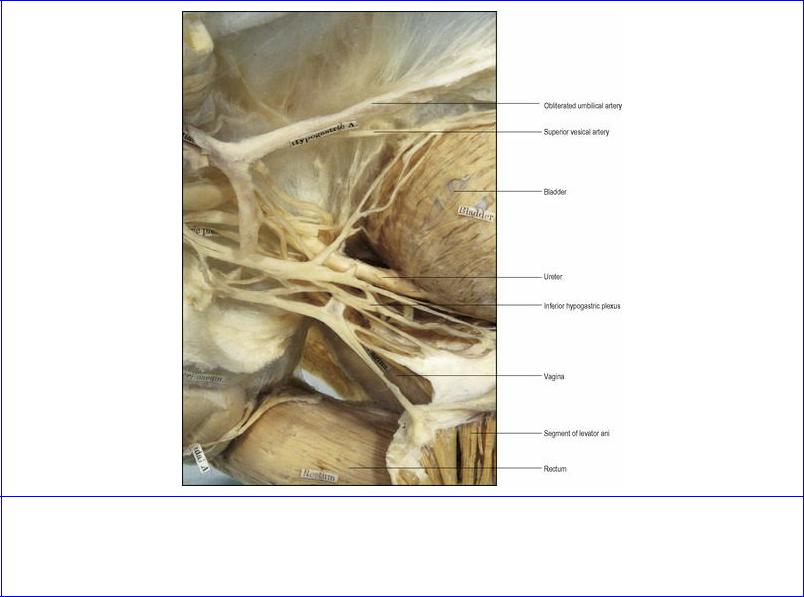
plexus is a rectangular, fenestrated plaque of nerves and ganglia, measuring nearly 5 cm anteroposteriorly and 2 cm vertically (Fig. 5.66).
Figure 5.66 Lateral aspect of the right inferior hypogastric plexus in an exhibit of the viscera and autonomic nerves from a female pelvis in the Anatomy Museum of the Royal College of Surgeons of England.
Its sympathetic components are derived from the superior hypogastric plexus (see p. 281), via the hypogastric nerve (Fig. 5.47) and from the sacral sympathetic ganglia. Preganglionic parasympathetic fibres join the plexus from S2, 3 and 4 nerves; these are the pelvic splanchnic nerves. About half the fibres in the hypogastric nerves are myelinated (preganglionic) and they relay in the ganglia of the inferior hypogastric plexus. The remaining sympathetic fibres and all the parasympathetic fibres pass through without relay. The parasympathetic motor and secretomotor fibres relay in the walls of the viscera.
Visceral branches of the inferior hypogastric plexus accompany visceral branches and tributaries of the internal iliac artery and vein as neurovascular bundles. In general it appears that the muscles of the bladder (detrusor muscle) and rectum are innervated by parasympathetic nerves from the pelvic splanchnics, the smooth muscle of the bladder neck, prostate, seminal vesicle and vas deferens by sympathetic nerves from the superior hypogastric plexus, and the smooth muscle of the internal sphincter of the anal canal by branches from the sacral sympathetic ganglia; all these nerves emerge from the inferior hypogastric plexus. Normal sensations of distension of bladder and rectum probably
pass through the pelvic splanchnic nerves; pain fibres are carried by both parasympathetic and sympathetic nerves.
As well as the pelvic viscera the pelvic splanchnic nerves supply the colon from the splenic flexure distally. These branches run up from the inferior hypogastric plexuses to the superior hypogastric plexus, or more often to its left, and then ascend with branches of the inferior mesenteric artery or as independent retroperitoneal nerves.
Thus the pelvic parasympathetics are motor to the emptying muscle of the bladder, and of the gut from splenic flexure to rectum. They are also secretomotor to the gut and vasodilator to the erectile tissue in the perineum. The sympathetics are motor to the visceral muscle of the bladder neck and internal anal sphincter. They are motor, too, to the vas deferens, seminal vesicles and prostatic muscle. The sympathetics also have a facilitating function in relation to uterine muscle.
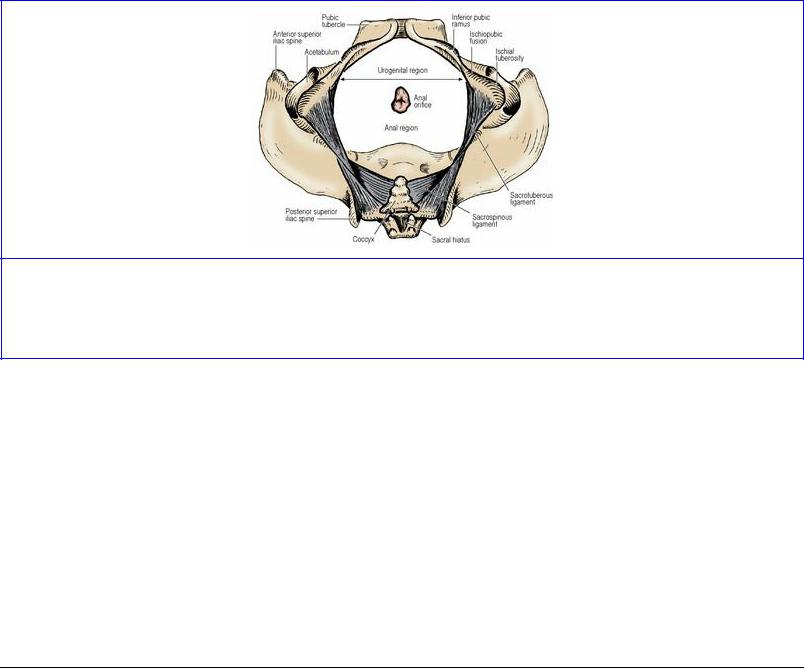
Part eighteen. Perineum
The perineum consists of that part of the trunk of the body caudal to the pelvic diaphragm (levator ani and coccygeus). A line joining the anterior parts of the ischial tuberosities divides this diamondshaped area into a larger posterior anal region and a smaller anterior urogenital region (Fig. 5.67). The anal region contains the anal canal and the ischioanal fossae with their contents. Its sides are formed by the sacrotuberous ligaments (covered by the lower border of gluteus maximus) and its base is formed by the line between the anterior parts of the ischial tuberosities; its contents are the same in each sex. The urogenital region lies in front of the line and is bounded laterally by the conjoined ischiopubic rami; in each sex it contains the external genitalia.
Figure 5.67 Urogenital and anal regions of the pelvic outlet. The anterior urogenital region is triangular and contains the external genitalia. The anal region is a pentagon and contains the anal canal and ischioanal fossae.
Cutaneous nerves
The skin of each side of the anal region is supplied by the inferior rectal nerve (S3, 4), the perineal branch of S4 and some twigs from the coccygeal plexus (S5).
In the urogenital region, the ilioinguinal nerve (L1) supplies the anterior third of the scrotum (labium majus). The skin of the penis (clitoris) is mainly supplied by the dorsal nerve (S2), a branch of the pudendal nerve. The posterior two-thirds of the scrotum (labium majus) is supplied laterally by the perineal branch of the posterior femoral cutaneous nerve and medially (labium minus) by scrotal (labial) branches of the perineal branch of the pudendal nerve (S3). A pudendal nerve block will therefore not anaesthetize the whole vulva; the anterior and lateral parts must be locally infiltrated to supplement the main nerve block.
Anal region
Anal canal
The anal canal is the last 4 cm of the alimentary tract; it is usually shorter in females. Like the rest of the gut it is a tube of muscle but the fibres are all circular, consisting of the internal and external anal sphincters, which are composed of visceral and skeletal muscle respectively. These sphincters,
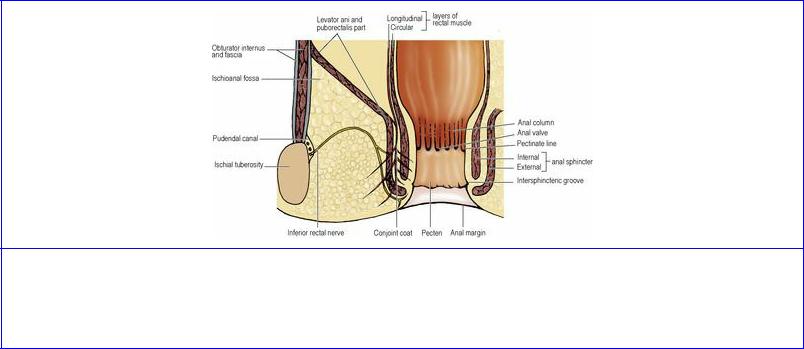
assisted by the configuration of the mucous membrane, hold it continually closed except for the temporary passage of flatus and faeces. The junction of rectum and anal canal is at the pelvic floor, i.e. at the level where the puborectalis part of levator ani clasps the gut and angles it forwards (Fig. 5.68). From this angled junction with the rectum, 2.5 cm in front of the tip of the coccyx, the anal canal passes downwards and somewhat backwards to the skin of the perineum.
The muscles of the anal canal can be regarded as forming ‘a tube within a funnel’ (Parks) ( Fig. 5.69). The sides of the upper part of the funnel are the levator ani muscles, and the stem of the funnel is the external sphincter which is continuous with levator ani. The tube inside the stem of the funnel is the internal sphincter which is a thickened continuation of the inner circular layer of rectal muscle. Internally lies the submucosa and mucous membrane.
Figure 5.69 Coronal section of the anal canal and the right ischioanal (ischiorectal) fossa. For clarity only the inferior rectal nerve is shown leaving the pudendal nerve in the pudendal canal; the corresponding vessels pursue a similar course across the fossa.
External anal sphincter
The sphincter has been described in the past as having deep, superficial and subcutaneous parts, based largely on the attachments of the middle (superficial) part, but the parts blend with one another to form a continuous tube.
At its upper (rectal) end, circular skeletal muscle fibres of this sphincter blend with the puborectalis part of levator ani (Fig. 5.69), except of course in the midline at the front where there are no levator ani fibres; here the sphincter fibres alone complete the ring. The region where puborectalis fuses with the external sphincter (which is also the level of the upper end of the internal sphincter) is termed the anorectal ring, and is palpable on rectal examination (see p. 295). Fibromuscular strands from the middle part of the external sphincter pass backwards to the posterior surface of the coccyx, contributing to an anococcygeal ligament. A retrosphincteric space occupied by fibrofatty tissue lies between these fibres and the muscular raphe formed by the iliococcygeal part of levator ani (Fig. 5.68). The multilayered fibromuscular anococcygeal ligament, with its external sphincter, iliococcygeus and pubococcygeus components, together with the overlying superior fascia of the pelvic diaphragm is also termed the postanal plate, on which lies the rectum (see p. 291). Anteriorly there is some intermingling of external sphincter muscle fibres with the transverse perinei and
bulbospongiosus muscles at the perineal body; this is less evident in the male so that a surgical plane of cleavage can be established between the external sphincter and the perineal body. In the female, the external sphincter is shorter and the deep fibres are deficient anteriorly. The lowest part of the external sphincter curves inwards to lie below the lower end of the internal sphincter (Fig. 5.69). This submucosal apposition of the two sphincters is at the site of the palpable intersphincteric groove in the lower part of the anal canal. When operating on the anaesthetized patient, however, the internal sphincter is often found to extend to the anal orifice.
Internal anal sphincter
This is the thickened downward continuation of the inner circular muscle of the rectum which, in cadaveric prosections, is usually found to extend along three-quarters of the length of the anal canal. (Fig. 5.69). At the anorectal junction the outer longitudinal layer of rectal muscle becomes fibroelastic and, together with some striated muscle fibres of puborectalis, forms the conjoint longitudinal coat which runs down between the two sphincters. Strands from this sheet penetrate the internal sphincter and the lower part of the external sphincter; some reach the fat of the ischioanal fossa and the perianal skin; others pass through the internal sphincter to the mucosa of the anal canal, particularly at the pectinate line (see below), where strands tethering the mucous membrane were named the mucosal suspensory ligament by Parks. It is possible that the puckering of perianal skin is due to the attachment of these fibroelastic strands to perianal skin, but some investigators describe separate smooth muscle fibres in this region, forming the so-called corrugator cutis ani muscle.
Mucous membrane
In the upper third of the anal canal the mucous membrane shows 6 to 10 longitudinal ridges, the anal columns. They are prominent in children. At their lower ends adjacent columns are joined together by small horizontal folds, the anal valves; the pockets so formed above the valves are the anal sinuses, into which open mucus-secreting anal glands. About half the anal glands are submucosal and the rest penetrate through the internal sphincter. Infection in these glands results in anal abscesses and fistulae. The level of the anal valves is the pectinate line (also called the dentate line) below which is a pale, smooth-surfaced area, the pecten, which extends down to the intersphincteric groove. Below the groove is a truly cutaneous area, continuous at the anus (anal margin) with the skin of the buttock. Histologically the lining below the groove is typical skin with keratinized stratified squamous epithelium, hair follicles, sebaceous glands and sweat glands. The lining of the pecten is nonkeratinized stratified squamous epithelium, with no hair follicles, sebaceous glands or sweat glands. The anal column area, being continuous with rectal mucosa, has typical columnar intestinal cells and tubular glands. But immediately above and below the pectinate line there is a zone of variable, often mixed epithelial structure, so that there is no abrupt line of change from the single-layered gut type to multilayered pecten type. (This contrasts with the gastro-oesophageal junction where there is an abrupt change from stratified squamous to columnar epithelium.)
Small submucous masses, comprising fibroelastic connective tissue, smooth muscle, dilated venous spaces and arteriovenous anastomoses, form anal cushions at left lateral (3), right posterior (7) and right anterior (11 o'clock) positions in the upper anal canal. Smaller cushions may be located in between. By their apposition these anal cushions assist the sphincter in maintaining watertight closure of the canal. Excessive straining at stool may cause enlargement of these cushions and the formation of haemorrhoids (piles).
The lining of the upper part of the anal canal is embryologically derived from the cloaca, i.e. it is endodermal; the lower part is from the proctodeum or anal pit and is ectodermal (see p. 29). The dividing line between these territories is usually considered to be at the pectinate line.
Blood supply
Branches of the superior rectal artery supply the upper end of the canal, their terminations lying within the anal columns. A small part of the muscular wall is supplied by the median sacral arteries, while the lower end, including its mucous membrane, receives the ends of the inferior rectal vessels which have crossed the ischioanal fossae. Within the walls there is good anastomosis between the various vessels.
The veins correspond to the above arteries and are continuous with the rectal venous plexuses (see p. 294). The upper part of the canal and plexus drains via the superior rectal and inferior mesenteric veins to the portal system, whereas the lower end drains to the internal iliac veins through the inferior and middle rectal veins. The anal canal is thus a site of portal–systemic anastomosis (see p. 267), the union being in the region of the anal columns.
Lymph drainage
The lymph drainage shows a watershed corresponding to the vascular pattern. The upper canal drains upwards to join the lymphatics of the rectum (see p. 294) whereas lymph from the lower end passes to the (palpable) superficial inguinal group.
Nerve supply
The inferior rectal branches of the pudendal nerves supply the external sphincter; they also provide the sensory supply from 1–2 cm above the pectinate line downwards, where the lining of the anal canal is highly sensitive. The motor fibres originate from Onuf's nucleus, situated mainly in the anterior horn of S2 segment, which also innervates the sphincter urethrae. The puborectalis and the deep part of the external anal sphincter have a high proportion of slow twitch fibres and function as tonic muscles, showing constant electromyographic activity even in sleep and under light anaesthesia. Autonomic nerves pass to the internal sphincter and the upper part of the canal. Sympathetic fibres from the pelvic plexus, with preganglionic cell bodies in the first two lumbar segments of the cord, cause contraction of the internal sphincter, and pelvic splanchnic (parasympathetic) nerves relax it. Afferent fibres from the upper end of the canal are carried by both sympathetic and parasympathetic nerves.
The anal reflex is described on page 17.
Defecation
Several factors contribute to normal anal continence: contraction of puborectalis and the external sphincter, maintenance of the angle between rectum and anal canal with abdominal pressure flattening the lower anterior rectal wall over the upper end of the canal, and the presence of mucosal cushions in the canal. The internal sphincter, although assisting closure, can only maintain continence if there is no distension (which causes relaxation of the sphincter). The rectum can accommodate itself to receive a certain amount of colonic content without any significant increase in pressure. There are no specialized receptors in the rectal wall, but they are present in the anal canal where gas, fluid and

solid can be distinguished by the cerebral cortex, and there are also stretch receptors in levator ani and the perirectal tissues. When increasing rectal pressure causes faeces to enter the upper anal canal, the external sphincter contracts and forces the contents back into the rectum. If only gas enters, its presence can be tested by a slight conscious increase of abdominal pressure which will let it escape. Defecation is allowed to occur by release of the cortical inhibition that developed during childhood training. Abdominal pressure is increased, puborectalis relaxes and the anorectal angle straightens with relaxation of the external sphincter and contraction of the lower colon and rectum (via their parasympathetic supply).
Incontinence may follow damage to the external sphincter or pudendal nerve (e.g. in obstetrics and perineal operations). In cerebral or spinal cord lesions there may be loss of cortical control.
Ischioanal (ischiorectal) fossa
The ischioanal (ischiorectal) fossa is a wedge-shaped space filled with fat lateral to the anal canal (Figs 5.69 and 5.71). The base of each fossa lies on the skin over the anal region of the perineum. The external sphincter of the anal canal and the sloping levator ani muscles form the medial wall of each fossa, while the lateral wall is formed by the ischial tuberosity below with obturator internus (covered by its fascia) above. The apex of the wedge is where the medial and lateral walls meet (where levator ani is attached to its tendinous origin over the obturator fascia). At the base the anterior boundary is the posterior border of the perineal body and muscles of the urogenital diaphragm (see p. 317), and the posterior boundary is the sacrotuberous ligament overlapped by the lower border of gluteus maximus. The fat in the lower part of the fossa adjacent to the skin is in small lobules, while the fat in the upper reaches of the fossa is in large lobules.
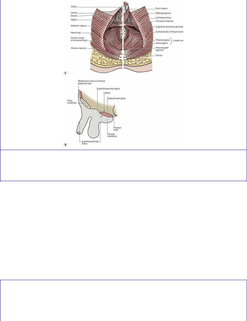
Figure 5.71 Muscles of the perineal region and the perineal pouches: A perineal muscles, female on the left and male on the right of the picture; B diagrammatic representation of the perineal pouches in the male.
Each fossa has an anterior recess that passes forwards above the perineal membrane, potentially as far as the posterior surface of the body of the pubis. The recesses of the two sides do not communicate across the midline. Posteriorly, however, the two fossae communicate with one another, low down through the fibrofatty tissue of the retrosphincteric space within the anococcygeal ligament (see p. 313), providing a horseshoe-shaped path for the spread of infection from one fossa to the other.
The pudendal canal (of Alcock) is a connective tissue tunnel in the lower lateral wall of the fossa, overlying obturator internus and the medial side of the ischial tuberosity. The canal is formed by a splitting of the obturator fascia above the falciform process of the sacro-tuberous ligament. It contains the pudendal nerve and internal pudendal vessels (Fig. 5.70), which it conducts from the lesser sciatic notch to the deep perineal pouch above the perineal membrane (see p. 317).
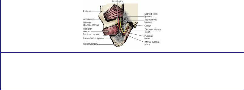
Figure 5.70 Lateral wall of the left ischioanal fossa from behind, with the connective tissue of the pudendal canal removed to show the pudendal nerve and vessels running forwards on the medial side of the ischial tuberosity. The middle part of the sacrotuberous ligament has been removed and the venae comitantes of the internal pudendal artery are not shown.
The pudendal nerve and internal pudendal vessels leave the pelvis through the greater sciatic foramen, passing beneath the lower border of piriformis to reach the buttock. Their course in the buttock is short. They turn and enter the lesser sciatic foramen, the vessels passing over the tip of the spine of the ischium, the nerve more medially over the sacrospinous ligament.
Running transversely across the ischioanal fossa from the pudendal canal towards the anal canal are the inferior rectal branches of the pudendal nerve and internal pudendal vessels. Their course is not straight across the base of the fossa, but arches convexly upwards through the fat towards the apex and then downwards to the anal canal. Incisions to drain ischioanal abscesses usually do not interfere with them. Accompanied by the vessels, the nerve breaks up into several branches which supply the external sphincter, mucous membrane of the lower anal canal and perianal skin.
At the front of the fossa the posterior scrotal (labial) nerves and vessels (from the pudendals) pass superficially into the urogenital region. At the back of the fossa the perineal branch of S4 nerve and the perforating cutaneous nerve traverse the fossa.
Perineal body
The perineal body, also called the central tendon of the perineum, is a midline fibromuscular mass to which a number of muscles gain attachment, and within which they decussate. It is attached to the posterior border of the perineal membrane (see below). It lies between the anal canal and the vagina (Fig. 5.64) or bulb of the penis (Fig. 5.71A). The rectovaginal septum blends into it above. The muscles running into it include the external anal sphincter, pubovaginalis (puboprostaticus) part of levator ani, bulbospongiosus, and the superficial and deep transverse perineal muscles. Its position and connections provide a stabilizing influence for pelvic and perineal structures. Injury to it during childbirth may weaken the pelvic floor and contribute to prolapse of the vagina and uterus.
Part nineteen. Male urogenital region
In the male urogenital region two layers of fascia, the superior and inferior fasciae of the urogenital diaphragm, enclose the sphincter urethrae and the deep transverse perineal muscles; together these fasciae and muscles are referred to as the urogenital diaphragm. The superior fascia is thin and illdefined; the inferior fascia is thicker and termed the perineal membrane. These fascia enclose between them a deep perineal space (pouch) which contains the membranous part of the urethra, the internal pudendal vessels, the dorsal nerve of penis and perineal nerve at the sides, and the paired bulbourethral glands, in addition to the sphincter urethrae and deep transverse perineal muscles.
The perineal membrane is an unyielding sheet of fibrous tissue which forms the base upon which the penis and penile musculature are fixed (Fig. 5.71A). It is attached on either side to the ischiopubic rami from just behind the subpubic angle back to the level of the anterior part of the ischial tuberosities. Its anterior border forms the transverse perineal ligament, and there is a small gap between this and the arcuate pubic ligament through which the deep dorsal vein of the penis passes to reach the vesicoprostatic plexus. Its posterior border fuses centrally with the perineal body. When standing upright, the perineal membrane lies approximately horizontally. Above the membrane lies the membranous urethra surrounded by the urethral sphincter, below the apex of the prostate. The membrane is pierced by the urethra, the ducts of the bulbourethral glands, and by nerves and vessels.
After leaving the prostate just in front of the apex, the prostatic urethra (see p. 299) becomes the membranous urethra which passes down for about 1.5 cm and pierces the perineal membrane, 2.5 cm behind the pubic symphysis, to become the penile urethra. The membranous urethra is the shortest and least dilatable part of the urethra. In the upper part of its wall there are some smooth muscle fibres continuous with those of the prostatic urethra.
The sphincter urethrae, often called clinically the external urethral sphincter, is roughly pearshaped. The thinner upper end of the ‘pear’ extends upwards out of the deep perineal pouch to surround the lower part of the prostatic urethra; the lower more bulbous part is below the apex of the prostate and above the perineal membrane. Some of its fibres arise from the pubic rami and pass as U-shaped loops in front of and behind the urethra, some run from the transverse perineal ligament to the perineal body, and some completely encircle the urethra. Although consisting of striated muscle, the sphincter fibres are of small diameter and are of the slow twitch variety. They are supplied by the perineal branch of the pudendal nerve (see p. 321). Above the posterior part of the perineal membrane the deep transverse perineal muscle extends from the ischial ramus to the perineal body, where its fibres decussate with those of the contralateral muscle and the external anal sphincter. It merges anteriorly with the sphincter urethrae and shares the same nerve supply.
The bulbourethral glands (of Cowper) lie one on each side of the membranous urethra in the deep perineal pouch, i.e. above (deep to) the perineal membrane, covered by the urethral sphincter. They are about 1 cm in diameter. The single duct from each, about 2.5 cm long, pierces the perineal membrane to open into the bulb of the penile urethra. The glands contribute a small amount to seminal fluid.
Deep to the skin of the urogenital region is the superficial perineal fascia (of Colles), a continuation into the perineum of the membranous fascia (of Scarpa) from the anterior abdominal wall (see p. 179). It is attached to the ischiopubic rami and the posterior margin of the perineal membrane, thus
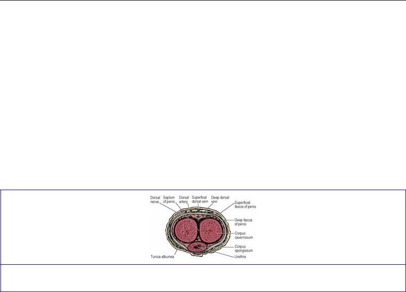
closing in a subfascial space, the superficial perineal space (pouch), that is in continuity with the space deep to the membranous (Scarpa's) fascia of the anterior abdominal wall (see Fig. 4.1, p. 180). From its marginal attachments in the urogenital region, this fascia is projected into a bulbous scrotal expansion and a cylindrical penile expansion (Fig. 5.71B), the distal end of the latter being attached round the corona of the glans penis. Rupture of the penile urethra permits extravasation of urine beneath Colles' fascia whence the collection distends the tissues of the scrotum and penis and can then pass upwards over the anterior abdominal wall beneath Scarpa's fascia. A deep perineal fascia intimately surrounds the cavernous bodies of the penis and clitoris and the superficial perineal muscles associated with them.
Penis
The penis has a root and a body. The root of the penis is attached to the inferior surface of the perineal membrane and consists of the (central) bulb of the penis with a crus on each side. Each crus is attached to the angle between the perineal membrane and the everted margin of the ischiopubic ramus, receives the deep artery of the penis near its anterior end, and continues forwards to become the corpus cavernosum. The bulb is the posterior end of the corpus spongiosum. At the front of the root area, below the subpubic angle, the two corpora cavernosa are bound together side by side with the corpus spongiosum behind them (when the penis is dependent, but ventral to them when erect) to form the body of the penis (Fig. 5.72). The penile urethra runs through the whole length of the corpus spongiosum from the bulb at the back to its expanded external end which is the glans penis. The glans forms the tip of the penis, overlapping the distal ends of the corpora cavernosa. The urethra enters the bulb from above near its front so that most of the bulge of the bulb is behind and below the urethra. The bulb has a slight (palpable) midline notch on its under surface and extends back towards the perineal body. The arteries of the bulb enter it near the urethra, which in this region receives the ducts of the bulbourethral glands.
Figure 5.72 Cross-section of the body of the penis.
The corpus spongiosum and the two corpora cavernosa (Fig. 5.72) are each surrounded by a tough fibrous sheath, the tunica albuginea of the corpus (not to be confused with the tunica albuginea of the testis); that of the corpus spongiosum enlarges distally to enclose the glans. From the tunica fibrous trabeculae pass into the corpora dividing their substance into numerous endothelial cell-lined cavernous spaces into which helicine arteries open (see p. 321). Between the corpora cavernosa there is a connective tissue septum which is partly divided into comb-like strands. The fibrous sheaths of the corpora are encircled by the deep fascia of the penis, an extension of the deep perineal fascia. This fascia is attached to the front surface of the pubic symphysis by a triangular sheet of
fibrous tissue, the suspensory ligament of the penis. The midline deep dorsal vein, with a dorsal artery on each side and more laterally a dorsal nerve, lies deep to the deep fascia of the penis.
The skin is hairless and prolonged forwards in a fold, the prepuce, which partly overlaps the glans and doubles back to be attached to the neck of the glans. Beneath the skin is the superficial fascia of the penis (Buck's fascia), a cylindrical prolongation of Colles' fascia (see p. 318), and in the midline is the superficial dorsal vein which is accompanied by lymphatics from the skin and the anterior part of the urethra. On the inferior aspect of the glans, a fold of skin, the frenulum, passes from the prepuce to the ventral end of the urethral orifice.
Blood supply
The penis receives three pairs of arteries which are branches of the internal pudendals (see p. 309). The artery to the bulb supplies the corpus spongiosum, including the glans. The deep artery of the penis supplies the corpus cavernosum. The dorsal artery supplies skin, fascia and glans. There is anastomosis, via the continuity of corpus spongiosum and glans, between the artery of the bulb and the dorsal artery; the deep arteries supply the corpora cavernosa only. The skin of the penis is also supplied by the superficial external pudendal branches of the femoral arteries.
Venous return from the corpora is partly by way of veins that accompany the arteries and join the internal pudendal veins, but mostly by the deep dorsal vein which pierces the suspensory ligament, passes above the perineal membrane and enters the vesicoprostatic venous plexus. The superficial dorsal vein drains the dorsal skin of the penis and divides to join the superficial external pudendal tributaries of the great saphenous veins.
Lymph drainage
Lymphatics from the penile skin pass to superficial inguinal nodes, but the glans and the corpora drain to deep inguinal nodes.
Nerve supply
The skin of the penis is supplied by the pudendal nerves (see p. 311) via the posterior scrotal and dorsal nerves; the latter supply the glans. The dermatome mainly involved is S2. A small area of skin on the dorsum of the proximal penis is supplied by the ilioinguinal nerve (L1). The bulbocavernosus and ischiocavernosus muscles (see below) which contract spasmodically during ejaculation are supplied by the perineal nerve (from the pudendal, S2, 3). The sympathetic nerves necessary for the initial stages of ejaculation (see p. 322) are derived from L1 segment of the spinal cord via the superior and inferior hypogastric plexuses. The pelvic splanchnic nerves (S2, 3) provide the parasympathetic supply to the cavernous tissue of all three corpora and allow increased blood flow for erection (see p. 322).
Circumcision
Ritual circumcision for religious or racial reasons is probably the oldest operation in the world. In children or adults circumcision may be required for a tightly constricting prepuce (phimosis). The prepuce is incised on the dorsum from the tip towards the base of the glans, dissecting away any adhesions, and then the incision is carried circumferentially, followed by suture of the skin edges. Bleeding from a vessel in the frenulum needs to be controlled.

Superficial perineal muscles
The bulb and each crus of the penis are provided with overlying muscles: bulbospongiosus and ischiocavernosus, respectively (Fig. 5.71A). In addition there is a transverse pair (superficial transverse perineal) along the posterior border of the perineal membrane. The three superficial perineal muscles of each side thus form a triangular pattern when viewed in the lithotomy position.
Bulbospongiosus arises from the perineal body and in front of that from a median raphe that joins the pair together. Its posterior fibres are directed forwards and laterally over the bulb to be inserted into the perineal membrane and a dorsal fibrous expansion on the penis; the more posterior of these fibres clasp the corpus spongiosum, while the more anterior extend on to the corpora cavernosa. Bulbospongiosus acts to empty the urethra at the end of micturition, assists in erection (by compressing the deep dorsal vein of the penis) and contracts during ejaculation.
Ischiocavernosus arises from the posterior part of the perineal membrane and from the ischial ramus and is inserted by an aponeurosis on to the surface of the corpus cavernosum. Its function is to assist in the support of, and move slightly, the erect penis.
The superficial transverse perineal muscle arises from the ischial tuberosity and is inserted into the perineal body. It helps to stabilize the perineal body.
Nerve supply. All three muscles are supplied by the perineal branch of the pudendal nerve (S2, 3).
Male urethra
The urethra consists of prostatic, membranous and spongy parts, with a total length of about 20 cm. The prostatic and membranous parts have already been described (see pp. 299 and 317).
The spongy or penile urethra, about 15 cm long, is within the corpus spongiosum of the penis and can be divided into bulbous and pendulous parts. The posterior part of the corpus, attached to the undersurface of the perineal membrane, is enlarged as the bulb. After piercing the perineal membrane the urethra enters the bulb and at once takes a right-angled curve forwards within the bulb. The urethra continues through the corpus spongiosum beyond the root of the penis into its body. Just proximal to the external urethral meatus at the tip of the glans there is a short dilated region, the navicular fossa. The lining here is stratified squamous epithelium, in contrast to the whole of the rest of the urethra which possesses the transitional epithelium typical of the urinary tract. The urethral mucosa displays some very small blind-ending pockets (lacunae) and there are numerous mucous urethral glands (of Littre). The empty urethra is horizontal in cross-section but the meatus is a vertical slit, hence the spiral stream of urine.
The urethra is narrowest at the external meatus, and shows dilatations in the prostatic part, bulb and navicular fossa. When passing a catheter the 90° change of direction in the bulbar part before the membranous urethra is entered has to be kept in mind. On account of a particularly large lacuna on the roof of the navicular fossa, any instruments being passed through the external meatus should initially be pointed towards the floor of the fossa.
Blood supply
As with many tubular structures, there is no single ‘artery to the urethra’; the blood supply is from any

adjacent vessels as it passes through the prostate, sphincter urethrae and corpus spongiosum.
Nerve supply
The mucous membrane of the penile part receives a branch from the perineal nerve, with filaments from the inferior hypogastric plexuses reaching more proximal parts. The urethral sphincters and the control of micturition have been described on page 297–298.
Development
The part of the urethra proximal to the openings of the ejaculatory ducts and prostatic utricle is formed, like the trigone of the bladder, from the lower ends of the mesonephric ducts and ureters that are absorbed into the bladder wall (see p. 29). The rest is from the pelvic and phallic parts of the urogenital sinus and the genital tubercle, supplemented ventrally in the penile part by the fusion of the urogenital folds (see p. 29). The epithelium of the navicular fossa (ectoderm from the genital tubercle) is a solid column that becomes canalized; if it fails to do so, and there is accompanying failure of complete fusion of the urogenital folds, the result is hypospadias—a midline opening on the ventral surface.
Scrotum
The scrotum is a pouch of skin containing the testes and spermatic cords. The subcutaneous tissue has no fat, but contains the dartos muscle which sends a sheet into the midline fibrous septum of the scrotum (Fig. 5.12). The rugosity of the skin is due to contraction of the dartos. The dartos is smooth muscle, and is supplied by sympathetic fibres probably carried by the genital branch of the genitofemoral nerve.
Blood supply
The blood supply of the skin is from the superficial and deep external pudendal arteries (from the femoral) and from scrotal branches of the perineal artery (from the internal pudendal). Venous drainage is mainly by external pudendal veins, superficial and deep, to the great saphenous vein (see Fig. 3.3, p. 114).
Lymph drainage
Lymph drainage is to the superficial inguinal nodes.
Nerve supply
The anterior axial line (see p. 14) crosses the scrotum. The anterior one-third of the scrotal skin is supplied by the ilioinguinal nerve (L1) and the genital branch of the genitofemoral nerve (L1). The posterior two-thirds is supplied by scrotal branches of the perineal nerve (S3), reinforced laterally by the perineal branch of the posterior femoral cutaneous nerve (S2).
Development
The scrotum develops from the labioscrotal swellings and the urogenital folds (see p. 29). Where the urogenital folds meet is marked by a midline cutaneous raphe on the scrotum, which continues on to the inferior surface of the penis. (In the female, the labioscrotal folds form the labia majora, and the urogenital folds remain separate as the labia minora.)
Perineal vessels and nerves
The internal pudendal artery (see p. 309) enters the deep perineal pouch from the anterior end of the pudendal canal and passes forwards along the ischiopubic ramus above the perineal membrane, with the dorsal nerve of penis above it and the perineal nerve below it. The perineal branch of the artery pierces the posterior angle of the perineal membrane and gives origin to posterior scrotal and transverse perineal branches. Further forward another branch, artery to the bulb, pierces the membrane alongside the urethra to enter the corpus spongiosum. It gives branches to the cavernous tissue of this corpus and passes forwards to supply the glans penis.
Near the anterior margin of the perineal membrane the internal pudendal artery divides into its terminal branches, the deep and dorsal arteries of the penis. The deep artery pierces the membrane to enter the crus of the penis and supplies, by its helicine branches, the erectile cavernous tissue of the corpus cavernosum. The dorsal artery pierces the membrane and passes between the crus and the pubic symphysis to pierce the suspensory ligament and run forward, alongside the median deep dorsal vein and with the dorsal nerves laterally, between the deep fascia of the penis and the fibrous sheaths of the corpora cavernosa. The arteries pass to the glans where they anastomose with the terminal branches of the arteries to the bulb.
The deep dorsal vein of the penis drains most of the blood from the corpora. It runs proximally in the midline, pierces the suspensory ligament and passes upwards in the gap between the pubic symphysis (arcuate pubic ligament) and the perineal membrane to enter the pelvis and join the vesicoprostatic plexus.
The pudendal nerve (see p. 311) divides within the pudendal canal into its terminal branches, the dorsal nerve of the penis and the perineal nerve. Both enter the deep perineal pouch, running forwards respectively above and below the internal pudendal artery. The dorsal nerve of the penis appears to be the direct continuation of the pudendal nerve. It pierces the anterior angle of the perineal membrane and then accompanies the dorsal artery on its lateral side. It supplies the skin of the penis and glans and gives branches to the corpus cavernosum. It has no branches in the deep perineal pouch.
The perineal nerve, which is the larger terminal branch of the pudendal, gives muscular branches to the superficial and deep perineal muscles and to the sphincter urethrae. The branch to bulbospongiosus supplies sensory fibres to the mucous membrane of the urethra. Either before or just after entering the deep perineal pouch the perineal nerve gives off the posterior scrotal branches which run forwards superficial to the perineal membrane to supply scrotal skin.
Erection and ejaculation
The neural controls of erection and ejaculation are different; erection is parasympathetic and ejaculation is sympathetic and somatic. Impulses in the genital branches of the pelvic splanchnic nerves (parasympathetic) lead to vasodilatation of the helicine arteries of the erectile tissue of the corpora cavernosa (hence the old name of nervi erigentes). As the spongy tissue becomes engorged there may be some compression of the draining veins, but erection (whether reflex from physical stimulation or psychogenic) occurs mainly by increased arterial flow following relaxation of smooth muscle in the trabeculae of the corpora, which is dependent on the production of nitrous oxide and cyclic GMP.
The sympathetic outflow from segments T11 to L2 to the epididymis, vas deferens, seminal vesicle, ejaculatory duct, prostate, superficial trigonal muscle of the bladder and circular muscle of the bladder neck causes contraction of the smooth muscle of those structures, so causing the flow of seminal fluid into the prostatic urethra (emission). The internal urethral opening of the bladder neck becomes constricted so that there is no retrograde flow into the bladder. Rhythmic contraction of bulbospongiosus (supplied by the perineal nerve) compresses the penile urethra and expels the fluid. Orgasmic sensations run in the spinothalamic tract and are abolished by cord transection, but in transections above the lower thoracic segments ejaculation can still occur.