

Part nine. Spleen
The spleen, the largest of the lymphoid organs (see p. 9), lies under the diaphragm on the left side of the abdomen (Fig. 5.26), and although not part of the alimentary tract it drains to the portal venous system.
The odd numbers 1, 3, 5, 7, 9, 11 summarize some splenic statistics. It measures 1 × 3 × 5 inches, weighs 7 oz and lies deep to the left 9th to 11th ribs (H. A. Harris). These are average measurements; the size of the spleen varies considerably.
Being developed in the dorsal mesogastrium (see below), the spleen projects into the greater sac surrounded by peritoneum of the original left leaf of the dorsal mesogastrium. It lies at the left margin of the lesser sac (Fig. 5.49) below the diaphragm, and its diaphragmatic surface is moulded into a reciprocal convexity. Its hilum lies in the angle between the stomach and left kidney, each of which impresses a concavity alongside the attached splenic vessels (Fig. 5.41). Its long axis lies along the line of the tenth rib, and its lower pole does not normally project any further forward than the midaxillary line. A small colic area lies in contact with the splenic flexure and the phrenicocolic ligament. Its anterior border is notched, a relic of the fusion of the several ‘splenules’ from which the organ arises in the embryo.
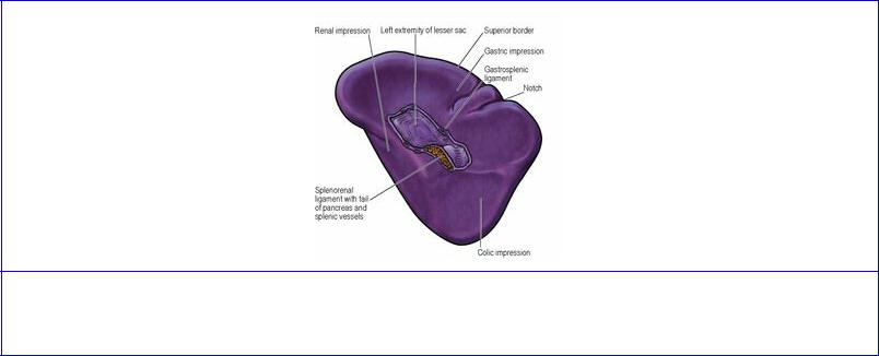
Figure 5.41 Visceral surface of the spleen, showing the impressions for adjacent viscera and the two layers of peritoneum that form the splenorenal and gastrosplenic ligaments.
Its visceral peritoneum, or serous coat, invests all surfaces (gastric, diaphragmatic, colic and renal). The two leaves of the greater omentum pass from the hilum forwards to the greater curvature of the stomach (the gastrosplenic ligament) and backwards to the front of the left kidney (the splenorenal ligament) (Fig. 5.49). The hilum of the spleen makes contact with the tail of the pancreas, which lies within the splenorenal ligament.
If the spleen enlarges, its long axis extends downwards and forwards in the direction of the umbilicus, and its anterior border approaches the costal margin to the left of the greater curvature of the stomach. (A kidney enlarging downwards does so in the direction of the iliac fossa.) The spleen must at least double its normal size before its anterior border passes beyond the left costal margin. A palpable spleen is identified by the notch in its anterior border. In some diseases the spleen is grossly enlarged
and may extend across the upper abdomen beyond the umbilicus towards the right iliac fossa. Whatever the degree of enlargement the spleen glides in contact with the diaphragm and anterior abdominal wall in front of the splenic flexure, which remains anchored by the phrenicocolic ligament, and no colonic resonance is found on percussion over the organ. Retroperitoneal tumours (e.g. of the left kidney) do not displace the overlying colon and they are crossed by a band of colonic resonance.
The structures that are related to the surfaces of the spleen can be identified by holding the convexity (diaphragmatic surface) of the detached organ in the hollow of the left hand, and rotating it until the notched anterior border lies to the front, near the thumb (Fig. 5.41). The concavity between the notched anterior border and the hilum is the gastric impression. Behind the hilum is the concave renal surface, while at the lower pole (at the tip of the little finger of the left hand) is the small colic impression.
The structure of the spleen has been referred to on page 9.
Blood supply
The splenic artery passes between the layers of the splenorenal ligament and at the hilum divides into two or three main branches, from which five or more branches enter the spleen. Veins accompany the arteries and unite to form the splenic vein. Based on the vascular arrangement, it is possible that the spleen consists of two or three segments, with intersegmental vessels being small and scanty, but the evidence for such segmentation is not conclusive.
Lymph drainage
Lymph drains into several nodes lying at the hilum and thence, by way of the pancreaticosplenic nodes, to the coeliac nodes.
Nerve supply
The spleen is supplied from the coeliac plexus with sympathetic fibres only.
Development
The spleen begins to develop in the sixth week as several condensations of mesodermal cells in the dorsal mesogastrium which, because of the splenic presence, becomes divided into the splenorenal and gastrosplenic ligaments (Fig. 5.21B). The spleen thus comes to lie at the left margin of the lesser sac. The original condensations become aggregated into a single organ; the splenic notch may represent a region where there is incomplete fusion. ‘Accessory spleens’ are the result of lack of fusion; one or several may be found, usually along the splenic vessels or in the peritoneal attachments. They occur in up to 20% of the population and are rarely larger than 2 cm in diameter.
Surgical approach
Removal of the spleen (splenectomy) essentially involves cutting its two ‘pedicles’, the splenorenal and gastrosplenic ligaments. In an emergency after rupture with haemorrhage, the left or posterior layer of the splenorenal ligament is incised and the spleen turned medially so that the splenic vessels can be dissected away from the tail of the pancreas and ligated (arteries before veins). The short gastric vessels and the gastrosplenic ligament are then divided and removal completed. For an elective procedure it is usual to enter the lesser sac by dividing the gastrosplenic ligament and its
vessels and then to deal with the splenic vessels and the splenorenal ligament. The stomach must not be perforated when ligating the short gastric vessels, and damage to the tail of the pancreas and splenic flexure of the colon must be avoided.
Part ten. Posterior abdominal wall
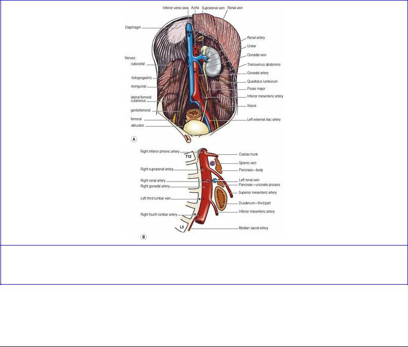
The five lumbar vertebrae project forwards into the abdominal cavity as the lumbar spine has a normal lordosis (forward convexity). The midline forward projection is enhanced by the inferior vena cava and aorta, which lie in front of the bodies of the vertebrae (Fig. 5.42A). To each side of this convexity lie deep paravertebral gutters. They are floored in by the psoas and quadratus lumborum muscles and, below the iliac crest, by the iliacus muscle. The crura and adjacent parts of the diaphragm (see p. 184) are also part of the posterior abdominal wall. The kidneys lie high up in the paravertebral gutters.
Figure 5.42 Posterior abdominal wall: A great vessels and nerves; B diagrammatic representation of the aorta as seen from the right side.
The lumbar vertebrae are separated from each other by thick intervertebral discs, which unite them very strongly. A broad ribbon, the anterior longitudinal ligament, is attached anteriorly and crosses the lumbosacral prominence to become fused with the periosteum on the front of the upper sacrum.
Muscles
Psoas major
This muscle lies in the gutter between the bodies and transverse processes of the lumbar vertebrae. Its vertebral attachment is to the discs above the five lumbar vertebrae, the adjoining parts of the bodies of the vertebrae, to fibrous arches that span the concavities of the sides of the upper four vertebral
bodies and to the medial ends of all the lumbar transverse processes. Thus there is one continuous attachment from the lower border of T12 to the upper border of L5 vertebrae. The muscle passes downwards along the pelvic brim (Fig. 5.42A) and then beneath the inguinal ligament into the thigh, where its tendon is attached to the lesser trochanter of the femur. The lumbar plexus (see p. 278) is embedded within the muscle, and part of the external vertebral venous plexus is behind it (in front of the transverse processes). The genitofemoral nerve emerges from the front of the muscle, the iliohypogastric, ilioinguinal, lateral femoral cutaneous and femoral nerves from its lateral border, and the obturator nerve and the lumbosacral trunk from its medial border. The four lumbar arteries (see p. 276) and veins pass backwards medial to the four arches and run laterally behind the psoas muscle.
The strong psoas fascia (part of the iliac fascia) invests the surface of the muscle, attached to the vertebral bodies, the fibrous arches, and the transverse processes, and to the iliopubic eminence (see p. 165) as the muscle extends along the pelvic brim. It retains the pus of a psoas abscess, and spinal tuberculosis may present as a cold abscess in the groin. The sheath is not part of the lumbar fascia (see p. 274), but the lateral edge blends with the anterior layer of that fascia (over quadratus lumborum).
There is a thickening in the psoas fascia curving obliquely from the body of L1 (or L2) vertebra to the transverse process of L1 vertebra. This is the medial arcuate ligament, from which fibres of the diaphragm arise in continuity alongside the crus. The part of the psoas above this ligament is above the diaphragm, i.e. in the thorax. The sympathetic trunk passes from thorax to abdomen beneath this ligament.
Nerve supply. By the first three lumbar nerves, mainly L2.
Action. Its action on the hip joint is described on pages 118 and 129. Being attached to the sides of lumbar vertebrae it is a lateral flexor of the vertebral column. When acting from below with iliacus and their fellows of the opposite side, it assists in the flexion of the trunk produced by anterior abdominal wall muscles (especially rectus abdominis), psoas acting on the lumbar column and iliacus on the hip bone.
Psoas minor, present in only two out of every three individuals, is a slender muscle lying on the surface of psoas major. Its short belly arises from T12 and L1 vertebrae and its long tendon flattens out to blend with the psoas fascia and gain attachment to the iliopubic eminence. It is supplied by L1 nerve and is a weak flexor of the lumbar spine.
Quadratus lumborum
This is a flat sheet lying deep in the paravertebral gutter, edge to edge with psoas medially and transversus abdominis laterally (Fig. 5.42A). It lies in the anterior compartment of the lumbar fascia. It arises from the stout transverse process of L5 vertebra, from the strong iliolumbar ligament and from a short length of the adjoining iliac crest. Its fibres pass upwards to the transverse processes (lateral to psoas) of the upper four lumbar vertebrae and, more laterally, to the inferior border of the medial half of the twelfth rib. Its lateral border slopes upwards and medially and so crosses the lateral border of the iliocostalis component of erector spinae (see p. 429), which slopes upwards and laterally. Its anterior surface is covered by the anterior layer of the lumbar fascia. A thickening in front of this fascia passing from the first lumbar transverse process to the twelfth rib constitutes the
lateral arcuate ligament. The fibres of the diaphragm arise in continuity from the ligament. The subcostal neurovascular bundle (vein, artery, nerve from above downwards) emerges from the thorax beneath the ligament and slopes down across the lumbar fascia. The muscle represents the innermost of the three muscular layers of the body wall and is in series with the diaphragm and the transversus thoracis muscle group.
Nerve supply. By T12 and the upper three or four lumbar nerves.
Action. Its chief action is to prevent the diaphragm from elevating the twelfth rib during inspiration. By depressing the twelfth rib it aids descent of the contracting diaphragm. Additionally it is a lateral flexor of the lumbar spine.
Iliacus
This muscle arises from the upper two-thirds of the iliac fossa up to the inner lip of the iliac crest and from the anterior sacroiliac ligament. It is triangular in shape and its fibres converge medially towards the lateral margin of psoas and pass out of the iliac fossa beneath the lateral part of the inguinal ligament. It is inserted into the psoas tendon and the adjacent part of the femur below the lesser trochanter.
The iliacus muscle is covered by the strong iliac fascia; this is attached to bone at the margins of the muscle, and to the inguinal ligament. The fascia is continuous with the psoas fascia, forms a floor to the abdominal cavity and serves for the attachment of parietal peritoneum. Apart from its prolongation into the femoral sheath (see p. 118) it does not extend into the thigh.
Nerve supply. By the femoral nerve (L2, 3) in the iliac fossa.
Action. It acts, with psoas, on the hip joint (see p. 118).
Fascia
Each muscle of the posterior abdominal wall (quadratus lumborum, psoas and iliacus) is covered with a dense and unyielding fascia. The fasciae over adjacent muscles blend at their margins. The psoas fascia and the iliac fascia have been described above.
Lumbar fascia. This is the lumbar part of the thoraco-lumbar fascia. In the lumbar part of the trunk of the body, three layers of fascia enclose two muscular compartments. The anterior and middle layers occupy only the lumbar region, but the posterior layer extends above this to the lower part of the neck and below to the dorsal surface of the sacrum. Quadratus lumborum occupies the anterior compartment, while erector spinae fills the posterior compartment. The anterior layer extends from the front of the iliolumbar ligament and adjoining iliac crest to the lower border of the twelfth rib. Medially it is attached to the front of each lumbar transverse process, adjoining the attachment of the psoas fascia. Laterally it blends with the middle layer along the lateral border of quadratus lumborum; here transversus abdominis and internal oblique take origin. The middle layer extends from the back of the iliolumbar ligament and adjoining iliac crest up to the twelfth rib. Medially it is attached to the tips of the lumbar transverse processes. Laterally it blends with both anterior and posterior layers. The latter line of fusion is along the lateral border of erector spinae. The posterior layer lies over the whole erector spinae mass of muscle. It is attached medially to the spinous
processes and supraspinous ligaments of all the sacral, lumbar and thoracic vertebrae. Its lateral margin traced from below upwards extends along the transverse tubercles of the sacrum to the posterior part of the iliac crest, from where it slopes outwards to the twelfth rib. Above the twelfth rib its attachment is to the angles of all the ribs; its lateral border over the thoracic cage thus slopes up medially. In the thorax this single posterior layer constitutes the thoracic part of the thoracolumbar fascia; it is only below the thorax where there are no ribs that the thoracolumbar fascia is in three layers. The posterior layer is thick and strong over the lumbar region, being here reinforced by fusion of the aponeurotic origin of latissimus dorsi. Over the thorax it gradually becomes thinner and it fades out above the first rib over the extensor muscles of the neck, where it is replaced by the splenius muscle.
Vessels
The central vascular features of the posterior abdominal wall are the abdominal part of the aorta and the inferior vena cava, with the vein lying on the right side of the artery.
Abdominal aorta
The thoracic aorta becomes the abdominal aorta on passing behind the median arcuate ligament and between the crura of the diaphragm, on the front of the body of T12 vertebra (Fig. 5.43). It passes downwards behind the peritoneum on the bodies of the lumbar vertebrae, inclining slightly to the left, with the left sympathetic trunk at its left margin. On the body of L4 it divides into the two common iliac arteries.
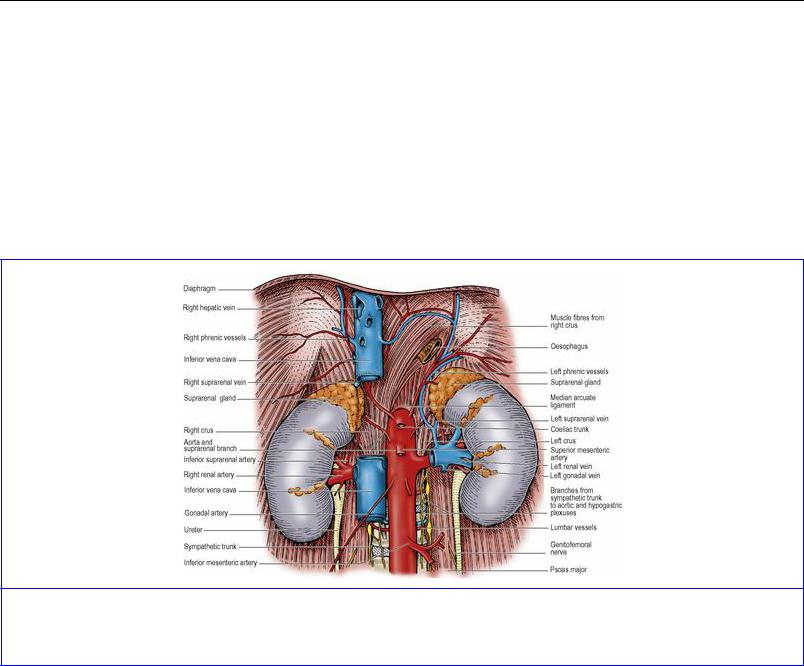
Figure 5.43 Abdominal aorta, inferior vena cava, kidneys and suprarenal glands. (The sympathetic plexus on the front of the aorta has been omitted.)
Between the origins of the coeliac trunk and the superior mesenteric artery the aorta is crossed by the splenic vein and the body of the pancreas (Fig. 5.42B). Between the superior and inferior mesenteric origins lie the left renal vein, the uncinate process of the pancreas and the third part of the duodenum.
The surface marking of the abdominal aorta is from 2.5 cm above the transpyloric plane in the midline to a point 1 to 2 cm below and to the left of a normally situated umbilicus, level with the highest points of the iliac crests.
Branches
The main branches of the abdominal aorta fall into three groups: single ventral arteries to the gut and its derivatives (coeliac, superior and inferior mesenteric), paired branches to other viscera (suprarenal, renal and gonadal arteries) and paired branches to the abdominal wall (inferior phrenic and lumbar arteries). A small posterior branch, the median sacral artery, leaves the aorta a little above its bifurcation, and runs in the midline over the sacral promontory into the hollow of the sacrum. It anastomoses with the lateral sacral arteries and gives minute branches to the rectum. The gut branches have already been described (see p. 242 onwards); the remainder are considered below and where appropriate with the viscera concerned.
The inferior phrenic arteries are the first branches of the abdominal aorta, and may arise by a common stem just above the coeliac trunk. Each slopes upwards over the crus of the diaphragm, which they supply (Fig. 5.43). The left artery passes behind the oesophagus; the right passes behind the inferior vena cava. They give off small suprarenal branches.
The suprarenal arteries arise from the aorta between its inferior phrenic and renal branches (Fig. 5.43). They run laterally across the crus of the diaphragm. The right artery passes behind the inferior vena cava, and the left behind the posterior wall of the lesser sac, to reach the suprarenal glands.
The renal arteries are large vessels arising at right angles from the aorta at the level of L2 vertebra (Fig. 5.43). The left artery is shorter than the right; it crosses the left crus and psoas, behind the left renal vein, both being covered by the tail of the pancreas and the splenic vessels. The longer right artery crosses the right crus and psoas behind the inferior vena cava and the short right renal vein; these structures separate the artery from the head of the pancreas and bile duct and from the second part of the duodenum. Each artery approaches the hilum of the kidney and divides into branches to supply the renal segments, as described on page 285 (Fig. 5.50).
Each renal artery gives off small suprarenal and ureteric branches. One or two accessory renal arteries arise frequently from the aorta, above or below the main artery.
The gonadal arteries have a similar origin and course in both sexes. The testicular or ovarian arteries arise from near the front of the aorta, below the renal arteries but well above the origin of the inferior mesenteric. They slope steeply downwards over psoas and genitofemoral nerve (the right artery first crossing the inferior vena cava, Fig. 5.43) crossing the ureter and supplying its middle portion, and being themselves crossed by the colic vessels. The right artery is also crossed by the third part of the duodenum and the root of the mesentery; the left artery is crossed by the inferior mesenteric vein. They reach the pelvic brim about halfway between the sacroiliac joint and the inguinal ligament, after which their course is different in the two sexes. The testicular artery runs along the pelvic brim above the external iliac artery, enters the deep inguinal ring and passes in the spermatic cord to the testis (Fig. 5.7). The ovarian artery crosses the pelvic brim and the external iliac vessels and enters the suspensory ligament to pass to the ovary and uterine tube (Fig. 5.62).

Figure 5.62 Bladder, deferent ducts, seminal vesicles and prostate: posterosuperior aspect.
The subcostal arteries arise from the lowest part of the thoracic aorta and each enters the abdomen beneath the lateral arcuate ligament. It runs between the subcostal nerve and vein on the anterior surface of the lumbar fascia over quadratus lumborum behind the kidney, and passes laterally into the neurovascular plane of the anterior abdominal wall (between internal oblique and transversus).
The lumbar arteries, four in number, leave the abdominal aorta opposite the bodies of the upper four vertebrae. Hugging the bone they pass behind the lumbar sympathetic trunks and the fibrous arches in the psoas (Fig. 5.43). On the right side the inferior vena cava overlies the lower two and the right crus the upper two lumbar arteries; the left crus overlies the uppermost left artery. Each artery gives off posterior and spinal branches and passes laterally behind the psoas muscle. The three upper arteries pass laterally behind quadratus lumborum into the neurovascular plane between transversus abdominis and internal oblique. The fourth lumbar artery, like the subcostal, passes across to the neurovascular plane in front of quadratus lumborum, along the upper margin of the iliolumbar ligament. There is no fifth lumbar artery; its place is taken by the lumbar branch of the iliolumbar artery (from the internal iliac, see p. 308). It ascends from the pelvis in front of the lumbosacral trunk and passes laterally behind the obturator nerve and psoas muscle. The lumbar branch supplies psoas and quadratus lumborum and gives a spinal branch which enters the L5–S1 intervertebral foramen. The iliac branch runs into the iliac fossa, supplying iliacus and the ilium, and ends in the anastomosis at the anterior superior iliac spine.
The common iliac arteries are formed by the bifurcation of the aorta, on the body of L4 vertebra but to the left of the midline, so that the right artery is longer than the left (Fig. 5.47). Each passes to the front of the sacroiliac joint where the bifurcation into external and internal iliac arteries occurs; the ureter lies in front of this bifurcation or the very beginning of the external iliac. The left common iliac is crossed by the inferior mesenteric (superior rectal) vessels lying beside the ureter behind the apex of the mesocolon (see p. 238). Each is also crossed by the sympathetic contributions to the superior hypogastric plexus, the sympathetic trunk itself passing behind the artery.
The external iliac artery continues in the line of the common iliac, along the pelvic brim on psoas, and passes beneath the inguinal ligament to enter the femoral sheath as the femoral artery. Its two branches are given off just above the ligament (Fig. 5.8). The inferior epigastric artery ascends along the medial margin of the deep inguinal ring, and the deep circumflex iliac artery runs above the
inguinal ligament (see p. 225).
The surface marking of the common and external iliac arteries is along a line from the aortic bifurcation (see above) to the midpoint between the anterior superior iliac spine and the pubic symphysis. The common iliac artery corresponds to the upper third and the external iliac artery to the lower two-thirds of this line. The bifurcation of the common into the external and internal iliacs is 3 cm from the midline, level with the tubercles of the iliac crests (the intertubercular plane).
The internal iliac arteries enter the pelvis and are described on page 308.
Inferior vena cava
The inferior vena cava has a longer course than the aorta in the abdomen. It begins opposite L5 vertebra by the confluence of the two common iliac veins behind the right common iliac artery (Fig. 5.42A). It runs upwards on the right of the aorta, grooves the bare area of the liver, and pierces the central tendon of the diaphragm on a level with the body of T8 vertebra. It lies on the bodies of the lumbar vertebrae and the right crus of the diaphragm, overlapping the right sympathetic trunk, and crossing the right renal, suprarenal and inferior phrenic arteries (Fig. 5.43). It also partly overlaps the right suprarenal gland and the coeliac ganglion.
In the infracolic compartment the inferior vena cava lies behind the peritoneum of the posterior abdominal wall; it is crossed by the root of the mesentery, the right gonadal artery and the third part of the duodenum. In the supracolic compartment it lies at first behind the portal vein, head of the pancreas and bile duct, then behind the peritoneum that forms the posterior wall of the epiploic foramen. Above this it is behind the bare area of the liver, into which it excavates a deep groove ( Fig. 5.32B).
With the exception of the gonadal (especially testicular) veins, the inferior vena cava and its tributaries do not have valves.
The surface marking of the inferior vena cava is a vertical line 2.5 cm to the right of the midline from the intertubercular plane to the sixth costal cartilage.
Tributaries
The vena caval tributaries are not identical with the branches of the abdominal aorta. In particular there is none corresponding to the three ventral branches to the gut. The blood from the alimentary tract, pancreas and spleen is collected by the portal venous system, and only after passing through the liver does it reach the vena cava via the hepatic veins, the vena cava's highest tributaries. Above its formation by the union of the two common iliac veins, the other tributaries (in ascending order) are the fourth and third lumbar veins of both sides, right gonadal, both renal, right suprarenal and both inferior phrenic veins.
Each common iliac vein is formed in front of the sacroiliac joint by the union of the internal iliac vein from the pelvis (see p. 309) and the external iliac vein, the continuation of the femoral vein, which enters the abdomen on the medial side of its corresponding artery and runs along the pelvic brim. The two common iliac veins continue upwards to unite to form the vena cava behind the right common iliac artery (Fig. 5.42A). Because the vena cava is to the right of the midline, the left common iliac vein is longer than the right. It is posteromedial to its artery and joins its fellow almost at a right angle

after bulging forwards across the body of L5 vertebra. It may be compressed by the overlying artery. The right common iliac vein is almost vertical and lies posterior and then lateral to its artery. Each common iliac vein receives iliolumbar and perhaps lateral sacral veins, while the left usually receives the median sacral vein which lies on the right of the corresponding artery.
The lumbar veins accompany the lumbar arteries and drain the lateral and posterior abdominal walls, with anastomotic connections anteriorly with the epigastric veins and posteriorly with the vertebral venous plexuses. The fourth and third empty into the vena cava; those from the left pass behind the aorta, and all are expected to lie behind the sympathetic trunks but occasionally one or more may be in front. The second and first do not usually reach the vena cava but join the ascending lumbar vein, which connects common iliac, iliolumbar and lumbar veins and passes vertically upwards behind psoas and in front of the lumbar transverse processes. Each ascending lumbar vein joins the subcostal vein to form the azygos (on the right) and hemiazygos (on the left) veins respectively (see p. 211). These veins enter the thorax, on the right through the aortic opening of the diaphragm, and on the left by perforating the left crus.
The gonadal veins accompany the arteries (testicular or ovarian) and each is usually paired. As they run up on psoas the two venae comitantes unite. On the right the vein usually enters the vena cava just below the renal vein at an acute angle; but the right testicular vein may occasionally join the renal vein. The left gonadal vein invariably joins the left renal vein at a right angle. The testicular veins usually have valves at their terminations; they may be present in the ovarian veins.
The renal veins lie in front of the renal arteries and behind the pancreas, and join the vena cava at right angles, at the level of L2 vertebra (Fig. 5.44). Each emerges from the hilum of the kidney as five or six tributaries which soon unite. The left renal vein is three times as long as the right (7.5 cm compared with 2.5 cm) and usually crosses in front of the aorta. It receives the left suprarenal vein and left gonadal vein and possibly a left inferior phrenic vein. In contrast the shorter right renal vein usually drains only its own kidney.
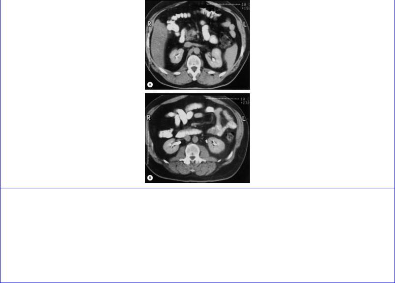
Figure 5.44 CT scans of the upper abdomen (viewed by convention from below). In A, the left renal vein crosses in front of the aorta to enter the inferior vena cava. The right crus of the diaphragm is seen beside the aorta. In B, the pelvis of the right kidney is indicated by the white opacity (representing oral contrast medium which has been absorbed and excreted by the kidney), while on the left side the ureter, similarly outlined, lies immediately medial to the lower pole of the kidney. The psoas major and quadratus lumborum muscles are seen on either side of the lumbar vertebra.
The left renal vein may have to be ligated and divided during surgery for aortic aneurysm. Provided that this is done to the right of the point of entry of the gonadal and suprarenal veins the kidney is not harmed. Rarely, the left renal vein may be double, with one vein passing anterior and one posterior to the aorta, or only a posterior vein may be present.
While the left suprarenal vein runs downwards and medially to the left renal vein, the right suprarenal vein is a very short vessel that passes horizontally to the posterior aspect of the inferior vena cava behind the bare area of the liver (Fig. 5.43)—an arrangement that complicates the surgery of the right gland.
The inferior phrenic veins accompany the arteries on the lower surface of the diaphragm and normally join the vena cava just below the liver, but the left vessel may join the left renal or suprarenal vein, or even be double with different destinations.
The three main hepatic veins (right, central and left) and several accessory hepatic veins enter the vena cava as it lies in its groove on the back of the liver; they are further described on page 262.
Lymph nodes and lymph trunks
From nodes which drain the alimentary tract, liver, biliary tract, spleen and pancreas (see p. 247), lymphatics pass back along the coeliac, superior and inferior mesenteric arteries to preaortic nodes
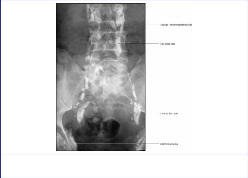
situated around the origins of these three vessels. Similarly, lymphatics pass back along the paired branches of the aorta, both visceral and somatic, to para-aortic nodes which lie alongside the aorta at the origins of the paired vessels. The lymph drainage of any viscus follows its artery back to the aorta. Lymph from some pelvic viscera drains through nodes along the internal iliac arteries, to nodes along the common iliac arteries. These nodes also receive lymph from the lower limb via the inguinal (superficial and deep) nodes and external iliac nodes. The common iliac nodes drain up to the paraaortic nodes (Fig. 5.45).
Figure 5.45 Lymphangiogram showing lymph nodes on the pelvic brim and posterior abdominal wall.
From the highest of these aortic groups, a variable number of intestinal and lumbar lymph trunks (and smaller lymphatics from the posterior ends of the lower intercostal spaces) join to form the elongated, sac-like cisterna chyli (though frequently the single cisterna is replaced by a confluence of lymph trunks). It is situated under cover of the right crus, in front of the bodies of L1 and L2 vertebrae, between the aorta and azygos vein. Its upper end becomes continuous with the thoracic duct (see p. 210).
Some of the fat absorbed by intestinal cells enters blood capillaries and so reaches the liver directly by the portal vein, but other processed lipid molecules enter the lacteals (lymph vessels) of the intestinal villi as chylomicrons, so producing the milky-looking lymph which enters the bloodstream via the cisterna chyli and thoracic duct.

Nerves
Somatic nerves
The anterior rami of the upper four lumbar spinal nerves at their emergence from the intervertebral foramina necessarily pass into the substance of psoas major. They give segmental branches of supply to psoas and quadratus lumborum and then break up into anterior and posterior divisions, which reunite to form the branches of the lumbar plexus within the substance of psoas. Most of the branches are for the supply of the lower limb; others innervate the lower anterior abdominal wall and give sensory branches to the parietal peritoneum. The branches of the lumbar plexus are summarized on page 325.
The segmental outflow from the spinal cord continues in series with the intercostal nerves of the thoracic wall. T12 (the subcostal nerve) and all five lumbar nerves emerge serially from the intervertebral foramina, but they do not all share in the supply of the anterior abdominal wall. Only T12 and L1 do so, in fact. L2, 3 and 4, after each giving a branch to the muscles of the posterior abdominal wall (psoas and quadratus lumborum) participate, through the lumbar plexus, in the formation of nerves for the flexor and extensor compartments of the thigh. The nerves to the thigh are the obturator (for the adductor compartment, a derivative of the flexor compartment of the thigh), and the femoral and lateral femoral cutaneous nerves (extensor compartment). A part of L4 and all L5 pass down as the lumbosacral trunk to the sacral plexus for distribution to the lower limb (see p. 310).
Nerves for the supply of the anterior abdominal wall cross the anterior surface of quadratus lumborum. They are the subcostal and the iliohypogastric and ilioinguinal nerves (Fig. 5.42A).
The subcostal nerve (T12) passes from the thorax behind the lateral arcuate ligament, where it lies below the vein and artery. The neurovascular bundle slopes down (parallel with the twelfth rib) across the front of the anterior layer of the lumbar fascia behind the kidney. The subcostal nerve passes through transversus abdominis to reach the neurovascular plane and continues around the anterior abdominal wall, whose muscles it supplies, ending by supplying the lower part of rectus abdominis, the pyramidalis muscle and the skin over them. It has a collateral (muscular) branch, and its lateral cutaneous branch pierces the oblique muscles and descends over the iliac crest to supply the skin of the anterior part of the buttock between the iliac crest and the greater trochanter.
The iliohypogastric and ilioinguinal nerves lie in front of quadratus lumborum at a lower level. They both arise from the anterior ramus of L1. The ilioinguinal nerve represents the collateral branch of the iliohypogastric. The nerves emerge from the lateral border of psoas major as a common stem (which then divides) or as separate nerves. As they slope across quadratus lumborum, behind the kidney, they pierce the anterior layer of the lumbar fascia and pass in front of it through transversus abdominis, to continue downwards and forwards, above the iliac crest, in the neurovascular plane.
The iliohypogastric nerve gives a lateral cutaneous branch which pierces the oblique muscles above the iliac crest to supply skin of the upper part of the buttock behind the area supplied by the subcostal nerve. The nerve then continues downwards in the neurovascular plane and pierces the internal oblique above the anterior superior iliac spine. It pierces the external oblique aponeurosis about 2.5 cm above the superficial inguinal ring and ends by supplying suprapubic skin.
The ilioinguinal nerve runs parallel with the iliohypogastric at a lower level. Piercing the lower border of internal oblique, it enters the inguinal canal and emerges through the superficial inguinal ring covered by the external spermatic fascia, which it pierces to become subcutaneous. It supplies the anterior one-third of the scrotum (mons pubis and labium majus), the root of the penis (clitoris), and the upper and medial part of the groin. Before it perforates the lower border of the internal oblique muscle it gives motor branches to those muscle fibres of internal oblique and transversus which are inserted into the conjoint tendon and are thereby of importance in maintaining the integrity of the inguinal canal.
The lateral femoral cutaneous nerve is formed by union of fibres from the posterior divisions of the anterior rami of L2 and L3. It emerges from the lateral border of psoas major and passes across the iliac fossa on the surface of the iliacus muscle (Fig. 5.42A) deep to the iliac fascia, posterior to the caecum on the right and the descending colon on the left. The nerve passes below or perforates the inguinal ligament a centimetre from the anterior superior iliac spine and so enters the thigh (see Fig. 3.2, p. 113). The nerve supplies the parietal peritoneum of the iliac fossa. Its distribution in the thigh is described on page 113.
The femoral nerve is formed in the substance of psoas major by union of branches from the posterior divisions of the anterior rami of L2–4. It emerges from the lateral border of psoas in the iliac fossa and runs down deep in the gutter between psoas and iliacus behind the iliac fascia. It gives branches to iliacus (L2 and 3) and leaves the iliac fossa by passing beneath the inguinal ligament to the lateral side of the femoral sheath, on the iliacus muscle (see Fig. 3.1, p. 112). Its course in the thigh is described on page 119.
The above nerves emerge from the lateral margin of psoas. The genitofemoral and obturator nerves have each a different pathway.
The genitofemoral nerve is formed in the substance of psoas major by union of branches from L1 and 2. It emerges from the anterior surface of psoas major and runs down on the muscle deep to the psoas fascia. In front of the fascia the left nerve is overlaid by the ureter, gonadal vessels, ascending branch of the left colic artery and the inferior mesenteric vein. The right nerve is overlaid by the ureter, gonadal vessels, the ileocolic artery and the root of the mesentery of the small intestine. Just above the inguinal ligament it perforates the psoas fascia and divides into genital and femoral branches (see Fig. 3.1, p. 112). The genital branch is composed of L2 and the femoral branch of L1 fibres.
The genital branch passes through the deep ring and enters the inguinal canal. It supplies motor fibres to the cremaster muscle, and sensory fibres to the spermatic fasciae, tunica vaginalis of the testis, and a small area of skin of the scrotum in males and the mons pubis and labium majus in females. The femoral branch passes down behind the inguinal ligament with the femoral artery, pierces the femoral sheath and the fascia lata, and supplies the skin over the upper part of the femoral triangle.
The obturator nerve and the lumbosacral trunk emerge from the medial border of psoas to enter the pelvis (see p. 310).
Autonomic nerves
The abdomen receives both sympathetic and parasympathetic nerves. The sympathetic supply is twofold, by the lumbar part of the sympathetic trunk and by the coeliac plexus which receives fibres from the thoracic part of the trunk. The coeliac plexus is wholly visceral; it supplies all the abdominal organs, including the gonads. The ganglionated lumbar trunk supplies somatic branches for the lower abdominal wall and the lower limb, but its visceral branches supply only the pelvic organs. The parasympathetic supply is provided by the vagus from above and the pelvic splanchnic nerves from below; it is wholly visceral. The vagus gives a branch to the coeliac plexus and the pelvic splanchnics join the inferior hypogastric plexus.
Sympathetic nerves
The lumbar part of the sympathetic trunk brings preganglionic fibres descending from the lower thoracic trunk, and it receives a further input of preganglionic fibres (white rami) from the first and second lumbar nerves. Its ganglia give off somatic and visceral branches, and the trunk passes down across the pelvic brim, behind the common iliac vessels, to become the sacral part of the trunk (see p. 311).
The lumbar part of the sympathetic trunk enters the abdomen by passing behind the medial arcuate ligament on the front of psoas major. It runs down behind the peritoneum on the vertebral bodies along the medial margin of the psoas muscle (Fig. 5.46). As elsewhere it lies in front of the segmental vessels, the lumbar arteries and veins, but some lumbar veins may pass in front of the trunk.
Figure 5.46 Sympathetic rami at L1 or L2 vertebral levels. An accessory ganglion in psoas major relays directly back to the segmental nerve; the remainder of L1 and L2 outflow passes into the sympathetic trunk for local or distant relay.
The left lumbar trunk lies beside the left margin of the aorta (Fig. 5.43) with para-aortic lymph nodes in front of it, while the right trunk lies behind the inferior vena cava.
The lumbar ganglia are usually four, but fusion of ganglia may reduce the number. White rami communicantes from the first two lumbar nerves join the trunk and relay in the lumbar and sacral ganglia. Grey rami communicantes from the lumbar ganglia accompany the lumbar arteries around the sides of the vertebral bodies, medial to the fibrous arches, to join the anterior rami of lumbar nerves, for distribution to the body wall and lower limb, through the branches of the lumbar plexus. Vasoconstrictor fibres for the femoral artery and its branches travel in the femoral nerve.
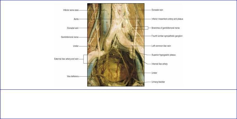
Lumbar splanchnic nerves arise from all lumbar ganglia. Those from the first and second ganglia pass to plexuses in front of the aorta; those from the third and fourth ganglia pass respectively in front of and behind the common iliac arteries. They join with each other and with fibres from the aortic plexuses to form the superior hypogastric plexus (Fig. 5.47), which comprises both preand postganglionic sympathetic fibres. This plexus lies anterior to the aortic bifurcation and the left common iliac vein and between the common iliac arteries, in front of L5 vertebra and the sacral promontory. It was previously known as the presacral nerve—a misnomer as it is prelumbar and a plexus, not presacral and a single nerve. It lies behind the parietal peritoneum which can be stripped off its anterior aspect surgically, an avascular areolar tissue plane lying between them. The plexus often lies a little to the left of the midline, close to the apex of the attachment of the sigmoid mesocolon. The plexus divides in the manner of an inverted Y into the right and left hypogastric nerves (which may not be single but a bundle) which run down into the pelvis to join the inferior hypogastric plexuses (see p. 311). There are only a few ganglion cells in the superior hypogastric plexus; preganglionic white fibres in the hypogastric nerves pass through to relay in ganglia in the inferior hypogastric plexuses.
Figure 5.47 Prosection of posterior abdominal wall and pelvis in the Anatomy Museum of the Royal College of Surgeons of England. The abdominal and pelvic viscera have been removed except for the urinary bladder. Red and blue resin has been injected into arteries and veins.
The coeliac plexus lies around the origin of the coeliac trunk above the upper border of the pancreas. The greater and lesser splanchnic nerves (see p. 211) pierce the crura of the diaphragm and enter the two large coeliac ganglia which lie in front of the crura, the right one behind the inferior vena cava and the left behind the splenic artery. The splanchnic nerves are almost all preganglionic (white) and many relay in the coeliac ganglia. The least splanchnic nerve relays in a small renal ganglion behind the renal artery. This is merely an offshoot of the main coeliac ganglion itself. Separated masses of ganglion may lie on the aorta at the superior and even the inferior mesenteric artery origins.
Postganglionic fibres from the coeliac ganglia and preganglionic splanchnic fibres form a network on the aorta, the relevant parts of which are the coeliac, superior mesenteric, intermesenteric (abdominal

aortic) and inferior mesenteric plexuses, in accordance with their relation to the origin of the midline gut arteries. The fibres from these plexuses supply all the abdominal viscera, which they reach by streaming along the visceral branches of the aorta. Those passing to the kidney pick up the branches of the renal ganglion to form the renal plexus behind the renal artery. Testis and ovary are supplied by a sympathetic plexus that accompanies each gonadal artery.
The sympathetic fibres are vasomotor, motor to sphincters (e.g. pyloric), inhibitory to peristalsis, and carry sensory fibres from all the viscera supplied.
Pre-ganglionic fibres from the greater splanchnic nerve pass without relay to the cells of the suprarenal medulla (these cells share a common origin from neural crest ectoderm with the cell bodies of sympathetic ganglia). These preganglionic fibres cause the suprarenal medulla to pour forth adrenaline. The vasomotor supply to the suprarenal gland reaches it by postganglionic fibres which have relayed in the coeliac ganglion.
Lumbar sympathectomy
Surgical removal of the third and fourth lumbar ganglia employs an extraperitoneal approach through a transverse muscle-cutting incision in the anterior abdominal wall, or the flank, on the appropriate side. The intact peritoneum is stripped off the deep surface of transversus abdominis and the posterior abdominal wall as far as the vertebral column, carefully avoiding damage to the gonadal vessels, ureter and genitofemoral nerve overlying psoas major. Exposure of the right sympathetic trunk requires careful retraction of the inferior vena cava, which lies in front of it. On the left the trunk is beside the aorta and more easily accessed. Although the lumbar vessels usually lie behind the sympathetic trunks, some lumbar veins may pass in front of the trunks. Lumbar ganglia may also be destroyed by injecting them with phenol via a posterior paravertebral approach under radiographic or ultrasonic guidance.
Parasympathetic nerves
Both vagi, intermixed in the vagal trunks (see p. 250), contribute fibres to the coeliac plexus. Without relay they accompany postganglionic sympathetic fibres to the gut as far as the distal transverse colon. They also enter the renal plexus and pass into the kidney.
The pelvic splanchnic nerves, from cell bodies in the lateral horn of sacral segments 2 to 3 or 4 (see p. 20), arise from the anterior rami of those sacral nerves distal to the anterior sacral foramina. They join the inferior hypogastric plexus for distribution to pelvic viscera. Some fibres rise up out of the pelvis, usually passing to the left of the superior hypogastric plexus, occasionally through it, to ascend behind the peritoneum and supply the large intestine from the splenic flexure downwards.
The vagus is motor and secretomotor to the gut and its glands down to the transverse colon; the pelvic splanchnics fulfil this function from the splenic flexure distally. The vagus is inhibitory to the pyloric sphincter. Sensory fibres are present in the vagus and pelvic splanchnics.