

Pediatrics(2)
.pdf-- Surgical repair generally before 1 year if possible
-3.4.2. Cyanotic Heart Diseases
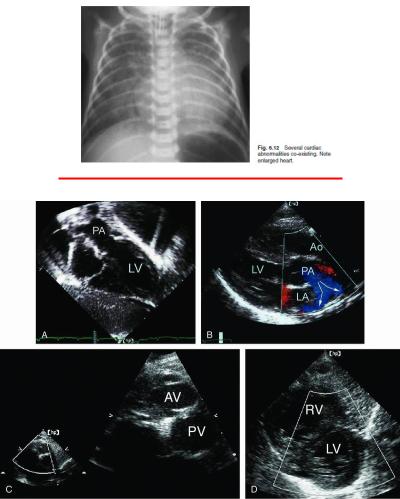
An example of D-transposition of the great arteries in an infant is shown. A: From the subcostal view, the pulmonary artery (PA) can again be seen to arise from the anatomic left ventricle (LV). B: By demonstrating
bifurcation of the great artery that arises from the posterior left ventricle, ventriculoarterial discordance is confirmed. C: A short-axis view at the base of the heart demonstrates the parallel course of the great arteries with an anterior aortic valve (AV). D: The right ventricle (RV) is seen anterior and rightward of the left ventricle. It is dilated and hypertrophied. Ao, aorta; LA, left atrium.
-Definition: Cyanotic heart disease is a heart defect, present at birth (congenital), that results in low blood oxygen levels (< 90 % even with oxygen).
-
-Common lesions
-decreased flow to the lungs (does not cause heart failure)
-• Tetralogy of fallot
-• Pulmonary atresia
-- Increased flow to the lungs (does cause heart failure and failure to thrive):
-• Transposition of great vessels (TGA)
-• Truncus arteriosus
-• Single ventricle / Tricuspid atresia
-Tetralogy of Fallot
-
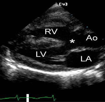
-Long-axis image from a patient with tetralogy of Fallot demonstrates the overriding aorta (Ao) and a large subaortic ventricular septal defect (asterisk). Right ventricular hypertrophy is also present. LA, left atrium; LV, left ventricle; RV, right ventricle.
-Definition: Tetralogy of Fallot refers to a type of congenital heart defect comprising of:
-- Large ventricular septal defect
-- Narrowing of the pulmonary outflow tract (pulmonary stenosis)
-- Overriding aorta
-- Right ventricular hypertrophy
-Signs and Symptoms
-- Progressive cyanosis with pulmonary systolic murmur
-- digital clubbing occurs after long time
-- Hallmark: Paroxysmal hyper cyanotic attacks (blue spells) with the following manifestations
-• Hyperpnea and restlessness
-• Increased cyanosis
-• Gasping respiration
-• Syncope or convulsions
-•Spontaneous squatting position is frequent (in older children)
-• Heart murmur disappears
-Complications
-- delayed development/growth
-- Polycythemia
-- Hypercyanotic attack, sometimes associated with seizures and death
-Infective endocarditis
-Brain abscess
-Investigations
-- Chest x-ray
-- Complete blood count (CBC)
-- Echocardiogram
-- Electrocardiogram (ECG)
-Management
-- Avoid dehydration and stress
-- Propanolol 0.5-1mg/kg every 6 hours to prevent hypercyanotic attacks
-- Iron 5mg/kg /day to prevent microcytosis
-- Surgical repair, urgent as soon as spells begin
-- In case of Hypercyanotic attacks
-• Squatting position (hold the infant with the legs flexed on the abdomen)
-• Oxygen 6l/min with mask
-• Diazepam 0.3mg/kg IV or 0.5mg PR if convulsing
-• normal saline 10-20ml/kg/ 30 minutes
-• Sodium bicarbonate 8.5% 1ml/kg to correct acidosis
-• Morphine 0.1mg/kg IV if persistent attacks (but risk of
-respiratory depression)
-• Propranolol IV 0.1 – 0.2 mg/kg slowly then continue oral maintenance to relax the infundibular spasms
-Recommendations
-- All children with cyanotic heart diseases who come with diarrhea and vomiting should be admitted for closer observation.
-Furosemide is contra-indicated.
-- All new born babies with suspected cyanotic heart disease should be referred to a cardiologist/tertiary hospital immediately.
-- Common causes of heart failure in Neonates:
-
-3.5. Acquired Heart Diseases
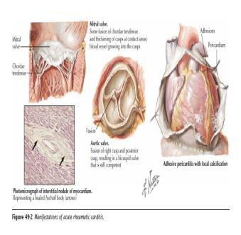
-3.5.1. Acute Rheumatic fever
-
-
-
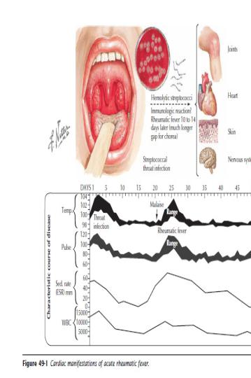
-Definition: This is an acute, systemic connective tissue disease in
-children related to an immune reaction to untreated group A Beta
-haemolytic streptococcus infection of the upper respiratory tract. The initial attack of acute rheumatic fever occurs in most cases between the ages of 3 and 15 years.
-Cause
-- Auto-immune disease
-Signs and Symptoms (Revised Jones Criteria)