

Pediatrics(2)
.pdf-
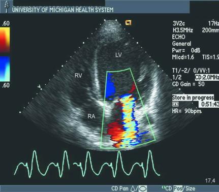
- A small aortic valve vegetation (arrow) is shown during diastole (A) and systole (B). C:
Color Doppler demonstrates severe aortic regurgitation. Ao, aorta; LA, left atrium; LV, left ventricle; RV, right ventricle.
-Definition: Infection of the endothelial surface of the heart. Suspectc infective endocarditis in all children with persistent fever and underlying heart disease.
-Causes/predisposing factors
-- Rheumatic valvular disease
-- Congenital heart disease
-Signs and Symptoms
-- Persistent low grade fever without an obvious underlying cause
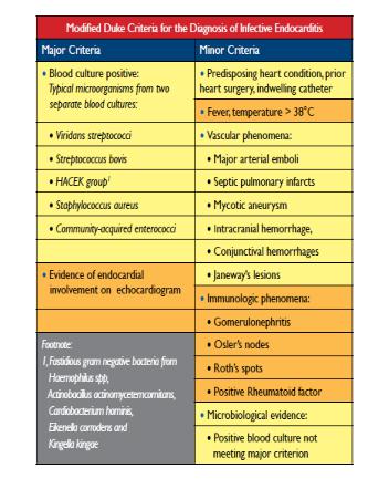
-- Fatigue, joint pain, new murmurs, clubbing, splenomegaly and haematuria
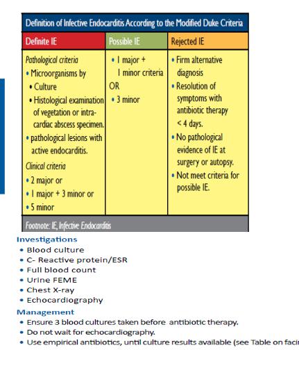
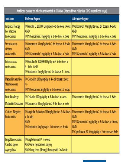
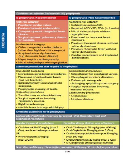
-DUKE CRITERIA IN CHILDREN
-
3.6. Cardiomyopathies
3.6.1. Dilated Cardiomyopathy
Apical four-chamber view recorded in a patient with a dilated cardiomyopathy and functional mitral regurgitation that is the result of lateral displacement of the papillary muscles. Note the mitral regurgitation jet, with the color flow Doppler signal filling approximately
40% of the left atrial area. LV, left ventricle; RA, right atrium; RV, right ventricle.
Definition: dilated cardiomyopathy refers to a group of conditions of diverse etiology in which both ventricles are dilated with reduced contractility.
Classification
Classification based on the predominant structural and functional abnormalities
-dilated Cardiomyopathy: primarily systolic dysfunction
-Hypertrophic Cardiomyopathy: primarily diastolic dysfunction
-Restrictive Cardiomyopathy: primarily diastolic but often combined with systolic dysfunction
Causes
-Infections (e.g. Viral+++, Rickettsia, Chagas disease)
-Neuromuscular disorders (e.g. duchenne dystrophy, Becker dystrophy)
-Endocrine, metabolic and nutritional (e.g. hyperthyroidism, Fatty acid oxidation disorders, beriberi, kwashiorkor)
-diseases of coronary arteries (e.g. Kawasaki, Aberrant Left Coronary Artery - ALCAPA)
-Autoimmune diseases (e.g. Rheumatic carditis, juvenile rheumatoid arthritis, systemic lupus erythematosus, dermatomyositis, systemic lupus erythematosus)
-drugs toxicity (e.g. doxorubicin,cyclophosphamide, IPECA)
-Hematologic diseases (e.g. anemia, Sickle cell anemia, hypereosinophilic syndrome Löffler syndrome)
Signs and Symptoms (See signs of congestive heart failure)
Diagnosis
-ECG: proeminent P wave, LV or RV hypertrophy, nonspecific T-wave abnormalities
-Chest X-ray: cardiomegaly, pulmonary edema
-Echocardiogram: confirm diagnosis and shows LA and LV dilation, poor contractility
-FBC, Urea and creatinine, Electrolytes (Na, K)
-Myocardial biopsy, PCR
Management
- Treatment: (Refer to principles and medication of congestive heart failure)
3.6.2. Hypertrophic Cardiomyopathy
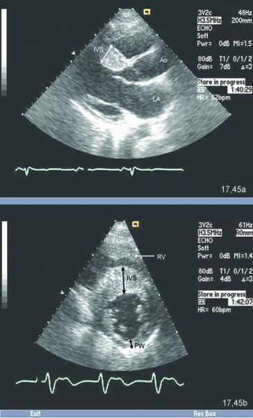
Parasternal long-axis (A) and short-axis (B) views recorded in a patient with classic hypertrophic cardiomyopathy. In both the long-axis and short-axis views, note the marked thickening of the interventricular septum and the normal thickness of the posterior wall (PW). In the short axis view, note that there is a spectrum of hypertrophy of the left ventricle, with maximum hypertrophy in the septum, no hypertrophy of the true posterior wall, and intermediate hypertrophy of the lateral and true inferior wall. Ao, aorta; IVS, interventricular septum; LA, left atrium; RV, right ventricle.
Causes
-Left ventricle obstruction (Coartation of aorta, hypertension, aortic stenosis)
-Secondary (infants of diabetic mothers, corticosteroids in premature infants)
-Metabolic (Glycogen storage disease type II (Pompe disease)
-Familiar hypertrophic cardiomyopathy
-Syndroms (Beckwith - Wiedman syndrom, Friedereich, ataxia)
Signs and Symptoms
-Weakness
-Fatigue
-dyspnea on effort
-Palpitations
-Angina pectoris