

Kluwer - Handbook of Biomedical Image Analysis Vol
.2.pdfxxii |
Contents |
|
9. |
Accurate Lumen Identification, Detection, and Quantification in MR |
|
|
Plaque Volumes . . . . . . . . . . . . . . . . . . . . . . . . . . . . . . . . . . . . . . . . . . . . . . |
451 |
|
Jasjit Suri, Vasanth Pappu, Olivier Salvado, Baowei Fei, |
|
|
Swamy Laxminarayan, Shaoxiong Zhang, Jonathan Lewin, |
|
|
Jeffrey Duerk, and David Wilson |
|
10. |
Hessian-Based Multiscale Enhancement, Description, and Quantification |
|
|
of Second-Order 3-D Local Structures from Medical Volume Data . . . . . . |
531 |
|
Yoshinobu Sato |
|
11. |
A Knowledge-Based Scheme for Digital Mammography . . . . . . . . . . . . . . |
591 |
|
Sameer Singh and Keir Bovis |
|
12. |
Simultaneous Fuzzy Segmentation of Medical Images . . . . . . . . . . . . . . . . |
661 |
|
Gabor T. Herman and Bruno M. Carvalho |
|
13. |
Computer-Aided Diagnosis of Mammographic Calcification Clusters: |
|
|
Impact of Segmentation . . . . . . . . . . . . . . . . . . . . . . . . . . . . . . . . . . . . . . . . |
707 |
|
Maria Kallergi, John J. Heine, and Mugdha Tembey |
|
14. |
Computer-Supported Segmentation of Radiological Data . . . . . . . . . . . . . . |
753 |
|
Philippe Cattin, Matthias Harders, Johannes Hug, Raimundo Sierra, |
|
|
and Gabor Szekely |
|
The Editors . . . . . . . . . . . . . . . . . . . . . . . . . . . . . . . . . . . . . . . . . . . . . . . . . . . . . . |
799 |
|
Index . . . . . . . . . . . . . . . . . . . . . . . . . . . . . . . . . . . . . . . . . . . . . . . . . . . . . . . . . . |
805 |
|

Chapter 1
Model-Based Brain Tissue Classification
Koen Van Leemput,1 Dirk Vandermeulen,2 Frederik Maes,2
Siddharth Srivastava,2 Emiliano D’Agostino,2 and Paul Suetens2
1.1 Introduction
Several neuropathologies of the central nervous system such as multiple sclerosis (MS), schizophrenia, epilepsy, Alzheimer, and Creutzfeldt–Jakob disease (CJD) are related to morphological and/or functional changes in the brain. Studying such diseases by objectively measuring these changes instead of assessing the clinical symptoms is of great social and economical importance. These changes can be measured in three dimensions in a noninvasive way using current medical imaging modalities. Magnetic resonance imaging (MRI), in particular, is well suited for studying diseases of the nervous system due to its high spatial resolution and the inherent high soft tissue contrast.
Manual analysis of MRI by a trained human expert is a tedious and difficult task, because the structures of interest show complex edge configurations in 3D and may lack clearly visible anatomical borders. In clinical trials, the number of MR images is often so large that manual analysis by human experts is too time-consuming. Furthermore, it is not clear how a human rater combines information obtained from different channels when multispectral MR data are examined. Also, the intraand interobserver variability associated with manual delineations complicates the analysis of the results. Therefore, there is a need for fully automated methods for MR brain image quantification that can analyze
1 Department of Radiology, Helsinki University Central Hospital, Finland
2 Medical Image Computing (Radiology -ESAT/PSI), Faculties of Medicine and Engineering, Katholieke Universiteit Leuven, Belgium
1
2 |
Leemput et al. |
large amounts of multispectral MR data in a reproducible way that correlates well with expert analyses.
A key component in image analysis and interpretation is image segmentation, defined as the delineation of anatomical structures and other regions of interest. In this chapter we will present a framework for the accurate segmentation of brain tissues (Gray matter, white matter, CSF) from multispectral MR images of the brain. We will also discuss how this framework can be used to quantify pathology-related abnormalities (mostly in intensity but also in morphology) within these tissues.
The overall strategy is to build statistical models for normal brain MR images, with emphasis on accurate intensity models. Signal abnormalities are detected as model outliers, i.e., voxels that cannot be well explained by the model. Special attention is paid to automatically estimate all model parameters from the data itself and, hence, to eliminate subjective manual tuning and training.
1.1.1 Segmentation Methodology
The simplest image segmentation methods, such as region growing and edge detection, rely entirely on local image operators and a heuristic grouping of neighboring pixels with similar local photometric characteristics. These approaches are simple to understand and to implement, and are very generic since they do not assume specific knowledge about the objects to be analyzed. However, these methods ultimately fail when either or both of the image data and the object model (shape, context) are complex, as in cross-sectional images of the brain. Indeed, the complex 3-D shape of the brain and its affected areas, and ambiguities in the images induced by the imaging process, such as limited resolution, partial volume effects, noise, low contrast, intensity inhomogeneities, and other artifacts, make brain tissue segmentation difficult, even for human experts.
In order to effectively deal with this complex morphology and MR imaging ambiguity, brain image segmentation methods must incorporate models that describe prior knowledge about the expected geometry and intensity characteristics of the anatomical objects of interest in the images.
Intensity-based methods fit appropriate photometric models to the data. In these approaches, the objects are simple voxels with an associated
Model-Based Brain Tissue Classification |
3 |
scalar, such as the gray value, or vector of characteristic features, such as (ρ, T1, T2)-weighted intensity values in MRI. If the feature vectors are represented in a multidimensional feature space, the segmentation strategy then consists of partitioning this feature space in a number of nonoverlapping regions that separate different voxel types. Unclassified voxels then receive the label of their class in feature space. The boundaries between the regions in features space are obtained by optimizing any of a set of decision criteria, depending on the a priori assumptions about the feature space distributions. Parametric classification approaches assume that the distributions in feature space follow a certain parametric model. Typically, the multivariate Gaussian model is used. This model can be extended to explicitly include imaging artifacts such as the partial volume effect [1–3] and the intensity inhomogeneity present in MR images [4–6]. Parameter values for the distribution of each class can be learned from a representative set of samples in a supervised training phase, usually requiring cumbersome user interaction. Fully automated, unsupervised learning procedures estimate the distribution parameters from the image to be segmented itself.
Geometry-based methods use prior knowledge about the overall shape of an object to separate it from its surroundings in the image. Typically, a surface deforms under the influence of external image derived forces (attracting the surface to object edges, etc.) and internal elasticity constraints (e.g. surface continuity and smoothness) [7]. An extensive survey of these methods in medical image analysis is given in [8]; recent examples include [9–12]. Within the context of brain image analysis, deformable brain atlasguided approaches have been proposed. Here, prior knowledge about the image scene is represented iconically (i.e. an image-like representation). The image is segmented by matching it with the iconic atlas representation. The matching process must have sufficient degrees of freedom and robustness so as to cope with the biological variability and pathological abnormalities. However, even with nonlinear registration methods, accurate brain tissue segmentations are difficult due to anatomical variability of the cortical folding.
Intensity-based tissue classification and geometry-based atlas-driven meth-
ods are seemingly complementary segmentation strategies.
4 |
Leemput et al. |
The advantage of MR-intensity-based tissue classification is its ability to produce high-quality definitions of tissue boundaries. This is especially important for human brain tissue classification, where highly curved interfaces between tissues (such as between gray and white matter) are difficult to recover from finite resolution images. However, it is unsuccessful when different structures have overlapping feature distributions (e.g., brain tissue and extracranial tissue in T1-weighted MRI).
Geometry-based methods have been successfully applied to the localization of particular anatomical structures, where sufficient information on shape and context is available. These methods, however, often require accurate initialization and, more importantly, can fail in the case of highly variable structures such as the cerebral cortex and in the presence of abnormal anatomical structures.
Following Warfield et al. [13] and Collins et al. [14], who developed the idea that the inherent limitations of intensity-based classification can be alleviated by combining it with an elastically deformable atlas, we propose here to combine photometric and geometric models in a single framework. Tissues are classified using an unsupervised parametric approach by using a mixture of Gaussians as the feature space distribution model in an iterative expectation-maximization loop. Classifications are further initialized and constrained by iconic matching of the image to a digital atlas containing spatial maps of prior tissue probabilities.
Section 1.1.2 presents the standard intensity model and optimization approach that we will use throughout this chapter. Section 1.1.3 discusses the basic geometric model (a brain atlas) and its iconic matching to image data using linear and nonlinear registration algorithms. Section 1.1.4 summarizes the changes made to the basic intensity model in order to model the MR imaging process more faithfully and to make the segmentation procedure more robust in the presence of abnormalities.
1.1.2Intensity Model and the Expectation-Maximization (EM) Algorithm
The intensity model proposed here is the so-called mixture of normal distributions [15–17]. Let Y = {yj , j = 1, 2, . . . , J} be a C-channel MR image with a
Model-Based Brain Tissue Classification |
5 |
total of J voxels, where yj denotes the possibly multispectral intensity of voxel j. Suppose that there are K tissue types present in the imaged area, and let l j {1, 2, . . . , K } denote the tissue type to which voxel j belongs. In the mixture model, it is assumed that each tissue type k has a typical intensity µk in the image, with tissue-specific normally distributed intensity fluctuations in the voxels. In other words, the probability density that voxel j of tissue type l j has intensity yj is given by
f (yj | l j , ) = GΣl j (yj − µl j ) |
(1.1) |
Here GΣ(·) denotes a zero-mean normal distribution with covariance Σ, and
= {µk, Σk, k = 1, 2, . . . , K } represents the total set of model parameters. For notational convenience, let all the voxel labels l j be grouped in a label
image L = {l j , j = 1, 2, . . . , J}. It is assumed that the label l j of each voxel is drawn independently from the labels of the other voxels, with an a priori known probability πk, i.e.
f (L) = πl j (1.2)
j
The overall probability density for image Y given the model parameters is then given by
f (Y | ) = f (Y | L, ) f (L)
L
= f (yj | )
j |
|
|
|
with f (yj | ) = f (yj | l j = k, ) · πk |
(1.3) |
k
Equation (1.3) is the well-known mixture model (see Fig. 1.1). It models the histogram of image intensities as a sum of normal distributions, each distribution weighted with its prior probability πk.
Image segmentation aims to reconstruct the underlying tissue labels L based on the image Y. If an estimation of the model parameters is somehow available, then each voxel can be assigned to the tissue type that best explains its intensity. Unfortunately, the result depends largely on the model parameters used. Typically, these are estimated by manually selecting representative points in the image of each of the classes considered. However, once all the voxels are classified, the model parameter estimation can in its turn automatically be improved based on all the voxels instead of on the subjectively selected ones alone.

6 |
Leemput et al. |
histrogram white matter gray matter CSF
total mixture model
(a) |
(b) |
Figure 1.1: The mixture model fitted to a T1-weighted brain MR image: (a) the intracranial volume and (b) its intensity histogram with a mixture of normal distributions overlayed. The normal distributions correspond to white matter, gray matter, and CSF.
Intuitively, both the segmentation and the model parameters can be estimated in a more objective way by interleaving the segmentation with the estimation of the model parameters.
The expectation-maximization (EM) algorithm [18] formalizes this intuitive
ˆ
approach. It estimates the maximum likelihood (ML) parameters
ˆ |
max log |
( |
Y | |
) |
= arg |
|
f |
|
by iteratively filling in the unknown tissue labels L based on the current parameter estimation , and recalculating that maximizes the likelihood of the so-called complete data {Y, L}. More specifically, the algorithm interleaves two steps:
Expectation step: find the function
Q( | (m−1)) = EL[log f (Y, L | ) | Y, (m−1)]
Maximization step: find
(m) = arg max Q( | (m−1))
with m the iteration number. It has been shown that the likelihood log f (Y | ) is guaranteed to increase at each iteration for EM algorithms [19].
Model-Based Brain Tissue Classification |
7 |
With the image model described above, the expectation step results in a statistical classification of the image voxels
f (l |
|
Y, (m−1)) |
|
f (yj | l j , (m−1)) · πl j |
|
j | |
= k f (yj | l j = k, (m−1)) · πk |
||||
|
|
||||
and the subsequent maximization step involves
|
|
(m) |
|
|
|
j f (l j = k | Y, (m−1)) · y(jm−1) |
|
|
||||||||||
|
|
µk |
|
= |
|
j f (l j = k | Y, (m−1)) |
|
|
|
|||||||||
|
|
j f (l j |
|
|
k |
|
(m 1) |
) |
|
(m−1) |
(m) |
) |
(m−1) |
(m) |
t |
|||
(m) |
= |
= |
| |
Y, |
− |
(yj |
|
|
− µk |
· (yj |
− µk ) |
|
||||||
k |
|
|
|
j f (l j· |
|
k |
|
Y, (m−1)) |
|
|
|
|||||||
|
|
|
|
|
|
|
|
|
|
= |
|
| |
|
|
|
|
|
|
(1.4)
(1.5)
(1.6)
Thus, the algorithm iteratively improves the model parameters by interleaving two steps (see Fig. 1.2): classification of the voxels based on the estimation of the normal distributions (Eq. 1.4) and estimation of the normal distributions based on the classification (Eqs. 1.5 and 1.6). Upon convergence, Eq. (1.4) yields the final classification result.
classify
update mixture model
Figure 1.2: Estimating the model parameters of the mixture model with an expectation-maximization algorithm results in an iterative two-step process that interleaves classification of the voxels with reestimation of the normal distributions.

8 |
Leemput et al. |
1.1.3 Geometry Model and the MMI Algorithm
The iterative model parameter estimation algorithm described above needs to be initialized with a first estimate of the parameters. One possibility to obtain such an estimate is to have the user manually select voxels that are representative for each of the classes considered. However, to eliminate the variability induced by such a preceding training phase, we avoid manual intervention by the use of a digital brain atlas that contains spatially varying prior probability maps for the location of white matter, gray matter, and CSF (see Fig. 1.3). These probability maps were obtained by averaging binary white matter, gray matter, and CSF segmentations of MR brain images from a large number of subjects, after normalization of all images into the same space using an affine transformation [20]. To apply this a priori information, the atlas is first normalized to the space of the study image by matching the study image to a T1 template associated with the atlas (see Fig. 1.3) by using the affine multimodality registration technique based on maximization of mutual information (MMI) of Maes et al. [21].
Mutual information (MI) is a basic concept from information theory, which is applied in the context of image registration to measure the amount of information that one image contains about the other. The MMI registration criterion postulates that MI is maximal when the images are correctly aligned. Mutual information does not rely on the intensity values directly to measure correspondence between different images, but on their relative occurrence in each of the
(a) |
(b) |
(c) |
(d) |
Figure 1.3: Digital brain atlas with spatially varying a priori probability maps for (a) white matter, (b) gray matter, and (c) CSF. High intensities indicate high a priori probabilities. The atlas also contains a T1 template image (d), which is used for registration of the study images to the space of the atlas. (Source: Ref. [23].)
Model-Based Brain Tissue Classification |
9 |
images separately and co-occurrence in both images combined. Unlike other voxel-based registration criteria, based on for instance intensity differences or intensity correlation, the MI criterion does not make limiting assumptions about the nature of the relationship between the image intensities of corresponding voxels in the different modalities, which is highly data-dependent, and does not impose constraints on the image content of the modalities involved. This explains the success of MMI for multimodal image registration in a wide range of applications involving various modality combinations [22]. It has furthermore been shown [21] that this registration criterion is fairly insensitive to moderate bias fields, such that it can be applied fully automatically and reliably to the MR images with a bias field inhomogeneity (see section 1.2). The properly registered and reformatted a priori tissue probability maps of the atlas provide an initial estimate of the classification from which initial values for the class-specific distribution parameters µk and Σk can be computed. This approach frees us from having to interactively indicate representative voxels of each class, which makes our method more objective and reproducible and allows the method to be fully automated.
The classification and intensity distribution parameters are then updated using the iterative scheme based on the EM procedure as outlined above. During iterations, the atlas is further used to spatially constrain the classification by assigning its prior probability maps to the a priori class probabilities πk. Thus, the voxels are not only classified based on their intensities, but also based on their spatial position. This makes the algorithm more robust, especially when the images are corrupted with a heavy bias field.
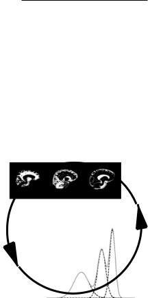
However, in the presence of gross morphological differences between atlas and patient images, intensity-based atlas-guided pixel classification as described above, using only affine iconic matching of atlas to image, may fail. Figure 1.4 (left) shows a cross section through the brain of a patient affected by periventricular leukomalacia (PVL). This brain presents gross morphological differences compared to a normal brain, especially around the ventricles, which are strongly enlarged. Brain tissue segmentation of such images using affine iconic matching of atlas and patient images will fail, if the gross morphological differences between atlas and patient images are not corrected for. Indeed, in this particular case, the affinely matched atlas labels large portions of the enlarged ventricles as white matter. The initial estimate of the tissue intensity parameters (i.e. mean and variance) is thus not reliable and it is therefore unlikely that the iterative