

Kluwer - Handbook of Biomedical Image Analysis Vol
.2.pdfSupervised Texture Classification for Intravascular Tissue Characterization |
101 |
(a) |
(b) |
(c) |
(d) |
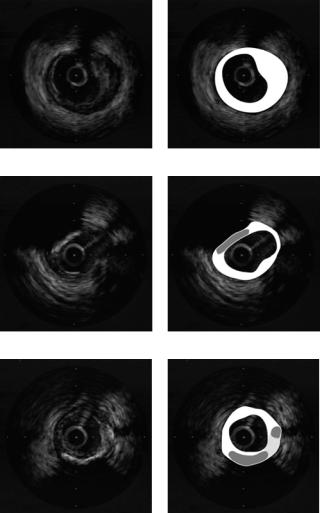
Figure 2.22: Boosting procedure for tissue characterization at different stages of its progress. (a) Expected hand classification by an expert. (b) First stage of the boosting procedure. (c) Classification with a five classifiers ensemble. (d) Classification with 10 “weaks” ensemble.
well as the ML classifier does not transform data in another feature space. This scheme is really well suited for real-time or near-real-time applications because of both time efficiency and reliability in the classification. This is, however, by no means the only near-real-time configuration available since accumulation local moments are computationally as fast as local binary patterns. However, the FLD dimensionality reduction hinders the process due to the complexity of the data in its original feature space. To overcome this problem, other classifiers can be used. The necessity to find reliable and fast classifiers lead us to boosting techniques. Boosting techniques allow a fast and simple classification procedure to improve its performance as well as maintaining part of its speed. To illustrate this fact Fig. 2.22 shows the evolution of the classification when more classifiers are added to the strong classifier. Figure 2.22(a) shows the expected hand classification by a physician. Figure 2.22(b) shows the base classification of a single “weak”. Figure 2.22(c) illustrates the result of the classification using an ensemble of five classifiers. Figure 2.22(d) shows the resulting classification after the addition of 10 weak classifiers to the ensemble. The error rates at different stages of the process are also shown in Table 2.6. These results are computed using a ML method as a weak classifier on the accumulation local moments space. The numbers show how the error rate is improved, and, though the raw classification error rate is nearly immutable, we can observe that there is a great change in the classification data points distribution in the image domain since the FP and FN rates drastically change. The postprocessing error rate gives better description of what is happening. The disposition of the error points in the classification image is more sparse and unrelated to their neighborhood, allowing better

102 |
Pujol and Radeva |
Table 2.6: Error rates using boosting methods with maximum likelihood with the accumulation local moments space
Ensemble no. |
RAW error |
FP |
FN |
Post error |
FP |
FN |
|
|
|
|
|
|
|
Base error |
34.86 |
28.20 |
6.98 |
41.94 |
40.33 |
1.10 |
Ensemble of 5 c. |
29.38 |
16.32 |
13.32 |
33.17 |
31.87 |
1.10 |
Ensemble of 10 c. |
31.44 |
7.36 |
23.37 |
7.92 |
3.22 |
4.76 |
|
|
|
|
|
|
|
postprocessing and classification rates. In this case, the classification rate is over 92% with a classifier as fast as applying 10 times a threshold. Therefore, using accumulation local moments and boosting techniques we have another fast and highly accurate scheme for real-time or near-real-time tissue characterization.
Up to this point, we have discussed the reliability of the soft plaque versus hard plaque discrimination process, which is our main concern, since the identification of calcium is reduced to the part of hard plaque with a large shadowing area. Using the method described in the former section, 99% of the calcium plaque is correctly identified. Figure 2.23 shows some results of the tissue characterization process. Figures 2.23(a) and 2.23(b) show the characterization of a soft plaque. In Figs. 2.23(c) and 2.23(d), there are two different kind of plaques detected, calcium (gray region) and soft plaque (white region). Figures 2.23(e) and 2.23(f) show the characterization of the three kind of plaques: fibrotic (light gray region), soft plaque (white region), and calcium (dark gray region).
2.4.3 Conclusions
Tissue characterization in IVUS images is a crucial problem for the physicians for studying the vascular diseases. However, this task is complex and suffers from multiple drawbacks (slow manual process, subjective interpretation, etc.) Therefore, automatic plaque characterization is a highly desirable tool.
However, automatic tissue characterization is a problem of high complexity. First of all, we need a unique and powerful description of the tissues to be classified. This is done by the feature extraction process, that in order to obtain complete and meaningful description, image features should be based on texture. Thus, a study of the most representable feature spaces is done, to conclude with some enlightening results. After analyzing the experimental results, we conclude that co-occurrence matrix measures, local binary patterns, and
Supervised Texture Classification for Intravascular Tissue Characterization |
103 |
(a) |
(b) |
(c) |
(d) |
(e) |
(f) |
Figure 2.23: Tissue characterization results: (b), (d), and (f) White labels soft plaque, dark gray areas are displayed where calcium plaques are located, and light gray areas labels hard plaque. (a), (c), and (e) Original images.
accumulation local moments are good descriptors of the different kind of plaque tissues. However, local binary patterns and accumulation local moments are also fast, in terms of low-time processing. On the other hand, the classification of the feature data is a critical step. Different approaches to the classification problem are described and proposed as candidates in our framework. We proved that
104 |
Pujol and Radeva |
k-nearest neighbor method gives the best performance as a classifier. But, ML and methods based on an ensemble of classifiers have high discrimination rate and lower classification time. Therefore, two real-time or near-real-time approaches are proposed: The first method combines local binary patterns with ML methods. The second method uses accumulation local moments and boosting techniques.
In conclusion, we have presented a general fully automatic and real-time or near-real-time framework with high accuracy plaque recognition rate for tissue characterization in IVUS images.
Questions
1.What is tissue characterization in IVUS images?
2.Why is automatic tissue characterization an important issue?
3.Why do we use texture-based descriptors?
4.Why do we use supervised classification?
5.What is the feature space?
6.Why is dimensionality reduction needed in the classification process?
7.What is the main idea under the boosting classification?
8.What is the segmentation of the plaque for?
9.Discuss the methodology for the tissue characterization framework.
10.Which are the most reliable frameworks for real-time classification?

Supervised Texture Classification for Intravascular Tissue Characterization |
105 |
Bibliography
[1]Wickline, S., Beyond intravascular imaging: Quantitative ultrasonic tissue characterization of vascular pathology, In: IEEE Ultrasonics symposium, 1994, pp. 1589–1597.
[2]Arul, P. and Amin, V., Characterization of beef muscle tissue using texture analysis of ultrasonic images, In: Proceedings of the Twelfth Southern Biomedical Engineering Conference, 1993, pp. 141–143.
[3]Mojsilovic, A. and Popovic, M., Analysis and characterization of
myocardial tissue with the wavelet image extension [US im-
ages], In: Image Processing, 1995 (Proceedings) Vol. 2, pp. 23–26,
1995.
[4]Jin, X. and Ong, S., Fractal characterization of kidney tissue sections, In: Engineering in Medicine and Biology Society, 1994. Engineering Advances: New Opportunities for Biomedical Engineers, Proceedings of the 16th Annual International Conference of the IEEE, Vol. 2, pp. 1136– 1137, 1994.
[5]Cohen, F. and Zhu, Q., Quantitative soft-tissue characterization in human organs using texture/attenuation models, In: Proceedings in Multidimensional Signal Processing Workshop, 1989, pp. 47–48.
[6]Mavromatis, S. and Boi, J., Medical image segmentation using texture directional features, In: Engineering in Medicine and Biology Society, 2001. Proceedings of the 23rd Annual International Conference of the IEEE, Vol. 3, pp. 2673–2676, 2001.
[7]Mavromatis, S., Mammographic mass classification using textural features and descriptive diagnostic data, In: Digital Signal Processing, DSP 2002. 14th International Conference on, Vol. 1, pp. 461–464, 2002.
[8]Donohue, K. and Forsberg, F., Analysis and classification of tissue with scatterer structure templates, IEEE Trans. Ultrasonics, Ferroelect. Frequency Control, Vol. 46, No. 2, pp. 300–310, 1999.
106 |
Pujol and Radeva |
[9]Ravizza, P., Myocardial tissue characterization by means of nuclear magnetic resonance imaging, In: Computers in Cardiology 1991 (Proceedings), pp. 501–504.
[10]Vandenberg, J., Arterial imaging techniques and tissue characterization using fuzzy logic, In: Proceedings of the 1994 Second Australian and New Zealand Conference on Intelligent Information Systems, 1994,
pp.239–243.
[11]Nailon, W. and McLaughlin, S., Comparative study of textural analysis techniques to characterize tissue from intravascular ultrasound, In: Proceedings of the IEEE International Conference of Image Processing, Switzerland, IEEE Signal Processing Society, Piscataway, NJ, 1996,
pp.303–305.
[12]Nailon, W. and McLaughlin, S., Intravascular ultrasound image interpretation, In: Proceedings of the International Conference on Pattern Recognition, Austria, IEEE Computer Society Press, Los Alamitos, CA, 1996, pp. 503–506.
[13]Nailon, W., Fractal texture analysis: An aid to tissue characterization with intravascular ultrasound, In: Proceedings 19th International Conference, IEEE/EMBS, 1997, pp. 534–537.
[14]Spencer, T., Characterization of atherosclerotic plaque by spectral analysis of 30mhz intravascular ultrasound radio frequency data, In: IEEE Ultrasonics Symposium, 1996, pp. 1073–1076.
[15]Dixon, K., Characterization of coronary plaque in intravascular ultrasound using histological correlation, In: 19th International Conference, IEEE/EMBS, pp. 530–533, 1997.
[16]Ahmed, M. and Leyman, A., Tissue characterization using radial transform and higher order statistics, In: Nordic Signal Processing Symposium, 2000, pp. 13–16.
[17]de Korte, C. L. and van der Steen, A. F. W., Identification of atherosclerotic plaque components with intravascular ultrasound elastography in vivo: A yucatan pig study, Circulation, Vol. 105, No. 14, pp. 1627–1630, 2002.
Supervised Texture Classification for Intravascular Tissue Characterization |
107 |
[18]Zhang, X. and Sonka, M., Tissue characterization in intravascular ultrasound images, IEEE Trans. Med. Imaging, Vol. 17, No. 6, pp. 889–899, 1998.
[19]Pujol, O. and Radeva, P., Automatic segmentation of lumen in intravascular ultrasound images: An evaluation of texture feature extractors, In: Proceedings for IBERAMIA, 2002, pp. 159–168.
[20]Pujol, O. and Radeva, P., Near real time plaque segmentation of ivus, In: Proceedings of Computers in Cardiology, 2003, pp. 159–168.
[21]Randen, T. and Husoy, J. H., Filtering for texture classification: A comparative study, Pattern Recogn., Vol. 21, No. 4, pp. 291–310, 1999.
[22]Haralick, R., Shanmugam, K., and Dinstein, I., Textural features for image classification, IEEE Trans. System, Man, Cybernetics, Vol. 3, pp. 610–621, 1973.
[23]Tuceryan, M., Moment based texture segmentation, Pattern Recogn. Lett., Vol. 15, pp. 659–668, 1994.
[24]Lindeberg, T., Scale-Space Theory in Computer Vision, Kluwer, Dordrecht, Netherlands, 1994.
[25]Jain, A. and Farrokhnia, F., Unsupervised texture segmentation using gabor filters, In: Systems, Man and Cybernetics, 1990 (Conference Proceedings), pp. 14–19.
[26]Mallat, S., A theory for multiresolution signal decomposition: The wavelet representation, IEEE Trans. Pattern Anal. Machine Intell., Vol. 11, No. 7, pp. 674–694, 1989.
[27]Mandelbrot, B., The Fractal Geometry of Nature, W. H. Freeman, New York, 1983.
[28]Ojala, T., Pietikainen, M., and Maenpaa, T., Multiresolution gray-scale and rotation invariant texture classification with local binary patterns, IEEE Trans. Pattern Anal. Machine Intell., Vol. 24, No. 7, pp. 971–987, 2002.
108 |
Pujol and Radeva |
[29]Julesz, B., Visual pattern discrimination, IRE Trans. Inf. Theory, Vol. IT-8, pp. 84–92, 1962.
[30]Ohanian, P. and Dubes, R., Performance evaluation for four classes of textural features, Pattern Recogn., Vol. 25, No. 8, pp. 819–833, 1992.
[31]Martinez, J. and Thomas, F., Efficient computation of local geometric moments, IEEE Trans. Image Process., Vol. 11, No. 9, pp. 1102–1111, 2002.
[32]Caelli, T. and Oguztoreli, M. N., Some tasks and signal dependent rules for spatial vision, Spatial Vision, No. 2, pp. 295–315, 1987.
[33]Chaudhuri, B. and Sarkar, N., Texture segmentation using fractal dimension, IEEE Trans. Pattern Anal. Machine Intell., Vol. 17, No. 1, pp. 72–77, 1995.
[34]Lindeberg, T., Scale-space theory: A basic tool for analysing structures at different scales, J. Appl. Stat., Vol. 21, No. 2, pp. 225–270, 1994.
[35]Rao, R. and Ballard, D., Natural basis functions and topographic memory for face recognition, In: Proceedings of International Joint Conference on Artificial Intelligence, 1995, pp. 10–17.
[36]Lumbreras, F., Segmentation, Classification and Modelization of Textures by means of Multiresolution Descomposition Techniques, PhD Thesis, Computer Vision Center, Universitat Autonoma` de Barcelona, 2001.
[37]Jain, A. and Farrokhnia, F., A multi-channel filtering approach to texture segmentation, In: Proceedings of Computer Vision and Pattern Recognition, CVPR’91, 1991, pp. 364–370.
[38]Fukunaga, K., Introduction to Statistical Pattern Recognition, Academic Press, New York, 1971.
[39]Belhumeur, P., Eigenfaces vs fisherfaces: Recognition using class specific linear projection, IEEE Pattern Analy. Machine Intell., Vol. 19, No. 7, pp. 711–720, 1997.
Supervised Texture Classification for Intravascular Tissue Characterization |
109 |
[40]Schapire, R. E., The boosting approach to machine learning. An overview, In: MSRI Workshop on Nonlinear Estimation and Classification, 2002.
[41]Viola, P. and Jones, M., Rapid object detection using a boosted cascade of simple features, In: Conference on Computer Vision and Pattern Recognition, 2001, pp. 511–518.
[42]Duda, R. and Hart, P., Pattern Classification, Wiley InterScience, New York, 2001. Second Edition.
[43]Sonka, M. and Zhang, X., Segmentation of intravascular ultrasound images: A knowledge-based approach, IEEE Trans. Med. Imaging, Vol. 17, No. 6, pp. 889–899, 1998.
[44]von Birgelen, C., Computerized assessment of coronary lumen and atherosclerotic plaque dimensions in three-dimensional intravascular ultrasound correlated with histomorphometry, Am. J. Cardiol., Vol. 78, pp. 1202–1209, 1996.
[45]Klingensmith, J., Shekhar, R., and Vince, D., Evaluation of threedimensional segmentation algorithms for identification of luminal and medial-adventitial borders in intravascular ultrasound images, IEEE Trans. Med. Imaging, Vol. 19, No. 10, pp. 996–1011, 2000.
[46]McInerney, T. and Terzopoulos, D., Deformable models in medical images analysis: A survey, Med. Image Anal., Vol. 1, No. 2, pp. 91–108, 1996.

Chapter 3
Medical Image Segmentation: Methods and
Applications in Functional Imaging
Koon-Pong Wong
3.1 Introduction
Detection, localization, diagnosis, staging, and monitoring treatment responses are the most important aspects and crucial procedures in diagnostic medicine and clinical oncology. Early detection and localization of the diseases and accurate disease staging can improve the survival and change management in patients prior to planned surgery or therapy. Therefore, current medical practice has been directed toward early but efficient localization and staging of diseases, while ensuring that patients would receive the most effective treatment.
Current disease management is based on the international standard of cancer staging using TNM classification, viz. size, location, and degree of invasion of the primary tumor (T), status of regional lymph node (N), and whether there is any distant metastasis (M). Over the century, histopathology retains its main role as the primary means to characterization of suspicious lesions and confirmation of malignancy. However, the pathologic interpretation is heavily dependent on the experience of the pathologist, and sampling errors may mean that there are insufficient amounts of tissue in the specimens, or the excised tissue does not accurately reflect tumor aggressivity. In addition, some lesions may return nondiagnostic information from the specimens, or they are difficult or too
1 Department of Electronic and Information Engineering, Hong Kong Polytechnic Univer-
sity, Hung Hom, Kowloon, Hong Kong
111