- •Contents
- •Foreword to the English translation
- •Preface
- •1 Introduction
- •1.1 Historical review
- •1.2 The birth of the concept of crystal growth
- •1.3 Morphology, perfection, and homogeneity
- •1.4 Complicated and complex systems
- •References
- •Suggested reading
- •2 Crystal forms
- •2.1 Morphology of crystals – the problems
- •References
- •Suggested reading
- •3 Crystal growth
- •3.1 Equilibrium thermodynamics versus kinetic thermodynamics
- •3.2 Driving force
- •3.3 Heat and mass transfer
- •3.4 Examples of mass transfer
- •3.6 Nucleation
- •3.7 Lattice defects
- •3.8 Interfaces
- •3.9 Spiral growth
- •3.10 Growth mechanism and morphology of crystals
- •3.11 Morphological instability
- •3.12 Driving force and morphology of crystals
- •3.13 Morphodroms
- •3.14 Element partitioning
- •3.15 Inclusions
- •References
- •Suggested reading
- •4 Factors determining the morphology of polyhedral crystals
- •4.1 Forms of polyhedral crystals
- •4.2 Structural form
- •4.3 Equilibrium form
- •4.4 Growth forms
- •4.4.1 Logical route for analysis
- •4.4.2 Anisotropy involved in the ambient phase
- •4.4.3 Whiskers
- •MAJOR FACTORS
- •METHODOLOGY
- •IMPURITIES
- •AMBIENT PHASES AND SOLVENT COMPONENTS
- •4.4.7 Factors controlling growth forms
- •References
- •Suggested reading
- •5 Surface microtopography of crystal faces
- •5.1 The three types of crystal faces
- •5.2 Methods of observation
- •5.3 Spiral steps
- •5.4 Circular and polygonal spirals
- •5.5 Interlaced patterns
- •5.6 Step separation
- •5.7 Formation of hollow cores
- •5.8 Composite spirals
- •5.9 Bunching
- •5.10 Etching
- •References
- •Suggested reading
- •6 Perfection and homogeneity of single crystals
- •6.1 Imperfections and inhomogeneities seen in single crystals
- •6.2 Formation of growth banding and growth sectors
- •6.3 Origin and spatial distribution of dislocations
- •References
- •7 Regular intergrowth of crystals
- •7.1 Regular intergrowth relations
- •7.2 Twinning
- •7.2.1 Types of twinning
- •7.2.2 Energetic considerations
- •7.2.4 Penetration twins and contact twins
- •7.2.5 Transformation twin
- •7.2.6 Secondary twins
- •7.3 Parallel growth and other intergrowth
- •7.4 Epitaxy
- •7.5 Exsolution, precipitation, and spinodal decomposition
- •References
- •Suggested reading
- •8 Forms and textures of polycrystalline aggregates
- •8.1 Geometrical selection
- •8.2 Formation of banding
- •8.3 Spherulites
- •8.4 Framboidal polycrystalline aggregation
- •References
- •Suggested reading
- •9 Diamond
- •9.1 Structure, properties, and use
- •9.2 Growth versus dissolution
- •9.3 Single crystals and polycrystals
- •9.4 Morphology of single crystals
- •9.4.1 Structural form
- •9.4.2 Characteristics of {111}, {110}, and {100} faces
- •9.4.3 Textures seen inside a single crystal
- •9.4.4 Different solvents (synthetic diamond)
- •9.4.5 Twins
- •9.4.6 Coated diamond and cuboid form
- •9.4.7 Origin of seed crystals
- •9.4.8 Type II crystals showing irregular forms
- •References
- •Suggested reading
- •10 Rock-crystal (quartz)
- •10.1 Silica minerals
- •10.2 Structural form
- •10.3 Growth forms
- •10.4 Striated faces
- •10.5 Growth forms of single crystals
- •10.5.1 Seed crystals and forms
- •10.5.2 Effect of impurities
- •10.5.3 Tapered crystals
- •10.6 Twins
- •10.6.1 Types of twins
- •10.6.2 Japanese twins
- •10.6.3 Brazil twins
- •10.7 Scepter quartz
- •10.8 Thin platy crystals and curved crystals
- •10.9 Agate
- •References
- •11 Pyrite and calcite
- •11.1 Pyrite
- •11.1.2 Characteristics of surface microtopographs
- •11.1.4 Polycrystalline aggregates
- •11.2 Calcite
- •11.2.1 Habitus
- •11.2.2 Surface microtopography
- •References
- •12 Minerals formed by vapor growth
- •12.1 Crystal growth in pegmatite
- •12.3 Hematite and phlogopite in druses of volcanic rocks
- •References
- •13 Crystals formed by metasomatism and metamorphism
- •13.1 Kaolin group minerals formed by hydrothermal replacement (metasomatism)
- •13.2 Trapiche emerald and trapiche ruby
- •13.3 Muscovite formed by regional metamorphism
- •References
- •14 Crystals formed through biological activity
- •14.1 Crystal growth in living bodies
- •14.2 Inorganic crystals formed as indispensable components in biological activity
- •14.2.1 Hydroxyapatite
- •14.2.2 Polymorphic minerals of CaCO3
- •14.2.3 Magnetite
- •14.3 Crystals formed through excretion processes
- •14.4 Crystals acting as possible reservoirs for necessary components
- •14.5 Crystals whose functions are still unknown
- •References
- •Appendixes
- •A.1 Setting of crystallographic axes
- •A.2 The fourteen Bravais lattices and seven crystal systems
- •A.3 Indexing of crystal faces and zones
- •A.4 Symmetry elements and their symbols
- •Materials index
- •Subject index
12.3 Hematite and phlogopite in druses of volcanic rocks 247
T
T
|
|
T |
|||
|
T |
||||
T |
|
|
|
|
T.B. |
|
|
|
|
|
|
T |
|
|
|
|
|
|
|
|
0.1 mm |
|
|
Figure 12.9. Spiral step pattern observed on both sides of a twin boundary on a {0001} face of a hematite crystal [7]. T.B. is the twin boundary, and indicate orientations. Phase contrast photomicrograph.
12.3Hematite and phlogopite in druses of volcanic rocks
In volcanic rocks formed by the solidification of magma near the Earth’s surface, small cavities of millimeter to centimeter order are often present, in which idiomorphic crystals of phlogopite and hematite occur. All these crystals grown from the vapor phase show typical spiral patterns. However, they grow in a much-reduced free space as compared to the case of pegmatite, and this characteristic is well represented on their surface microtopographs.
Figure 12.12 shows a step pattern on the {001} face of phlogopite; the step height is 1 nm, and the step separation is of order 10 m. The characteristic form of the spiral is five-sided, i.e. hexagonal with one edge truncated [10]. This characteristic form is due to the fact that the stacking of the unit layer, which has a hexagonal form, is in one direction. Since an interlacing pattern is not observed in this step pattern, this crystal is identified as a 1M polytype. On the surface of the (001) face shown in Fig. 12.12, a five-sided step pattern rotated by 120° and 180° appears in an island form on one surface. This indicates that several crystals agglutinate in rotated orientation, and that crystals attaining a certain size move around in the cavity and come together to form the crystal [11].
248 Minerals formed by vapor growth
Figure 12.10. Phase contrast photomicrograph of hematite from Vesuvius in Italy. Straight steps crossing curved growth steps, which have appeared due to the movement of dislocations after the cessation of growth [8], are shown.
Figure 12.11. Step patterns due to etching on hematite from the Azores Islands. All circular step patterns are depressions. Note steps of chopping waveform [9].
12.3 Hematite and phlogopite in druses of volcanic rocks 249
0.1 mm
Figure 12.12. Five-sided spiral steps observed on {001} face of phlogopite [10].
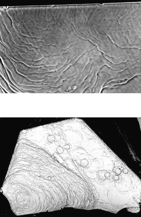
Another example showing the formation of many tiny crystallites in a small cavity, which move around and agglutinate, is found in hematite occurring in a similar way. Figure 12.13 is a surface microtopograph of the {0001} face of such a hematite crystal, and it indicates numerous hexagonal island-like portions on the terraces of step patterns. Around these islands, the step patterns of the host crystal are disturbed. This observation indicates that, as for phlogopite crystals, a large number of crystals with varying sizes are formed within a cavity, within which they move around and agglutinate.
250 Minerals formed by vapor growth
Figure 12.13. Surface microtopograph of hematite from a druse of ryolite. A large number of minute hematite crystals adhere and affect the advancement of the spiral growth layers on the host crystal.
References
1I. Sunagawa and A. Urano, Beryl crystals from pegmatites: morphology and mechanism of crystal growth, J. Gemmol., 26, 1999, 521–33
2M. D. Barton, Phase equilibria and thermodynamic properties of minerals in the BeO- Al2O3-SiO2-H2O system with petrologic applications, Am. Min., 71, 1986, 277–300
3 I. Sunagawa, Growth history of hematite, Am. Min., 45, 1959, 566–75
4I. Sunagawa, Mechanism of crystal growth, etching and twin formation of hematite,
Mineral. J., 3, 1960, 50–89
5 I. Sunagawa, Mechanism of growth of hematite, Am. Min., 47, 1962, 1138–55
6I. Sunagawa, Mechanism of natural etching of hematite crystals, Am. Min., 47, 1962, 1332–45
7Lu Taijing and I. Sunagawa, Origin of undulated growth steps on hematite crystals from Sasazawa, Japan, Mineral. J., 13, 1987, 409–23
8A. F. Seager and I. Sunagawa, Movement of screw dislocations in hematite, Min. Mag.,
33, 1962, 1–8
9I. Sunagawa, Growth and etch features of hematite crystals from the Azores Islands, Portugal, Surv. Geol. Portugal, Mem., 6, 1960, 1–44
10I. Sunagawa, Growth spirals on phlogopite crystals, Am. Min, 49, 1964, 1427–34
11I. Sunagawa and S. Tomura, Twinning in phlogopite, Am. Min., 61, 1976, 939–43