

книги студ / color atlas of physiology 5th ed[1]. (a. despopoulos et al, thieme 2003)
.pdf
|
Active motility (ability to move) is due to |
||
|
either the interaction of energy-consuming |
||
|
motor proteins (fueled by ATPase) such as my- |
||
|
osin, kinesin and dynein with other proteins |
||
|
such as actin or the polymerization and |
||
|
depolymerization of actin and tubulin. Cell di- |
||
Work |
vision (cytokinesis), cell migration (!p. 30), |
||
intracellular vesicular transport and cytosis |
|||
|
|||
Physical |
(!p. 12f.), sperm motility (!p. 306f.), axonal |
||
examples of cell and organelle motility. |
|||
|
transport (!p. 42), electromotility of hair cells |
||
|
(!p. 366), and ciliary motility (!p. 110) are |
||
Muscle, |
The muscles consist of cells (fibers) that |
||
contract when stimulated. Skeletal muscle is |
|||
|
|||
|
responsible for locomotion, positional change, |
||
and |
and the convection of respiratory gases. Car- |
||
diac muscle (!p. 190 ff.) is |
responsible for |
||
Nerve |
pumping the blood, and |
smooth muscle |
|
(!p. 70) serves as the motor of internal organs |
|||
|
|||
2 |
and blood vessels. The different muscle types |
||
|
Motility and Muscle Types |
||
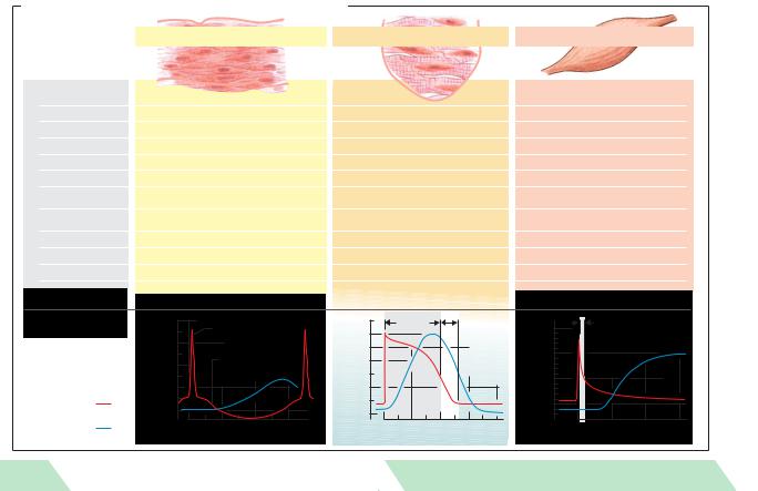
are distinguished by several functional characteristics (!A).
Motor Unit of Skeletal Muscle
Unlike some types of smooth muscle (singleunit type; !p. 70) and cardiac muscle fibers, which pass electric stimuli to each other through gap junctions or nexus (!A; p. 16f.), skeletal muscle fibers are not stimulated by adjacent muscle fibers, but by motor neurons. In fact, muscle paralysis occurs if the nerve is severed.
One motor neuron together with all muscle fibers innervated by it is called a motor unit (MU). Muscle fibers belonging to a single motor unit can be distributed over large portions (1 cm2) of the muscle cross-sectional area. To supply its muscle fibers, a motor neuron splits into collaterals with terminal branches (!p. 42). A given motor neuron may supply only 25 muscle fibers (mimetic muscle) or well over 1000 (temporal muscle).
|
Two types of skeletal muscle fibers can be |
|
|
distinguished: S – slow-twitch fibers (type 1) |
|
|
and F – fast-twitch fibers (type 2), including |
|
|
two subtypes, FR (2 A) and FF (2 B). Since each |
|
58 |
motor unit contains only one type of fiber, this |
|
classification also applies to the motor unit. |
||
|
Slow-twitch fibers are the least fatigable and are therefore equipped for sustained performance. They have high densities of capillaries and mitochondria and high concentrations of fat droplets (high-energy substrate reserves) and the red pigment myoglobin (short-term O2 storage). They are also rich in oxidative enzymes (!p. 72). Fast-twitch fibers are mainly responsible for brief and rapid contractions. They are quickly fatigued (FF ! FR) and are rich in glycogen (FF ! FR) but contain little myoglobin (FF"FR).
The fiber type distribution of a muscle depends on the muscle type. Motor units of the S type predominate in “red” muscles such as the soleus muscle, which helps to maintain the body in an upright position, whereas the F type predominates in “white” muscles such as the gastrocnemius muscle, which is involved in running activity. Each fiber type can also be converted to the other type. If, for example, the prolonged activation of fast-twitch fibers leads to a chronic increase in the cytosolic Ca2+ concentration, fast-twitch fibers will be converted to slow-twitch fibers and vice versa.
Graded muscle activity is possible because a variable number of motor units can be recruited as needed. The more motor units a muscle has, the more finely graded its contractions. Contractions are much finer in the external eye muscles, for example, which have around 2000 motor units, than in the lumbrical muscles, which have only around 100 motor units. The larger the number of motor units recruited, the stronger the contraction. The number and type of motor units recruited depends on the type of movement involved (fine or coarse movement, intermittent or persistent contraction, reflex activity, voluntary or involuntary movement, etc.). In addition, the strength of each motor unit can be increased by increasing the frequency of neuronal impulses, as in the tetanization of skeletal muscle (!p. 67 A).
Despopoulos, Color Atlas of Physiology © 2003 Thieme
All rights reserved. Usage subject to terms and conditions of license.
Usage .reserved rights All |
Atlas Color Despopoulos, |
terms to subject |
© Physiology of |
conditions and |
Thieme 2003 |
.license of |
|
A. Structure and function of heart, skeletal and smooth muscle |
|
|
|
|
|
|
|
|
|
|
||||||
Structure and |
|
|
Smooth muscle |
|
|
Cardiac muscle (striated) |
|
Skeletal muscle (striated) |
|
|||||||
|
|
|
|
|
|
|
|
|
|
|
|
|
|
|
|
|
function |
|
|
|
|
|
|
|
|
|
|
|
|
|
|
|
|
Motor |
|
|
|
|
|
|
|
|
|
|
|
|
|
|
|
|
end-plates |
None |
|
|
|
|
|
None |
|
|
|
|
|
Yes |
|
|
|
Fibers |
Fusiform, short (< 0.2 mm) |
|
|
Branched |
|
|
|
|
Cylindrical, long (< 15cm) |
|
||||||
|
|
|
– |
|
|
|
|
|
|
|
|
|
|
– |
|
|
Mitochondria |
Few |
|
|
|
|
|
Many |
|
|
|
|
|
Few (depending on muscle type) |
|
||
Nucleus per fiber |
1 |
|
|
|
|
|
1 |
|
|
|
|
|
Multiple |
|
|
|
Sarcomeres |
None |
|
|
|
|
|
Yes, length < 2.6 m |
|
|
Yes, length < |
3.65 m |
|
||||
|
|
|
|
|
|
|
|
|
– |
|
|
|
– |
|
|
|
Electr. coupling |
Some (single-unit type) |
|
|
|
Yes (functional syncytium) |
|
No |
|
|
|
||||||
Sarcoplasmic |
Little developed |
|
|
|
Moderately developed |
|
|
Highly developed |
|
|
||||||
reticulum |
|
|
|
|
|
|
|
|||||||||
|
|
|
|
|
|
|
|
|
|
|
|
|
|
|
|
|
Ca2+ “switch” |
Calmodulin/caldesmon |
|
|
|
Troponin |
|
|
|
|
Troponin |
|
|
|
|||
Pacemaker |
Some spontaneous rhythmic activity |
|
Yes (sinus nodes ca.1s–1) |
|
|
No (requires nerve stimulus) |
|
|||||||||
(1s–1 –1h–1) |
|
|
|
|
|
|
|
|||||||||
Response to stimulus |
Change in tone or rhythm frequency |
|
All or none |
|
|
|
|
Graded |
|
|
|
|||||
Tetanizable |
Yes |
|
|
|
|
|
No |
|
|
|
|
|
Yes |
|
|
|
Work range |
Length-force curve |
|
|
|
In rising |
|
|
|
|
|
At peak of |
|
|
|
||
|
is variable |
|
|
|
|
length-force curve |
(see 2.15E) |
|
length-force curve |
(see 2.15E) |
|
|||||
|
+ 20 |
|
|
|
|
|
Absolutely |
Relatively |
|
Absolutely |
|
|||||
Response |
|
|
“Spike” |
|
|
+ 20 |
refractory |
|
refractory |
+ 50 |
refractory |
|
||||
mV |
0 |
|
|
|
|
|
|
|
|
|
|
|
|
|||
|
|
|
mV |
0 |
|
|
|
|
|
|
|
|
|
|||
to stimulus |
|
|
Spontaneous |
|
|
|
|
|
|
mV 0 |
|
|
|
|||
– 20 |
|
– 20 |
|
|
|
|
|
|
|
|
||||||
|
|
|
|
|
|
|
|
|
|
|
||||||
|
fluctuation |
|
|
|
|
|
|
|
|
|
|
|
|
|||
|
|
|
|
|
|
|
|
|
|
|
– 50 |
|
|
|
||
|
– 40 |
|
|
|
– 60 |
|
|
|
|
|
|
|
|
|||
|
|
|
|
|
|
|
|
|
|
|
|
|
||||
Potential |
– 60 |
|
|
|
– 100 |
|
|
|
|
|
–100 |
|
|
|
||
|
|
|
|
|
|
|
|
|
|
|
|
|
||||
Muscle |
|
|
|
|
|
|
|
|
|
|
|
|
|
|
||
|
0 |
200 |
400 |
600 |
|
0 |
|
100 |
200 |
300 |
400 |
0 |
10 |
20 |
30 |
|
tension |
|
|
|
|||||||||||||
|
|
|
ms |
|
|
|
|
|
ms |
|
|
|
ms |
|
||
59
Plate 2.10 Muscle types, motor unit

2 Nerve and Muscle, Physical Work
60
Contractile Apparatus of Striated
Muscle
The muscle cell is a fiber (!A2) approximately 10 to 100 µm in diameter. Skeletal muscles fibers can be as long as 15 cm. Meat “fibers” visible with the naked eye are actually bundles of muscle fibers that are around 100 to 1000 µm in diameter (!A1). Each striated muscle fiber is invested by a cell membrane called the sarcolemma, which surrounds the sarcoplasm (cytoplasm), several cell nuclei, mitochondria (sarcosomes), substances involved in supplying O2 and energy (!p. 72), and several hundreds of myofibrils.
So-called Z lines or, from a three-dimen- sional aspect, Z plates (plate-like proteins; !B) subdivide each myofibril (!A3) into approx. 2 µm long, striated compartments called sarcomeres (!B). When observed by (two-di- mensional) microscopy, one can identify alternating light and dark bands and lines (hence the name “striated muscle”) created by the thick myosin II filaments and thin actin filaments (!B; for myosin I, see p. 30). Roughly 2000 actin filaments are bound medially to the Z plate. Thus, half of the filament projects into two adjacent sarcomeres (!B). The region of the sarcomere proximal to the Z plate contains only actin filaments, which form a so-called I band (!B). The region where the actin and myosin filaments overlap is called the A band. The H zone solely contains myosin filaments (ca. 1000 per sarcomere), which thicken towards the middle of the sarcomere to form the M line (M plate). The (actin) filaments are anchored to the sarcolemma by the protein dystrophin.
Each myosin filament consists of a bundle of ca. 300 myosin-II molecules (!B). Each molecule has two globular heads connected by flexible necks (head and neck = subfragment S1; formed after proteolysis) to the filamentous tail of the molecule (two intertwined α- helices = subfragment S2) (!C). Each of the heads has a motor domain with a nucleotide binding pocket (for ATP or ADP + Pi) and an actin binding site. Two light protein chains are located on each neck of this heavy molecule
(220 kDa): one |
is regulatory (20 kDa), the |
other essential |
(17 kDa). Conformational |
changes in the head–neck segment allow the myosin head to “tilt” when interacting with actin (sliding filaments; !p. 62).
Actin is a globular protein molecule (G- actin). Four hundered such molecules join to form F-actin, a beaded polymer chain. Two of the twisted protein filaments combine to form an actin filament (!B), which is positioned by the equally long protein nebulin.
Tropomyosin molecules joined end-to-end (40 nm each) lie adjacent to the actin filament, and a troponin (TN) molecule is attached every 40 nm or so (!B). Each troponin molecule consists of three subunits: TN-C, which has two regulatory bindings sites for Ca2+ at the amino end, TN-I, which prevents the filaments from sliding when at rest (!p. 62), and TN-T, which interacts with TN-C, TN-I, and actin.
The sarcomere also has another system of filaments (!B) formed by the filamentous protein titin (connectin). Titin is more than 1000 nm in length and has some 30 000 amino acids (Mr !3000 kDa). It is the longest known polypeptide chain and comprises 10% of the total muscle mass. Titin is anchored at its carboxyl end to the M plate and, at the amino end, to the Z plate (!p. 66 for functional description).
The sarcolemma forms a T system with several transverse tubules (tube-like invaginations) that run perpendicular to the myofibrils (!p. 63 A). The endoplasmic reticulum (!p. 10 ff.) of muscle fibers has a characteristic shape and is called the sarcoplasmic reticulum (SR; !p. 63 A). It forms closed chambers without connections between the intraand extracellular spaces. Most of the chambers run lengthwise to the myofibrils, and are
therefore |
called |
longitudinal |
tubules |
(!p. 63 A). |
The sarcoplasmic reticulum is |
||
more prominently developed in skeletal muscle than in the myocardium and serves as a Ca2+ storage space. Each T system separates the adjacent longitudinal tubules, forming triads (!p. 63 A, B).
Despopoulos, Color Atlas of Physiology © 2003 Thieme
All rights reserved. Usage subject to terms and conditions of license.

A. Ultrastructure of striated muscle fibers
|
|
Sarkomere |
|
Striated Muscle |
|
|
|
of |
|
|
|
|
|
|
100–1000 m |
10–100 m |
|
1 m |
Apparatus |
1 Bundle of fibers |
2 Muscle fiber (myocyte) |
3 Myofibril |
||
|
|
|
|
|
B. Sarcomere structure |
|
|
Contractile |
|
|
|
|
6 |
|
|
|
|
nm |
|
|
|
|
|
|
1.2 m |
Actin |
Tropomyosin |
|
2.11 |
|
Troponin |
|||
|
Sarcomere |
|||
|
|
|||
|
|
Actin filament |
Plate |
|
|
H zone |
|
||
|
|
|
||
|
|
|
|
|
Z disk |
Actin |
|
|
|
filament |
|
|
|
|
|
Titin |
|
|
|
|
Myosin |
|
|
|
|
filament |
|
Z disk |
|
|
M disk |
|
|
|
|
|
|
|
|
|
A band 1.6 m |
|
|
|
Myosin filament |
|
|
|
|
Myosin head |
|
|
|
|
Myosin molecule |
|
10nm |
|
|
|
|
I band |
6nm |
|
|
|
|
|
|
|
M disk |
|
|
|
C. Myosin II molecule |
|
|
|
|
Motor domain |
|
|
|
|
|
Actin- |
|
P |
|
binding |
|
|
domain |
|
|
|
|
|
|
|
|
Nucleotide- |
2 |
P |
|
|
nm |
|
(ATP or ADP) |
|
Regulatory light chain |
|
||
|
|
|
|
|
Essential light chain |
|
|
Shaft (150nm) |
Neck |
Head |
|
(flexible) |
61 |
||
|
20nm |
||
|
|
||
(After D.M.Warshaw)
Despopoulos, Color Atlas of Physiology © 2003 Thieme
All rights reserved. Usage subject to terms and conditions of license.

2 Nerve and Muscle, Physical Work
62
Contraction of Striated Muscle |
ATP (!p. 72) is essential for filament sliding |
|||||||
and, hence, for muscle contraction. Due to |
||||||||
|
|
|
|
|||||
Stimulation of muscle fibers. The release of |
their ATPase |
activity, |
the myosin |
heads |
||||
acetylcholine at the motor end-plate of skeletal |
(!p. 60) act as the motors (motor proteins) of |
|||||||
muscle leads to an end-plate current that |
this process. The myosin-II and actin filaments |
|||||||
spreads electrotonically and activates voltage- |
of a sarcomere (!p. 60) are arranged in such a |
|||||||
gated Na+ |
channels |
in the |
sarcolemma |
way that they can slide past each other. The |
||||
(!p. 56). This leads to the firing of action |
myosin heads connect with the actin filaments |
|||||||
potentials (AP) that travel at a rate of 2 m/s |
at a particular angle, forming so-called cross- |
|||||||
along the sarcolemma of the entire muscle |
bridges (!C1). Due to a conformational change |
|||||||
fiber, and penetrate rapidly into the depths of |
in the region of the nucleotide binding site of |
|||||||
the fiber along the T system (!A). |
myosin-II (!p. 61 C), |
the spatial extent of |
||||||
The conversion of this excitation into a con- |
which is increased by concerted movement of |
|||||||
traction is called electromechanical coupling |
the neck region, the myosin head tilts down, |
|||||||
(!B). In the skeletal muscle, this process |
drawing the thin filament a length of roughly |
|||||||
begins with the action potential exciting volt- |
4 nm (!C2). The second myosin head may also |
|||||||
age-sensitive |
dihydropyridine |
receptors |
move an adjacent actin filament. The head |
|||||
(DHPR) of the sarcolemma in the region of the |
then detaches and “tenses” in preparation for |
|||||||
triads. The DHPR are arranged in rows, and |
the next “oarstroke” when it binds to actin |
|||||||
directly opposite them in the adjacent mem- |
anew (!C3). |
|
|
|
||||
brane of the sarcoplasmic reticulum (SR) are |
Kinesin, another motor protein (!pp. 42 u. |
|||||||
rows of Ca2+ channels |
called ryanodine recep- |
58), independently advances on the micro- |
||||||
tors (type 1 in skeletal muscle: RYR1). Every |
tubule by incremental movement of its two |
|||||||
other RYR1 is associated with a DHPR (!B2). |
heads (8 nm increments), as in tug-of-war. In |
|||||||
RYR1 open when they directly “sense” by me- |
this case, fifty percent of the cycle time is |
|||||||
chanical means an AP-related conformational |
“work time” (duty ratio = 0.5). Between two |
|||||||
change in the DHPR. In the myocardium, on the |
consecutive interactions with actin in skeletal |
|||||||
other hand, each DHPR is part of a voltage- |
muscle, on the other hand, myosin-II “jumps” |
|||||||
gated Ca2+ channel of the sarcolemma that |
36 nm (or multiples of 36, e.g. 396 nm or more |
|||||||
opens in response to an action potential. Small |
in rapid contractions) to reach the next (or the |
|||||||
quantities of extracellular Ca2+ enter the cell |
11th) suitably located actin binding site (!C3, |
|||||||
through this channel, leading to the opening of |
jump from a to b). Meanwhile, the other my- |
|||||||
myocardial RYR2 (so-called trigger effect of |
osin heads working on this particular actin |
|||||||
Ca2+ or Ca2+ spark; !B3). Ca2+ ions stored in |
filament must make at least another 10 to 100 |
|||||||
the SR now flow through the opened RYR1 or |
oarstrokes of around 4 nm each. The duty ratio |
|||||||
RYR2 into the cytosol, increasing the cytosolic |
of a myosin-II head is therefore 0.1 to 0.01. This |
|||||||
Ca2+ concentration [Ca2+]i from a resting value |
division of labor by the myosin heads ensures |
|||||||
of ca. 0.01 µmol/L to over 1 µmol/L (!B1). In |
that a certain percentage of the heads will al- |
|||||||
skeletal muscle, DHPR stimulation at a single |
ways be ready to generate rapid contractions. |
|||||||
site is enough to trigger the coordinated open- |
When filament sliding occurs, the Z plates |
|||||||
ing of an entire group of RYR1, thereby increas- |
approach each other and the overlap region of |
|||||||
ing the reliability of impulse transmission. The |
thick and thin filaments becomes larger, but |
|||||||
increased cytosolic Ca2+ concentration satu- |
the length of the filaments remains un- |
|||||||
rates the Ca2+ binding sites on troponin-C, |
changed. This |
results |
in shortening |
of the |
||||
thereby canceling the troponin-mediated in- |
I band and H zone (!p. 60). When the ends of |
|||||||
hibitory effect of tropomyosin on filament |
the thick filaments ultimately bump against |
|||||||
sliding (!D). It is still unclear whether this |
the Z plate, maximum muscle shortening oc- |
|||||||
type of disinhibition involves actin–myosin |
curs, and the ends of the thin filaments overlap |
|||||||
binding or the detachment of ADP and Pi, as |
(!p. 67 C). Shortening of the sarcomere there- |
|||||||
described below. |
|
|
fore occurs at both ends of the myosin bundle, |
|||||
|
|
|
|
but in opposite directions. |
|
|||
!
Despopoulos, Color Atlas of Physiology © 2003 Thieme
All rights reserved. Usage subject to terms and conditions of license.

A. The sarcotubular system of myocytes (muscle fibers)
AP |
|
T system |
Sarcoplasmic |
|
|
|
|
(transverse tubules) |
reticulum |
|
|
|
|||
|
|
|
|
(longitudinal tubules) |
|
|
|
|
|
|
AP |
|
|
|
|
|
|
|
|
|
|
|
I |
|
|
|
|
|
Sarcolemma |
Muscle |
|
|
|
|
|
|
(cell membrane) |
||
|
|
|
|
|
Triads |
|
|
|
|
|
|
|
|
Striated |
|
|
|
|
|
|
Mitochondrion |
||
|
|
|
|
|
of |
||
|
|
|
|
|
|
|
|
|
|
|
|
|
(After Porter and |
Contraction |
|
|
|
|
|
|
Franzini-Armstrong) |
||
B. Ca2+ as mediator between electrical stimulus and contraction |
|
|
|||||
|
|
2.12 |
|||||
|
|
|
|
T system |
|
|
|
|
|
|
|
|
|
|
|
|
– 90 mV |
|
|
DHPR |
RYR1 |
|
Plate |
|
|
|
|
|
|||
|
Rest |
|
|
AP |
Sarco- |
||
|
|
|
Ca2+ |
plasmatic |
|
||
|
Low [Ca2+]i |
|
|
|
reticulum |
|
|
|
|
|
|
|
|
||
Stimulus |
[Ca2+]i |
|
|
2 Skeletal |
|
Cytosol |
|
|
|
|
|
|
|||
AP |
|
|
|
muscle |
|
|
|
|
|
|
Contraction |
DHPR |
|
|
|
|
|
|
|
|
|
|
|
0 |
10 |
20 |
30 ms |
with Ca2+ |
|
|
|
channel |
|
|
|
||||
|
|
|
|
RYR2 |
|
|
|
|
|
|
|
AP |
|
|
|
|
|
|
|
|
|
|
|
|
+30 mV |
|
|
Ca2+ |
|
|
|
|
High [Ca2+]i |
|
3 Myocardium |
|
|
|
|
|
1 Ca2+ release |
|
|
|
|
|
|
|
|
|
|
|
|
|
|
C. Sliding filaments
Myosin II
Pi
 a
a
Actin
1 Strong binding
|
|
Actin-myosinII binding |
|
|
|
Strong |
Weak |
Strong |
|
ATP |
|
|
|
Pi |
|
|
|
|
|
ADP |
|
ATP |
|
|
a |
a |
|
b |
|
4nm |
|
36nm or multiple |
|
63 |
2 Work phase |
3 Resting phase (ca. 90% of time; other |
|||
(ca.10% of time) |
myosin heads are meanwhile active) |
|
||
Despopoulos, Color Atlas of Physiology © 2003 Thieme
All rights reserved. Usage subject to terms and conditions of license.

2 Nerve and Muscle, Physical Work
64
!
Contraction cycle (!C and D). Each of the two myosin heads (M) of a myosin-II molecule bind one ATP molecule in their nucleotide binding pocket. The resulting M-ATP complex lies at an approx. 90! angle to the rest of the myosin filament (!D4). In this state, myosin has only a weak affinity for actin binding. Due to the influence of the increased cytosolic Ca2+ concentration on the troponin – tropomyosin complex, actin (A) activates myosin’s ATPase, resulting in hydrolysis of ATP (ATP !ADP + Pi) and the formation of an A-M-ADP-Pi complex (!D1). Detachment of Pi (inorganic phosphate) from the complex results in a conformational change of myosin that increases the actin–myosin association constant by four powers of ten (binding affinity now strong). The myosin heads consequently tilt to a 40! angle (!D2a), causing the actin and myosin filaments to slide past each other. The release of ADP from myosin ultimately brings the myosin heads to their final position, a 45! angle (!D2b). The remaining A-M complex (rigor complex) is stable and can again be transformed into a weak bond when the myosin heads bind ATP anew (“softening effect” of ATP). The high flexibility of the muscle at rest is important for processes such as cardiac filling or the relaxing of the extensor muscles during rapid bending movement. If a new ATP is bound to myosin, the subsequent weakening of the actin–myosin bond allows the realignment of the myosin head from 45! to 90! (!D3, 4), the position preferred by the M-ATP complex. If the cytosolic Ca2+ concentration remains "10– 6 mol/L, the D1 to D4 cycle will begin anew. This depends mainly on whether subsequent action potentials arrive. Only a portion of the myosin heads that pull actin filaments are “on duty” (low duty ratio; see p. 62) to ensure the smoothness of contractions.
The Ca2+ ions released from the sarcoplasmic reticulum (SR) are continuously pumped back to the SR due to active transport by Ca2+-ATPase (!pp. 17 A and 26), also called SERCA (!p. 16). Thus, if the RYR-mediated release of Ca2+ from the SR is interrupted, the cytosolic Ca2+ concentration rapidly drops below 10– 6 mol/L and filament sliding ceases (resting position; !D, upper left corner).
Parvalbumin, a protein that occurs in the cytosol of fast-twitch muscle fibers (!type F; p. 58), accelerates muscle relaxation after short contractions by binding cytosolic Ca2+ in exchange for Mg2+. Parvalbumin’s binding affinity for Ca2+ is higher than that of troponin, but lower than that of SR’s Ca2+-ATPase. It therefore functions as a “slow” Ca2+ buffer.
The course of the filament sliding cycle as described above mainly applies to isotonic contractions, that is, to contractions where muscle shortening occurs. During strictly isometric contractions where muscular tension increases but the muscle length remains unchanged, the sliding process tenses elastic components of a muscle, e.g. titin (!p. 66), and then soon comes to a halt. Afterwards, the A-M-ATP complex (!D3) probably transforms directly into A-M-ADP-Pi (!D1).
The muscle fibers of a dead body do not produce any ATP. This means that, after death, Ca2+ is no longer pumped back into the SR, and the ATP reserves needed to break down stable A-M complexes are soon depleted. This results in stiffening of the dead body or rigor mortis, which passes only after the actin and myosin molecules in the muscle fibers decompose.
Despopoulos, Color Atlas of Physiology © 2003 Thieme
All rights reserved. Usage subject to terms and conditions of license.
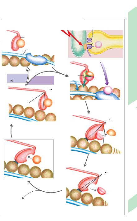
D. Work cycle of sliding filaments (isotonic contraction)
|
|
|
|
|
|
|
|
|
DHPR with (heart) and with- |
|||||
|
|
|
|
Action |
|
|
out (muscle) Ca2+channel |
|||||||
|
|
|
|
potential |
|
|
RYR |
|
|
|
||||
|
|
|
|
|
|
|
T system |
|
|
|
|
|
|
|
|
ATP |
Myosin |
|
|
|
|
|
|
|
|
|
Longitudinal |
||
|
|
|
|
|
|
|
|
|
|
|
||||
|
Tropomyosin |
|
|
|
|
|
|
|
|
Ca2+ |
tubule |
|||
|
|
|
|
|
|
|
|
|
||||||
|
|
|
|
|
|
|
|
|
|
|
|
|
|
|
|
Actin |
Troponin |
|
|
|
|
|
|
|
|
|
|
|
|
|
|
|
|
|
|
|
|
|
|
|
|
|
||
|
|
|
|
|
|
|
|
|
|
|
|
|
|
|
Resting position |
|
|
|
|
|
|
|
|
|
|
|
|
|
|
|
[Ca2+]i |
|
|
|
|
|
ATP- |
|
|
|
Pi |
|
|
|
|
|
|
|
|
|
|
|
|
|
|
|
|||
|
[Ca2+]i |
|
|
|
|
ase |
|
|
|
ADP |
|
|
|
|
|
1 mol/l |
mol/l |
|
|
|
|
|
|
|
|||||
|
= 1–10 |
|
|
|
|
|
|
|
|
|||||
|
|
|
|
|
|
|
|
|
|
|||||
|
|
|
|
|
|
|
|
|
|
|
|
|
|
|
|
|
|
45° 90° |
|
|
|
|
|
|
|
Ca2+ |
|
|
|
|
|
|
|
|
|
|
|
|
|
|
|
|
||
|
|
|
ATP |
|
|
|
1 Actin-myosin binding, |
|||||||
|
|
|
|
|
|
|
|
|
ATP cleavage |
|
|
|||
|
|
|
|
|
|
|
|
|
|
|
|
|
|
|
|
|
|
|
|
|
|
|
|
|
|
|
50° |
90° |
|
|
4 Loosening of actin–myosin |
|
|
|
|
|
|
|
|
|||||
|
bond (“softening” effect |
|
|
|
|
|
|
|
|
|||||
|
of ATP), myosin heads erect |
|
|
|
|
|
P |
|
|
|||||
|
|
|
|
|
|
|
|
|
|
|
|
|
|
|
|
|
|
|
|
|
|
|
|
|
|
|
i |
|
|
|
|
|
|
|
|
|
|
|
|
|
|
|
|
|
|
ATP |
|
|
2a Myosin heads tilt |
||
|
|
|
due to Pi release |
|||
|
|
|
|
|
|
|
|
|
|
|
|
45° 50° |
|
|
|
|
|
|
|
|
3 ATP binding |
With |
|
|
|
|
|
|
|
|
|
|
||
|
ATP |
|
|
|
|
|
|
Without |
|
|
ADP |
|
|
|
|
|
|
|
||
|
ATP |
|
|
|
|
|
|
|
|
|
|
||
Stable “rigor complex” |
|
|
|
|
|
|
persists |
|
|
|
|
|
|
|
|
|
2b |
Final head position |
||
(rigor mortis) |
|
|
||||
|
|
|
after ADP release |
|||
|
|
|
|
|
||
Despopoulos, Color Atlas of Physiology © 2003 Thieme
All rights reserved. Usage subject to terms and conditions of license.
Plate 2.13 Contraction of Striated Muscle II
65

2 Nerve and Muscle, Physical Work
66
Mechanical Features of Skeletal |
with the |
magnitude of depolarization. The |
||||
magnitude of contraction of tonus fibers is |
||||||
Muscle |
|
|
|
|||
|
|
|
regulated by variation of the cytosolic Ca2+ con- |
|||
|
|
|
|
|||
Action potentials generated in muscle fibers |
centration (not by action potentials!) |
|||||
increase the cytosolic Ca2+ |
concentration |
In contrast, the general muscle tone (reflex |
||||
[Ca2+]i, thereby triggering a contraction |
tone), or the tension of skeletal muscle at rest, |
|||||
(skeletal |
muscle; |
!p. 36 B; |
myocardium; |
is attributable to the arrival of normal action |
||
!p. 194). In skeletal muscles, |
gradation of |
potentials at the individual motor units. The |
||||
contraction force is achieved by variable re- |
individual contractions cannot be detected be- |
|||||
cruitment of motor |
units (!p. 58) and by |
cause the motor units are alternately (asyn- |
||||
changing the action potential frequency. A |
chronously) stimulated. When apparently at |
|||||
single stimulus always leads to maximum Ca2+ |
rest, muscles such as the postural muscles are |
|||||
release and, thus, to a maximum single twitch |
in this involuntary state of tension. Resting |
|||||
of skeletal muscle fiber if above threshold (all- |
muscle |
tone is regulated by reflexes |
||||
or-none response). Nonetheless, a single |
(!p. 318ff.) and increases as the state of atten- |
|||||
stimulus does not induce maximum shorten- |
tiveness increases. |
|||||
ing of muscle fiber because it is too brief to |
Types of contractions (!B). There are |
|||||
keep the sliding filaments in motion long |
different types of muscle contractions. In |
|||||
enough for the end position to be reached. |
isometric contractions, muscle force (“ten- |
|||||
Muscle shortening continues only if a second |
sion”) varies while the length of the muscle re- |
|||||
stimulus arrives before the muscle has |
mains constant. (In cardiac muscle, this also |
|||||
completely relaxed after the first stimulus. |
represents isovolumetric contraction, because |
|||||
This type of stimulus repetition leads to in- |
the muscle length determines the atrial or |
|||||
cremental mechanical summation or super- |
ventricular volume.) In isotonic contractions, |
|||||
position of the individual contractions (!A). |
the length of the muscle changes while muscle |
|||||
Should the frequency of stimulation become |
force remains constant. (In cardiac muscle, this |
|||||
so high that the muscle can no longer relax at |
also represents isobaric contraction, because |
|||||
all between stimuli, sustained maximum con- |
the muscle force determines the atrial or |
|||||
traction of the motor units or tetanus will |
ventricular pressure.) In auxotonic contrac- |
|||||
occur (!A). This occurs, for example, at 20 Hz |
tions, muscle length and force both vary simul- |
|||||
in slow-twitch muscles and at 60–100 Hz in |
taneously. An isotonic or auxotonic contrac- |
|||||
fast-twitch muscles |
(!p. 58). |
The muscle |
tion that builds on an isometric one is called an |
|||
force during tetanus can be as much as four |
afterloaded contraction. |
|||||
times larger than that of single twitches. The |
Muscle extensibility. A resting muscle con- |
|||||
Ca2+ concentration, which decreases to some |
taining ATP can be stretched like a rubber |
|||||
extent between superpositioned stimuli, re- |
band. The force required to start the stretching |
|||||
mains high in tetanus. |
|
action (!D, E; extension force at rest) is very |
||||
Rigor |
(!p. 2.13) |
as well as contracture, |
small, but increases exponentially when the |
|||
another state characterized by persistent |
muscle is under high elastic strain (see resting |
|||||
muscle shortening, must be distinguished |
tension curve, !D). A muscle’s resistance to |
|||||
from tetanus. Contracture is not caused by ac- |
stretch, which keeps the sliding filaments in |
|||||
tion potentials, but by persistent local depolari- |
the sarcomeres from separating, is influenced |
|||||
zation due, for example, to increased extra- |
to a small extent by the fascia (fibrous tissue). |
|||||
cellular K+ concentrations (K+ contracture) or |
The main factor, however, is the giant filamen- |
|||||
drug-induced intracellular Ca2+ release, e.g., in |
tous elastic molecule called titin (or connectin; |
|||||
response to caffeine. The contraction of so- |
1000 nm long, Mr = 3 to 3.7 MDa) which is in- |
|||||
called tonus fibers (specific fibers in the exter- |
corporated in the sarcomere (6 titin molecules |
|||||
nal eye |
muscles and in muscle spindles; |
per myosin filament). In the A band region of |
||||
!p. 318) is also a form of contracture. Tonus |
the sarcomere (!p. 61 B), titin lies adjacent to |
|||||
fibers do not respond to stimuli according to |
a myosin filament and helps to keep it in the |
|||||
the all-or-none law, but contract in proportion |
center of the sarcomere. Titin molecules in the |
|||||
!
Despopoulos, Color Atlas of Physiology © 2003 Thieme
All rights reserved. Usage subject to terms and conditions of license.
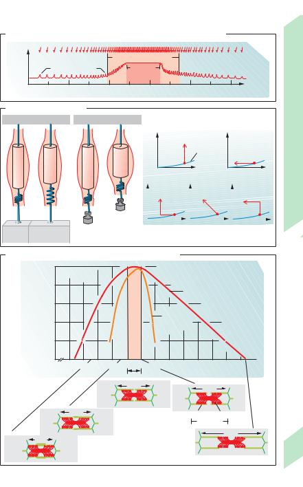
A. Muscle strength at increasing and decreasing stimulus frequencies
|
|
|
|
|
|
Stimulus |
Isometric muscleforce |
0 |
2 |
4 |
Range of summation |
8 |
10 Time (s) |
6 |
||||||
|
|
Single contractions |
|
Tetanus |
|
|
|
|
|
|
|
|
B. Types of contractions
Force |
Isometric |
Isotonic |
|
Resting |
|
|
|
tension |
00 |
|
curve |
Length |
|
Isotonic, then |
Auxotonic |
After- |
|||
isometric |
loaded |
||||
|
|
||||
|
|
|
|
|
|
Rest |
Isometric |
Rest |
Isotonic |
|
contraction |
contraction |
|||
|
|
C. Isometric muscle force relative to sarcomere length
|
100 |
|
|
|
|
|
|
|
|
|
|
|
|
force |
80 |
|
|
|
|
|
|
|
Skeletal muscle |
|
|||
|
|
|
|
|
|
|
|
|
|||||
muscleIsometric maximum)of(% |
|
|
|
|
forcemax.ofRange |
|
|
|
|
||||
60 |
|
|
|
|
|
|
|
|
|
|
|
||
|
|
|
|
|
|
|
|
|
|
|
|
|
|
|
|
|
|
|
|
|
|
Cardiac |
|
|
|
|
|
|
40 |
|
|
|
|
|
|
muscle |
|
|
|
|
|
|
20 |
|
|
|
|
|
|
|
|
|
|
|
|
|
0 |
1.4 |
1.6 |
1.8 |
2.0 |
2.2 |
2.4 |
2.6 |
2.8 |
3.0 |
3.2 |
3.4 |
3.6 |
|
1.2 |
||||||||||||
|
|
|
|
|
Lmax |
|
|
|
|
|
|
|
|
|
|
|
|
|
2.05 |
|
|
|
2.20 |
|
|
||
|
|
|
|
|
|
|
|
|
|
|
|
||
|
1.90 |
|
|
|
|
|
|
|
Actin |
Myosin |
|
||
|
|
|
|
|
|
|
|
|
|
Sarcomere |
|
||
|
|
m) |
3.65 |
1.50 |
Sarcomere length ( |
|
|
(skeletal muscle) |
|
|
|
|
|
|
(After Gordon et al.)
Plate 2.14 Mechanical Features of Skeletal Muscle I
67
Despopoulos, Color Atlas of Physiology © 2003 Thieme
All rights reserved. Usage subject to terms and conditions of license.