

книги студ / color atlas of physiology 5th ed[1]. (a. despopoulos et al, thieme 2003)
.pdf2 Nerve and Muscle, Physical Work
48
Propagation of Action Potentials in
Nerve Fiber
Electrical current flows through a cable when voltage is applied to it. The metal wire inside the cable is well insulated and has very lowlevel resistance, reducing current loss to a minimum. As a result, it can conduct electricity over long distances. Nerve fibers, especially unmyelinated ones (!p. 42), have a much greater internal longitudinal resistance (Ri) and are not well insulated from their surroundings. Therefore, the cable-like, electrotonic transmission of neural impulses dwindles very rapidly, so the conducted impulses must be continuously “refreshed” by generating new action potentials (!p. 46).
Propagation of action potentials: The start of an action potential is accompanied by a brief influx of Na+ into the nerve fiber (!A1a). The cell membrane that previously was inside negative now becomes positive ( + 20 to + 30 mV), thus creating a longitudinal potential difference with respect to the adjacent, still unstimulated nerve segments (internal –70 to –90 mV; !p. 44). This is followed by a passive electrotonic withdrawal of charge from the adjacent segment of the nerve fiber, causing its depolarization. If it exceeds threshold, another action potential is created in the adjacent segment and the action potential in the previous segment dissipates (!A1b).
Because the membrane acts as a capacitor, the withdrawal of charge represents a capacitating (depolarizing) flow of charge that becomes smaller and rises less steeply as the spatial distance increases. Because of the relatively high Ri of nerve fiber, the outward loops of current cross the membrane relatively close to the site of excitation, and the longitudinal current decreases as it proceeds towards the periphery. At the same time, depolarization increases the driving force (= Em – EK; !p. 32) for K+ outflow. K+ fluxing out of the cell therefore accelerates repolarization. Hence, distal action potentials are restricted to distances from which the capacitative current suffices to depolarize the membrane quickly and strongly enough. Otherwise, the Na+ channels will be deactivated before the threshold potential is reached (!p. 46).
Action potentials normally run forward (orthodromic) because each segment of nerve fiber becomes refractory when an action potential passes (!A1b and p. 46). If, however, the impulses are conducted backwards (antidromic) due, for example, to electrical stimulation of nerve fibers from an external source (!p. 50), they will terminate at the next synapse (valve-like function, !p. 42).
Although the continuous generation of action potentials in the immediately adjacent fiber segment guarantees a refreshed signal, this process is rather time-consuming (!B1). The conduction velocity, θ, in unmyelinated (type C) nerve fibers (!C) is only around 1 m/s. Myelinated (types A and B) nerve fibers (!C) conduct much faster (up to 80 m/s = 180 mph in humans). In the internode regions, a myelin sheath (!p. 42) insulates the nerve fibers from the surroundings; thus, longitudinal currents strong enough to generate action potentials can travel further down the axon (ca. 1.5 mm) (!A2). This results in more rapid conduction because the action potentials are generated only at the unmyelinated nodes of Ranvier, where there is a high density of Na+ channels. This results in rapid, jump-like passage of the action potential from node to node (saltatory propagation). The saltatory length is limited since the longitudinal current (1 to 2 nA) grows weaker with increasing distance (!B2). Before it drops below the threshold level, the signal must therefore be refreshed by a new action potential, with a time loss of 0.1 ms.
Since the internal resistance, Ri, of the nerve fiber limits the spread of depolarization, as described above, the axon diameter (2r) also affects the conduction velocity, θ (!C). Ri is proportional to the cross-sectional area of the nerve fiber (πr2), i.e., Ri !1/r2. Thick fibers therefore require fewer new APs per unit of length, which is beneficial for θ. Increases in fiber diameter are accompanied by an increase in both fiber circumference (2πr) and membrane capacity, K (K !r). Although θ decreases, the beneficial effect of the smaller Ri predominates because of the quadratic relationship.
Despopoulos, Color Atlas of Physiology © 2003 Thieme
All rights reserved. Usage subject to terms and conditions of license.

A. Continuous (1a, 1b) and saltatory propagation (2) of action potentials
|
AP |
AP |
Myelin sheath |
|
|
Na+ |
Na+ |
|
|
|
|
Action potential |
|
|
|
balance |
(AP) |
|
|
|
Depolarization |
Na+ |
+ |
|
Continuous |
Na+ |
Na+ AP |
Saltatory balancecharge current)(balancing |
Na |
Internode |
||||
|
charge |
|
Action |
|
|
|
Rest |
potential |
|
|
1a |
|
|
|
|
|
Refractory |
|
|
|
|
|
Depo- |
Node |
|
|
Depolarization |
larization |
|
|
1b |
|
2 |
|
B. Pulse propagation (action currents) in myelinated and unmyelinated nerve fibers
|
|
AP |
|
2 mm |
AP |
mm |
|
|
|
||
|
|
2 |
|
|
1nA |
|
1nA |
|
|
AP |
|
1 |
2 ms |
2 |
0.1ms 0.1ms |
C. Classification of nerve fibers (in humans)
Fiber type |
Function according to fiber type |
Diameter |
Conduction |
||
(Lloyd and Hunt types I–IV) |
( m) |
rate (m/s) |
|||
|
|||||
Aα |
Skeletal muscle efferent, afferents in muscle |
11–16 |
60 |
– 80 |
|
|
spindles (Ib) and tendon organs (Ib) |
|
|
|
|
Aβ |
Mechanoafferents of skin (II) |
6 –11 |
30 |
– 60 |
|
Aγ |
Muscle spindle efferents |
|
|
|
|
Aδ |
Skin afferents (temperature |
1– 6 |
2 |
– 30 |
|
|
and „fast“ pain) (III) |
|
|
|
|
B |
Sympathetic preganglionic; |
3 |
3 |
–15 |
|
|
visceral afferents |
|
|
|
|
C |
Skin afferents (“slow” pain); |
0.5–1.5 |
|
|
|
|
sympathetic postganglionic afferents (IV) |
(unmyelinated) |
0.25 |
– 1.5 |
|
|
|
|
|
|
|
(After Erlanger and Gasser)
Plate 2.4 Propagation of Action Potentials in Nerve Fiber
49
Despopoulos, Color Atlas of Physiology © 2003 Thieme
All rights reserved. Usage subject to terms and conditions of license.

|
|
Artificial Stimulation of Nerve Cells |
provide not only simple 1 : 1 connections, but |
|
|
|
also serve as switching elements for the |
||
|
|
|
|
|
|
|
When an electrical stimulus is applied to a |
nervous system. They can facilitate or inhibit |
|
|
|
nerve cell from an external source, current |
the neuronal transmission of information or |
|
|
|
flows from the positive stimulating electrode |
process them with other neuronal input. At the |
|
|
|
(anode) into the neuron, and exits at the nega- |
chemical synapse, the arrival of an action |
|
|
|
tive electrode (cathode). The nerve fiber below |
potential (AP) in the axon (!A1,2 and p. 48) |
|
|
|
the cathode is depolarized and an action |
triggers the release of the transmitter from the |
|
Work |
|
potential is generated there if the threshold |
presynaptic axon terminals. The transmitter |
|
|
potential is reached. |
then diffuses across the narrow synaptic cleft |
||
|
|
|||
Physical |
|
The conduction velocity of a nerve can be |
(ca. 30 nm) to bind postsynaptically to recep- |
|
|
distance from each other, then stimulating the |
the type of transmitter and receptor involved, |
||
|
|
measured by placing two electrodes on the |
tors in the subsynaptic membrane of a neuron |
|
|
|
skin along the course of the nerve at a known |
or of a glandular or muscle cell. Depending on |
|
Muscle, |
|
nerve (containing multiple neurons) and rec- |
the effect on the postsynaptic membrane may |
|
|
ording the time it takes the summated action |
either be excitatory or inhibitory, as is de- |
||
|
|
|||
|
|
potential to travel the known distance. The |
scribed below. |
|
and |
|
conduction velocity in humans is normally 40 |
Transmitters are released by regulated exo- |
|
|
to 70 m ! s– 1. Values below 40 m ! s– 1 are con- |
cytosis of so-called synaptic vesicles (!A1). |
||
Nerve |
|
sidered to be pathological. |
Each vesicle contains a certain quantum of |
|
|
Accidental electrification. Exposure of the |
neurotransmitters. In the case of the motor |
||
|
|
|||
2 |
|
body to |
high-voltage electricity, especially |
end-plate (!p. 56), around 7000 molecules of |
|
low-frequency alternating current (e.g., in an |
acetylcholine (ACh) are released. Some of the |
||
|
|
|||
|
|
electrical outlet) and low contact resistance |
vesicles are already docked on the membrane |
|
|
|
(bare feet, bathtub accidents), primarily affects |
(active zone), ready to exocytose their con- |
|
|
|
the conduction of impulses in the heart and |
tents. An incoming action potential functions |
|
|
|
|||
|
|
can cause ventricular fibrillation (!p. 200). |
as the signal for transmitter release (!A1,2). |
|
|
|
Direct current usually acts as a stimulus |
The higher the action potential frequency in |
|
|
|
only when switched on or off: High-frequency |
the axon the more vesicles release their con- |
|
|
|
alternating current (!15 kHz), on the other |
tents. An action potential increases the open |
|
|
|
hand, cannot cause depolarization but heats |
probability of voltage-gated Ca2+ channels in |
|
|
|
the body tissues. Diathermy works on this |
the presynaptic membrane (sometimes oscil- |
|
|
|
principle. |
|
lating), thereby leading to an increase in the |
|
|
|
|
cytosolic Ca2+ concentration, [Ca2+]i (!A1, 3 |
|
|
Synaptic Transmission |
and p. 36). Extracellular Mg2+ inhibits this |
|
|
|
process. Ca2+ binds to synaptotagmin (!A1), |
||
|
|
|
|
|
|
|
Synapses connect nerve cells to other nerve |
which triggers the interaction of syntaxin and |
|
|
|
cells (also applies for certain muscle cells) as |
SNAP-25 on the presynaptic membrane with |
|
|
|
well as to sensory and effector cells (muscle |
synaptobrevin on the vesicle membrane, |
|
|
|
and glandular cells). |
thereby triggering exocytosis of already |
|
|
|
Electrical synapses are direct, ion-conduct- |
docked vesicles (approximately 100 per AP) |
|
|
|
ing cell–cell junctions through channels (con- |
(!A1, 4). On the other hand, Ca2+ activates cal- |
|
|
|
nexons) |
in the region of gap junctions |
cium-calmodulin-dependent protein kinase-II |
|
|
(!p. 16f.). They are responsible for the con- |
(CaM-kinase-II; !A5, and p. 36), which acti- |
|
|
|
duction of impulses between neighboring |
vates the enzyme synapsin at the presynaptic |
|
|
|
smooth or cardiac muscle fibers (and some- |
terminal. As a result, vesicles dock anew on the |
|
|
|
times between neurons in the retina and in the |
active zone. |
|
|
|
CNS) and ensure also communication between |
Synaptic facilitation (= potentiation). If an |
|
|
|
neighboring epithelial or glial cells. |
action potential should arrive at the presynap- |
|
50 |
|
Chemical synapses utilize (neuro)transmit- |
tic terminal immediately after another AP (AP |
|
|
ters for the transmission of information and |
frequency ! approx. 30 Hz), the cytosolic Ca2+ |
||
!
Despopoulos, Color Atlas of Physiology © 2003 Thieme
All rights reserved. Usage subject to terms and conditions of license.
A. Chemical synapse
Na+ 
 AP 1
AP 1
|
Calmodulin |
Presynaptic |
|
|
|
|
|
Ca2+ |
|
|
ending |
|
Ca2+ |
|
|
Vesicle |
|
|
|
|
|
|
CaM- |
|
|
Synapto- |
kinase II |
Active zone |
|
tagmin |
|
|
|
|
|
|
|
|
Transmitter |
Ionotropic |
|
|
Synaptic |
receptor |
|
|
|
or |
|
cleft |
|
|
|
||
Metabotropic |
Cation |
|
|
|
receptor |
|
|
Postsynaptic cell |
channel |
|
|
|
|
||
5
|
Synapsin |
|
|
|
|
CaM- |
|
|
|
kinase II |
|
|
|
Vesicle |
|
|
|
preparation |
|
Active zone |
Docking |
|
|
|
|
||
K+ |
|
|
|
|
or |
|
|
Na+ (Ca2+) |
|
|
|
6 |
Signal chain |
K+ |
|
|
Ca2+ (Na+) |
||
|
7 |
||
|
0 |
2 |
|
|
|
Presynaptic |
|
|
action potential |
mV |
|
|
|
|
|
–80 |
|
|
|
3 |
Ca2+ influx |
0 |
ICa |
|
nA |
|
|
–0.5 |
|
|
|
4 |
|
|
Transmitter |
|
1 |
in cleft |
Transmitter |
mmol/L |
|
release |
|
|
|
|
|
|
0 |
|
|
|
(Partly after Llinás) |
|
mV |
Transmitter binding |
0 |
to receptors |
|
EPSP1 EPSP2 EPSP3 |
|
Summation
Postsynaptic |
–90 |
|
|
|
|
||
action potential |
|
|
(see plate B.) |
|
|
|
Plate 2.5 Synaptic Transmission I
51
Despopoulos, Color Atlas of Physiology © 2003 Thieme
All rights reserved. Usage subject to terms and conditions of license.

2 Nerve and Muscle, Physical Work
52
!
concentration will not yet drop to the resting value, and residual Ca2+ will accumulate. As a result, the more recent rise in [Ca2+]i builds on the former one. [Ca2+]i rises to a higher level after the second stimulus than after the first, and also releases more transmitters. Hence, the first stimulus facilitates the response to the second stimulus. Muscle strength increases at high stimulus frequencies for similar reasons (!p. 67 A).
Among the many substances that act as excitatory transmitters are acetylcholine (ACh) and glutamate (Glu). They are often released together with co-transmitters which modulate the transmission of a stimulus (e.g., ACh together with substance P, VIP or galanin; Glu with substance P or enkephalin). If the transmitter’s receptor is an ion channel itself (ionotropic receptor or ligand-gated ion channel; !A6 and F), e.g., at the N-cholinergic synapse (!p. 82), the channels open more often and allow a larger number of cations to enter (Na+, sometimes Ca2+) and leave the cell (K+). Other, so-called metabotropic receptors influence the channel via G proteins that control channels themselves or by means of “second messengers” (!A7 and F). Because of the high electrochemical Na+ gradient (!p. 32), the number of incoming Na+ ions is much larger than the number of exiting K+ ions. Ca2+ can also enter the cell, e.g., at the glutamate-NMDA receptor (!F). The net influx of cations leads to depolarization: excitatory postsynaptic potential (EPSP) (maximum of ca. 20 mV; !B). The EPSP begins approx. 0.5 ms after the arrival of an action potential at the presynaptic terminal. This synaptic delay (latency) is caused by the relatively slow release and diffusion of the transmitter.
A single EPSP normally is not able to generate a postsynaptic (axonal) action potential (APA), but requires the triggering of a large number of local depolarizations in the dendrites. Their depolarizations are transmitted electrotonically across the soma (!p. 48) and summed on the axon hillock (spatial summation; !B). Should the individual stimuli arrive at different times (within approx. 50 ms of each other), the prior depolarization will not have dissipated before the next one arrives, and summation will make it easier to reach
threshold. This type of temporal summation therefore increases the excitability of the postsynaptic neuron (!C).
Inhibitory transmitters include substances as glycine, GABA (γ-aminobutyric acid), and acetylcholine (at M2 and M3 receptors; !p. 82). They increase the conductance, g, of the subsynaptic membrane only to K+ (e.g., the metabotropic GABAB receptor.) or Cl– (e.g., the ionotropic glycine and GABAA receptors; !F). The membrane usually becomes hyperpolarized in the process (ca. 4 mV max.). Increases in gK occur when Em approaches EK (!p. 44). However, the main effect of this inhibitory postsynaptic potential IPSP (!D) is not hyperpolarization–which works counter to EPSP-related depolarization (the IPSP is sometimes even slightly depolarizing). Instead, the IPSP-related increase in membrane conductance short circuits the electrotonic currents of the EPSP (high gK or gCl levels). Since both EK and ECl are close to the resting potential (!p. 44), stabilization occurs, that is, the EPSP is cancelled out by the high K+ and Cl– shortcircuit currents. As a result, EPSP-related depolarization is reduced and stimulation of postsynaptic neurons is inhibited (!D).
Termination of synaptic transmission (!E) can occur due to inactivation of the cation channels due to a conformational change in the channel similar to the one that occurs during an action potential (!p. 46). This very rapid process called desensitization also functions in the presence of a transmitter. Other terminating pathways include the rapid enzymatic decay of the transmitter (e.g., acetylcholine) while still in the synaptic cleft, the re-up- take of the transmitter (e.g., noradrenaline) into the presynaptic terminal or uptake into extraneuronal cells (e.g., in glial cells of the CNS), endocytotic internalization of the receptor (!p. 28), and binding of the transmitter to a receptor on the presynaptic membrane (autoceptor). In the latter case, a rise in gK and a drop in gCa can occur, thus inhibiting transmitter release, e.g., of GABA via GABAB receptors or of noradrenaline via α2-adrenoceptors (!F and p. 86).
Despopoulos, Color Atlas of Physiology © 2003 Thieme
All rights reserved. Usage subject to terms and conditions of license.

B. Spatial summation of stimuli |
|
|
|
|
|
|||||
|
|
|
|
|
AP1 |
|
|
mV |
EPSP1 |
|
|
|
|
|
|
|
|
|
–70 |
|
|
|
|
|
|
|
|
AP2 |
|
–90 |
|
ms |
|
|
|
|
|
|
|
|
|
|
|
Dendrite |
|
|
|
|
|
–70 |
EPSP2 |
|
||
|
|
|
|
|
|
|
|
|
||
|
|
|
|
|
|
|
|
|
II |
|
|
|
|
|
|
|
|
|
|
|
|
|
|
Neuron (soma) |
|
AP3 |
–90 |
|
Transmission |
|||
|
|
|
|
|
|
|
|
–70 |
EPSP3 |
|
mV |
|
|
|
|
|
|
|
|
|
|
0 |
|
|
|
|
|
|
|
–90 |
|
|
Action potential |
|
|
|
Synaptic |
||||||
–10 |
|
|
|
|
||||||
(APA) |
|
|
|
|
|
|
|
|||
|
|
|
Axon |
|
|
|
|
|||
|
|
|
|
|
APA |
|
|
|
||
–30 |
|
|
|
|
hillock |
Electrotonic currents |
|
|||
|
|
|
|
|
|
|
(depolarizing) |
|
2.6 |
|
–50 |
|
|
|
|
Summed EPSP |
Axon |
|
|
|
|
|
|
|
|
|
|
|
|
Plate |
||
–70 |
|
|
|
|
|
|
|
|
|
|
–900 |
2 |
4 |
6 |
8 |
ms |
|
|
|
|
|
C. Temporal summation of stimuli |
|
|
|
|
|
|
|
||
|
|
AP1 |
|
Dendrite |
|
|
|
|
|
mV |
|
|
|
|
|
|
|
|
|
|
|
|
|
|
|
|
|
|
|
–70 |
EPSP1 |
|
|
|
|
|
|
|
|
–90 |
|
|
|
Neuron (soma) |
|
|
|
|
|
|
|
|
|
|
|
|
|
||
–70 |
EPSP2 |
AP2 |
|
mV |
|
|
|
|
|
|
|
0 |
|
|
Action potential |
||||
|
|
|
|
–10 |
|
|
|||
–90 |
|
|
|
|
|
(APA) |
|
||
|
|
|
|
|
|
|
|
|
|
|
|
|
|
–30 |
|
|
Summed EPSP |
||
Elapsed |
|
APA |
|
|
|
|
|
|
|
|
time |
Electrotonic currents |
–50 |
|
|
|
|
|
|
|
|
|
|
|
|
|
|||
|
|
|
|
|
|
|
|
|
|
|
|
(depolarizing) |
|
|
|
|
|
|
|
|
|
|
|
–70 |
|
|
|
|
|
|
|
|
|
–900 |
2 |
4 |
6 |
8 |
ms |
|
|
|
|
|
|
|
|
|
53 |
Despopoulos, Color Atlas of Physiology © 2003 Thieme
All rights reserved. Usage subject to terms and conditions of license.

|
D. Effect of IPSP on postsynaptic stimulation |
|
|
||
|
Excitatory |
|
APE |
|
|
|
|
|
|
|
|
|
transmitter |
|
mV |
|
|
|
|
|
|
|
|
|
|
|
–70 |
|
|
|
|
|
EPSP |
|
|
Work |
|
|
–90 |
API |
|
K+ |
|
ms |
|
||
|
|
|
|
||
Physical |
|
|
Inhibitory |
|
|
Na+ |
|
transmitter |
–70 |
|
|
Depolarization |
|
|
IPSP |
|
|
Muscle, |
|
|
|
||
Electrotonic transmission |
|
|
–90 |
|
|
|
|
|
|
|
|
and |
“Short-circuit” via |
|
|
|
|
K+- (and/or Cl–-) channels |
|
|
mV |
Summation |
|
Nerve |
|
|
|||
|
K+ |
|
–70 |
EPSP+IPSP |
|
|
|
|
|
|
|
2 |
Hyperpolarization |
|
|
||
|
–90 |
|
|||
|
|
|
|
|
|
|
Postsynaptic |
|
|
|
ms |
|
|
|
|
|
|
|
neuron |
|
Electrotonic currents |
|
|
|
|
|
hyperpolarize |
|
|
|
|
|
axon hillock |
To |
|
|
|
|
|
axon hillock |
|
|
E. Termination of transmitter action |
|
|
||
|
|
|
Extraneuronal |
|
|
|
|
|
uptake |
|
|
|
|
|
Reuptake |
Glial cell, etc. |
|
|
|
|
|
|
|
|
|
Inhibition |
|
|
|
|
Presynaptic |
of exocytose |
|
|
|
|
ending |
|
|
Diffusion |
|
|
|
|
|
|
|
|
gK |
gCa |
|
out of cleft |
|
|
|
|
|
||
|
Autoceptor |
|
|
|
|
|
Postsynaptic |
|
|
|
|
|
cell |
|
|
|
|
|
Enzymatic breakdown |
Rapid inactivation of |
|
|
|
|
of transmitter |
|
|
|
|
|
|
cation channel |
|
|
|
|
|
|
Internalization |
||
|
|
|
(desensitization) |
||
54 |
|
|
of receptor |
|
|
|
|
|
|
||
Despopoulos, Color Atlas of Physiology © 2003 Thieme
All rights reserved. Usage subject to terms and conditions of license.

F. Neurotransmitters in the central nervous system
Transmitter |
Receptor |
Receptor |
|
Effect |
|
|
|
|
subtypes |
types |
Ion conductance |
Second messenger |
|||
|
|
|
|
||||
|
|
|
|
Na+ |
K+ Ca2+ Cl– |
cAMP |
IP3/DAG |
Acetylcholine |
Nicotinic |
|
|
|
|
|
|
|
Muscarinic: |
|
|
|
|
|
|
|
M1, |
|
|
|
|
|
|
|
M2, M3 |
|
|
|
|
|
|
ADH |
V1 |
|
|
|
|
|
|
(= vasopressin) |
V2 |
|
|
|
|
|
|
CCK (= cholecystokinin) |
CCKA–B |
|
|
|
|
|
|
Dopamine |
D1, D5 |
|
|
|
|
|
|
|
D2 |
|
|
|
|
|
|
GABA |
GABAA,GABAC |
|
|
|
|
|
|
(= γ -aminobutyric acid) |
GABAB |
|
|
|
|
|
|
Glutamate (aspartate) |
AMPA |
|
|
|
|
|
|
|
Kainat |
|
|
|
|
|
|
|
NMDA |
|
|
|
|
|
|
|
m-GLU |
|
|
|
|
|
|
Glycine |
_ |
|
|
|
|
|
|
Histamine |
H1 |
|
|
|
|
|
|
|
H2 |
|
|
|
|
|
|
Neurotensin |
_ |
|
|
|
|
|
|
Norepinephrine, |
α1(A–D) |
|
|
|
|
|
|
α2(A–C) |
|
|
|
|
|
|
|
epinephrine |
|
|
|
|
|
|
|
|
β1–3 |
|
|
|
|
|
|
Neuropeptide Y (NPY) |
Y1–2 |
|
|
|
|
|
|
Opioid peptides |
µ, δ, κ |
|
|
|
|
|
|
Oxytocin |
_ |
|
|
|
|
|
|
Purines |
P1: A1 |
|
|
|
|
|
|
|
A2a |
|
|
|
|
|
|
|
P2X |
|
|
|
|
|
|
|
P2Y |
|
|
|
|
|
|
Serotonin |
5-HT1 |
|
|
|
|
|
|
(5-hydroxytryptamine) |
5-HT2 |
|
|
|
|
|
|
|
5-HT3 |
|
|
|
|
|
|
|
5-HT4–7 |
|
|
|
|
|
|
Somatostatin (= SIH) |
SRIF |
|
|
|
|
|
|
Tachykinin |
NK1–3 |
|
|
|
|
|
|
Amino acids |
|
|
|
|
|
Inhibits or |
promotes |
|
|
|
|
|
|
|
|
Catecholamines |
|
|
|
|
|
|
|
Peptides |
Ionotropic receptor |
Metabotropic receptor |
|
DAG |
|||
Others |
(ligand-gated |
(G protein-mediated |
cAMP |
PIP2 IP3 |
|||
ion channel) |
effect) |
|
|
ATP |
|||
|
|
|
|
||||
(Modified from F. E. Bloom)
Plate 2.7 u. 2.8 Synaptic Transmission III u. IV
55
Despopoulos, Color Atlas of Physiology © 2003 Thieme
All rights reserved. Usage subject to terms and conditions of license.

2 Nerve and Muscle, Physical Work
56
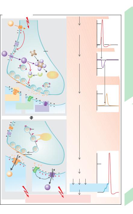
Motor End-plate
The transmission of stimuli from a motor axon to a skeletal muscle fiber occurs at the motor end-plate, MEP (!A), a type of chemical synapse (!p. 50ff.). The transmitter involved is acetylcholine (ACh, !cf. p. 82), which binds to the N(nicotinergic)-cholinoceptors of the subsynaptic muscle membrane (!A3). N-cholino- ceptors are ionotropic, that is, they also function as ion channels (!A4). The N-cholinocep- tor of the MEP (type NM) has 5 subunits (2α, 1", 1γ, 1δ), each of which contains 4 membranespanning α-helices (!p. 14).
The channel opens briefly (!B1) (for approx. 1 ms) when an ACh molecule binds to the two α-subunits of an N-cholinoceptor (!A4). Unlike voltage-gated Na+-channels, the openprobability po of the NM-cholinoceptor is not increased by depolarization, but is determined by the ACh concentration in the synaptic cleft
(!p. 50 ff.).
The channel is specific to cations such as Na+, K+, and Ca2+. Opening of the channel at a resting potential of ca. !90 mV leads mainly to an influx of Na+ ions (and a much lower outflow of K+; !pp. 32 ff. and 44). Depolarization of the subsynaptic membrane therefore occurs: endplate potential (EPP). Single-channel currents of 2.7 pA (!B1) are summated to yield a miniature end-plate current of a few nA when spontaneous exocytosis occurs and a vesicle releases a quantum of ACh activating thousands of NM-cholinoceptors (!B2). Still, this is not enough for generation of a postsynaptic action potential unless an action potential transmitted by the motor neuron triggers exocytosis of around a hundred vesicles. This opens around 200,000 channels at the same time, yielding a neurally induced end-plate current (IEP) of ca. 400 nA (!B3). End-plate current, IEP, is therefore dependent on:
the number of open channels, which is equal to the total number of channels (n) times the open-probability (po), where po is determined by the concentration of ACh in the synaptic cleft (up to 1 mmol/L);
the single-channel conductance γ (ca.
30 pS);
and, to a slight extent, the membrane potential, Em, since the electrical driving
“force” (= Em –ENa,K; !p. 32 ff.) becomes smaller when Em is less negative.
ENa,K is the common equilibrium potential for Na+ and K+ and amounts to approx. 0 mV. It is also called the reversal potential because the direction of IEP (= INa + IK), which enters the cell when Em is negative (Na+ influx " K+ outflow), reverses when Em is positive (K+ outflow " Na+ influx). As a result,
IEP # n ! po ! γ ! (Em – ENa, K) [A] |
[2.1] |
Because neurally induced EPPs in skeletal muscle are much larger (depolarization by ca. 70 mV) than neuronal EPSPs (only a few mV; !p. 50 ff.), single motor axon action potentials are above threshold. The EPP is transmitted electrotonically to the adjacent sarcolemma, where muscle action potentials are generated by means of voltage-gated Na+ channels, resulting in muscle contraction.
Termination of synaptic transmission in MEPs occurs (1) by rapid degradation of ACh in the synaptic cleft by acetylcholinesterase localized at the subsynaptic basal membrane, and
(2) by diffusion of ACh out of the synaptic cleft (!p. 82).
A motor end-plate can be blocked by certain poisons and drugs, resulting in muscular weakness and, in some cases, paralysis. Botulinum neurotoxin, for example, inhibits the discharge of neurotransmitters from the vesicles, and α-bungarotoxin in cobra venom blocks the opening of ion channels. Curare-like substances such as (+)-tubocurarine are used as muscle relaxants in surgical operations. They displace ACh from its binding site (competitive inhibition) but do not have a depolarizing effect of their own. Their inhibitory effect can be reversed by cholinesterase inhibitors such as neostigmine (decurarinization). These agents increase the concentration of ACh in the synaptic cleft, thereby displacing curare. Entry of anticholinesterase agents into intact synapses leads to an increase in the ACh concentration and, thus, to paralysis due to permanent depolarization. ACh-like substances such as suxamethonium have a similar depolarizing effect, but decay more slowly than ACh. In this case, paralysis occurs because permanent depolarization also permanently inactivates Na+ channels near the motor end-plate on the sarcolemma (!p. 46).
Despopoulos, Color Atlas of Physiology © 2003 Thieme
All rights reserved. Usage subject to terms and conditions of license.

A. Motor end-plate
Myelin sheath Motor axon
1 |
Motor end-plate |
Schwann cell
|
|
Mitochondrion |
Nerve |
|
|
|
Vesicle |
ending |
Finger |
2 |
|
Postsynaptic |
Basement |
|
|
|
|||
|
|
membrane |
|
|
|
|
folds |
|
|
|
|
|
|
|
|
|
|
Muscle fiber |
|
3 |
|
Acetylcholine |
|
|
|
|
|
|
|
|
|
vesicle |
Presynaptic membrane |
|
|
|
|
||
|
|
Nerve |
Synaptic cleft with basement membrane |
|
|
|
Postsynaptic membrane (sarcolemma) |
||
|
|
ending |
||
|
|
|
|
|
|
4 |
ACh |
K+ |
|
|
|
|
Active |
Cholinergic |
γ |
|
zone |
N-receptors |
|
|
|
α |
α |
|
|
Muscle fiber |
|
|
|
|
(Ca2+) Na+ |
|
|
(Partly after Akert and Peper) |
|
|
B. End-plate currents |
|
|
|
|
1 Quantum |
100-200 Quanta |
|
|
|
|
2.7pA |
|
|
|
4nA |
|
|
|
400nA |
0 |
1 |
2 |
3 |
0 |
1 |
2 |
3 |
0 |
1 |
2 |
3 |
|
|
|
Time (ms) |
|
|
|
Time (ms) |
|
|
Time (ms) |
|
1 Single-channel current |
2 Miniature end-plate |
|
3 Nerve-induced |
|
|||||||
|
|
|
|
|
current |
|
|
|
end-plate current |
|
|
(After Neher and Sakmann (1) and after Peper et al. (2))
Plate 2.9 Motor End-plate
57
Despopoulos, Color Atlas of Physiology © 2003 Thieme
All rights reserved. Usage subject to terms and conditions of license.