

книги студ / color atlas of physiology 5th ed[1]. (a. despopoulos et al, thieme 2003)
.pdfEndothelial Exchange Processes
|
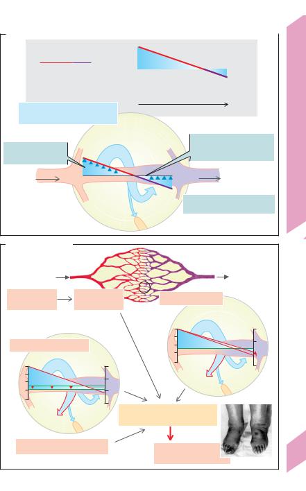
Nutrients and waste products are exchanged |
|||
|
across the walls of the capillaries and post- |
|||
|
papillary venules (exchange vessels; !p. 188). |
|||
|
Their endothelia contain small (ca. 2–5 nm) or |
|||
|
large (20–80 nm, especially in the kidneys and |
|||
|
liver) functional pores: permeable, intercellu- |
|||
|
lar fissures or endothelial fenestrae, respec- |
|||
|
tively. The degree of endothelial permeability |
|||
|
varies greatly from one organ to another. Vir- |
|||
|
tually all endothelia allow water and inorganic |
|||
System |
ions to pass, but most are largely impermeable |
|||
to blood cells and large protein molecules. |
||||
|
||||
|
Transcytosis and carriers (!p. 26f.) allow for |
|||
Cardiovascular |
passage of certain larger molecules. |
|||
Filtration and reabsorption. About 20 L/day |
||||
|
||||
|
of fluid is filtered (excluding the kidneys) into |
|||
|
the interstitium from the body’s exchange ves- |
|||
|
sels. About 18 L/day of this fluid is thought to be |
|||
|
reabsorbed by the venous limb of these vessels. |
|||
8 |
The remaining 2 L/day or so make up the lymph |
|||
flow and thereby return to the bloodstream |
||||
|
||||
|
(!A). The filtration or reabsorption rate Qf is a |
|||
|
factor of the endothelial filtration coefficient Kf |
|||
|
(= water permeability k · exchange area A) and |
|||
|
the effective filtration pressure Peff (Qf = Kf · Peff). |
|||
|
Peff is calculated as the hydrostatic pressure |
|||
|
difference P minus |
the oncotic pressure |
||
|
difference Δπ across the capillary wall (Star- |
|||
|
ling’s relationship; !A), where |
P = capillary |
||
|
pressure (Pcap) minus interstitial pressure (Pint, |
|||
|
normally ! 0 mmHg). At the level of the heart, |
|||
|
P at the arterial end of the systemic capillar- |
|||
|
ies is about 30 mmHg and decreases to about |
|||
|
22 mmHg at the venous end. Since Δπ (ca. |
|||
|
24 mmHg; !A) counteracts |
P, the initially |
||
|
high filtration rate (Peff = + 6 mmHg) is thought |
|||
|
to change into reabsorption whenever Peff be- |
|||
|
comes negative. (Since |
P is only 10 mmHg in |
||
the lungs, the pulmonary Peff is very low). Δπ occurs because the concentration of proteins (especially albumin) in the plasma is much higher than their interstitial concentration. The closer the reflection coefficient of the
plasma proteins (σprot) to 1.0, the higher Δπ and, consequently, the lower the permeability
of the membrane to these proteins (!p. 377).
According to Starling’s relationship, water reab-
208sorption should occur as long as Peff is negative. However, recent data suggest that a negative Peff re-
sults in only transient reabsorption. After several minutes it stops because the interstitial oncotic pressure rises due to “self-regulation”. Thus, a major part of the 18 L/d expected to be reabsorbed from the exchange vessels (see above) might actually be reabsorbed in the lymph nodes. Rhythmic contraction of the arterioles (vasomotion) may also play a role by decreasing Peff and thus by allowing intermittent capillary reabsorption.
In parts of the body below the heart, the effects of hydrostatic pressure from the blood column increase the pressure in the capillary lumen (in the feet !90 mmHg). The filtration rate in these regions therefore rise, especially when standing still. This is counteracted by two “self-regulatory” mechanisms:
(1) the outflow of water results in an increase in the luminal protein concentration (and thus Δπ) along the capillaries (normally the case in glomerular capillaries, !p. 152); (2) increased filtration results in an increase in Pint and a consequent decrease in P.
Edema. Fluid will accumulate in the interstitial space (extracellular edema), portal venous system (ascites), and pulmonary interstice (pulmonary edema) if the volume of filtered fluid is higher than the amount returned to the blood.
Causes of edema (!B):
Increased capillary pressure (!B1) due to precapil-
lary vasodilatation (Pcap"), especially when the capillary permeability to proteins also increases (σprot # and Δπ #) due, for example, to infection or anaphylaxis (histamine etc.). Hypertension in the portal vein leads to ascites.
Increased venous pressure (Pcap ", !B2) due, for example, to venous thrombosis or cardiac insufficiency (cardiac edema).
Decreased concentration of plasma proteins, especially albumin, leading to a drop in Δπ (!B3 and p. 379 A) due, for example, to loss of proteins (proteinuria), decreased hepatic protein synthesis (e.g., in liver cirrhosis), or to increased breakdown of plasma proteins to meet energy requirements (hunger edema).
Decreased lymph drainage due, e.g., to lymph tract compression (tumors), severance (surgery), obliteration (radiation therapy) or obstruction (bilharziosis) can lead to localized edema (!B4).
Increased hydrostatic pressure promotes edema formation in lower regions of the body (e.g., in the ankles; !B).
Diffusion. Although dissolved particles are dragged through capillary walls along with filtered and reabsorbed water (solvent drag; !p. 24), diffusion plays a much greater role in the exchange of solutes. Net diffusion of a substance (e.g., O2, CO2) occurs if its plasma and interstitial conc. are different.
Despopoulos, Color Atlas of Physiology © 2003 Thieme
All rights reserved. Usage subject to terms and conditions of license.
|
|
|
|
|
|
|
|
|
||||||||||||||||
A. Exchange of fluids via capillaries and venules |
|
|
|
|
|
|
|
|
||||||||||||||||
|
|
|
|
|
|
|
|
|||||||||||||||||
|
P (hydrostatic |
30 |
|
|
|
|
|
|
|
|
|
|
|
|
4.0 |
|
||||||||
|
|
|
|
|
mmHg |
|
|
|
|
|
|
|
|
|
Reabsorption |
|
kPa |
|
||||||
|
pressure difference) |
|
|
|
|
|
|
|
|
|
|
3.5 |
|
|||||||||||
|
Δπ (oncotic |
25 |
|
|
|
|
|
|
|
|
|
|
|
|
3.0 |
Processes |
||||||||
|
|
|
|
|
|
|
|
|
|
|
|
|
||||||||||||
|
|
|
|
|
|
|
|
|
|
|
|
|
|
|
|
|
|
|
|
|
|
|||
|
|
|
|
|
|
|
|
|
|
|
|
|
|
|
|
|
|
|
|
|
|
|||
|
|
|
|
|
|
|
|
|
|
|
|
|
|
|
|
|
|
|
|
|||||
|
|
|
|
|
|
|
|
|
|
|
|
|
|
|
|
|
|
|
|
|
||||
|
pressure difference) |
|
|
Filtration |
|
|
||||||||||||||||||
|
|
|
20 |
|
|
|
|
|
|
|
Path of exchange |
|
2.5 |
|
||||||||||
|
|
|
|
|
|
|
|
|
|
|
||||||||||||||
|
|
15 |
|
|
|
|
|
|
|
|
|
|||||||||||||
|
|
|
|
|
|
|
|
|
|
2.0 |
|
|||||||||||||
|
|
|
|
|
|
|
|
|
|
|
||||||||||||||
Filtration |
|
|
|
|
|
|
|
|
|
|
|
|
Exchange |
|||||||||||
|
|
|
|
|
|
|
|
|
|
|
|
|
|
|
|
|
|
|
|
|
|
|||
= Reabsorption + lymph drainage |
|
|
|
|
|
|
|
|
|
|
|
|
|
|||||||||||
|
|
|
|
|
|
|
|
|
|
|
|
|
|
|||||||||||
Peff (effective filtration |
|
|
|
|
|
|
|
|
|
|
|
|
|
|
|
|
|
|
Kf (filtration coefficient) |
Endothelial |
||||
|
|
|
|
|
|
|
|
|
Filtration |
|
|
= k (water permeability) |
||||||||||||
|
pressure) |
|
|
|
|
|
|
|
|
|
|
|
· A (exchange area) |
|
||||||||||
|
|
|
|
|
|
|
|
|
|
|
|
|
||||||||||||
|
|
|
|
|
|
|
|
|
|
|
|
|
|
|
|
|
|
|
|
|||||
= P Ð Δπ |
|
|
|
|
|
|
|
|
|
|
|
|
|
|
|
|
|
|
Venole |
|
|
|
||
Arteriole |
|
|
|
|
|
|
|
|
|
|
|
|
|
|
|
|
|
|
|
|
8.12 |
|||
|
|
Reabsorption |
|
|
|
|
|
|
|
|
|
|
|
Qf (Filtration/reabsorption rate) |
||||||||||
|
|
|
|
|
|
|
|
|
|
|
|
|
||||||||||||
|
|
|
|
|
|
|
|
|
|
|
|
|
||||||||||||
|
|
|
|
|
ca. 90% |
|
|
Plate |
||||||||||||||||
|
|
Interstitium |
|
|
|
|
||||||||||||||||||
|
|
|
|
|
|
= Peff ! Kf |
|
|
||||||||||||||||
|
|
|
|
|
|
|
|
|
||||||||||||||||
|
|
|
|
|
|
ca. 10% |
|
|
|
|
|
|
|
|
|
|
|
|
||||||
|
|
|
|
|
|
|
|
|
|
|
|
|
|
|
|
|
|
|
|
|||||
|
Lymph |
|
||
B. Causes of edema |
|
|
||
|
Arteriole |
Venule |
|
|
1 |
|
2 |
|
|
|
|
|
||
Precapillary |
Capillary |
Venous pressure rises |
|
|
vasodilatation |
pressure rises |
|
||
|
|
|||
|
|
mmHg |
kPa |
|
3 |
|
30 |
4.0 |
|
|
|
3.5 |
||
Decrease in plasma proteins |
25 |
|||
|
||||
mmHg |
kPa |
|
3.0 |
|
20 |
2.5 |
|||
30 |
4.0 |
|||
|
||||
25 |
3.5 |
Edema |
|
|
|
|
|
||
|
3.0 |
|
|
|
20 |
|
|
|
|
|
|
Filtration |
|
|
Edema |
|
> Reabsorption+lymph drainage |
|
|
4 |
|
|
|
|
Reduced lymph drainage |
Edema |
209 |
||
|
|
|||
|
|
(e.g., in ankles) |
||
Despopoulos, Color Atlas of Physiology © 2003 Thieme
All rights reserved. Usage subject to terms and conditions of license.

8 Cardiovascular System
210
Myocardial Oxygen Supply
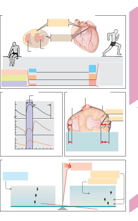
Coronary arteries. The blood flow to the myocardium is supplied by the two coronary arteries that arise from the aortic root. The right coronary artery (approx. 1/7th of the blood) usually supplies the greater portion of the right ventricle, while the left coronary artery (6/7th of the blood) supplies the left ventricle (!A). The contribution of both arteries to blood flow in the septum and posterior wall of the left ventricle varies.
.
Coronary blood flow (Qcor) is phasic, i.e., the amount of blood in the coronary arteries fluctuates during the cardiac cycle due to extremely high rises in extravascular tissue pressure during systole (!B, C). The blood flow in the epicardial coronary artery branches and subepicardial vessels remains largely unaffected by these pressure fluctuations. However, the subendocardial vessels of the left ventricle are compressed during systole when the extravascular pressure in that region (! pressure in left ventricle, PLV) exceeds the pressure in the lumen of the vessels (!C). Consequently, the left ventricle is mainly supplied during diastole (!B middle). The fluctuations in right ventricular blood flow are much less distinct because right ventricular pressure
(PRV) is lower (!B, C). .
Myocardial. O2 consumption (VO2) is defined as Qcor times the arteriovenous O2 concentration difference, (Ca–Cv)O2. The myocardial (Ca–Cv)O2 is relatively high (0.12 L/L blood), and oxygen extraction at rest ([Ca–Cv]O2/CaO2 = 0.12/ 0.21) is almost 60% and, thus, not able to. rise much further. Therefore, an increase in Qcor is practically. the only way to increase myocardial VO2 when the O2 demand rises (!D, right side).
Adaptation of the myocardial O2 supply according to need is therefore primarily achieved by adjusting vascular resistance (!D, left side). The (distal) coronary vessel resistance can normally be reduced to about 1/4 the resting value (.coronary reserve). The coronary blood flow Qcor (approx. 250 mL/min at rest) can therefore be increased as much as 4–5 fold. In other words, approx. 4 to 5 times more O2 can be supplied during maximum physical exertion.
Arteriosclerosis (atherosclerosis) of the coronary arteries leads to luminal narrowing and a resultant decrease in poststenotic pressure. Dilatation of the distal vessels then occurs as an autoregulatory response (see below). Depending on the extent of the stenosis, it may be necessary to use a fraction of the coronary reserve, even during rest. As a result, lower or insufficient quantities of O2 will be available to satisfy increased O2 demand, and coronary insufficiency may occur (!D)
Myocardial O2 demand increases with cardiac output (increased pressure–volume–work/time), i.e., in response to increases in heart rate and/or contractility, e.g., during physical exercise (!D, right).
It also increases as a function of mural tension (Tventr) times the duration of systole (tension–time index).
Since Tventr = Pventr · rventr/2w (Laplace’s law !Eq. 8.4b, p. 188), O2 demand is greater when the ventricular
pressure (Pventr) is high and the stroke volume small than when Pventr is low and the stroke volume high, even when the same amount of work (P ! V) is per-
formed. In the first case, the efficiency of the heart
is reduced. When the ventricular pressure Pventr is elevated, e.g., in hypertension, the myocardium there-
fore requires more O2 to perform the same amount of work (!D, right).
Since the myocardial metabolism is aerobic, an increased O2 demand quickly has to lead to vasodilatation. The following factors are involved in the coronary vasodilatation:
Metabolic factors: (a) oxygen deficiency since O2 acts as a vasoconstrictor; (b) Adenosine; oxygen deficiencies result in insufficient quantities of AMP being re-converted to ATP, leading to accumulation of adenosine, a degradation product of AMP. This leads to
A2 receptor-mediated vasodilatation; (c) Accumulation of lactate and H+ ions (from the anaerobic myocardial metabolism); (d) prostaglandin I2.
Endothelial factors: ATP (e.g., from platelets), bradykinin, histamine and acetylcholine are vasodilators. They liberate nitric oxide (NO) from the endothelium, which diffuses into vascular muscle cells to stimulate vasodilatation (!p. 279 E).
Neurohumoral factors: Norepinephrine released from sympathetic nerve endings and adrenal epinephrine have a vasodilatory effect on the distal
coronary vessels via !2 adrenoceptors.
Myocardial energy sources. The myocardium can use the available glucose, free fatty acids, lactate and other molecules for ATP production. The oxidation of each of these three energy substrates consumes a
certain fraction of myocardial O2 (O2 extraction coefficient); accordingly, each contributes approx. one-third of the produced ATP at rest. The myocardium consumes increasing quantities of lactate from the skeletal muscles during physical exercise (!A, !p. 72 and 282).
Despopoulos, Color Atlas of Physiology © 2003 Thieme
All rights reserved. Usage subject to terms and conditions of license.
A. Blood supply, O2 and substrate consumption of heart muscle
|
|
Right |
|
|
|
|
coronary artery |
|
|
|
Supply area |
|
|
|
|
|
Left |
|
|
|
|
coronary artery |
|
|
|
|
Left ventricle |
|
|
|
Right ventricle |
|
|
|
|
Rest Fraction of O2 consumed by substrate oxidation |
|||
|
|
(O2 extraction coefficient), 300g heart tissue |
||
|
250 |
|
· |
|
1/3 Glucose |
Coronary blood flow QCor(mL/min) |
|||
|
Arteriovenous O2 difference |
|
||
|
|
|
||
1/3 Free fatty acids |
0.12 |
(Ca –Cv)O2 (L/L Blut) |
|
|
1/3 Lactate |
30 |
· |
· |
(mL/min) |
O2 consumption VO2 |
=Qcor·(Ca –Cv)O2 |
|||
Exercise
|
1/7 Glucose |
600 |
1/5 Free fatty acids |
0.15 |
2/3 Lactate |
|
(from active |
90 |
skeletal muscles) |
|
|
B. Coronary blood flow |
|
|
|
||||
|
|
|
|
Systole |
|
|
|
|
|
|
|
(ejection phase) |
|
||
Aortic pressure (mmHg) |
120 |
|
|
Diastole |
|
|
|
100 |
|
|
Aorta |
|
|
|
|
|
|
|
|
|
|
||
80 |
|
|
|
|
|
|
|
|
|
|
|
|
|
|
|
|
100 |
|
|
|
|
|
|
cor |
60 |
|
|
Left |
|
|
|
· |
|
|
|
|
|
|
|
Blood flow Q (mL/min) |
20 |
|
|
|
|
|
|
|
|
coronary artery |
Berneand Levy) |
||||
0 |
|
|
Right |
|
|||
15 |
|
|
|
||||
|
|
coronary artery |
|||||
10 |
|
|
|
|
|
||
5 |
|
|
|
|
|
||
|
00 |
0.2 |
0.4 |
0.6 |
0.8 |
1.0 |
(After |
|
|
|
Time (s) |
|
|
||
|
|
|
|
|
|
||
C. Systolic pressures in heart |
|
||||
|
|
Aorta: 120 |
Left |
|
|
Right |
|
|
|
||
|
coronary |
|
|||
coronary |
|
artery: 120 |
|
||
artery: 120 |
|
|
|
|
|
|
|
|
|
Endocardium |
|
|
|
PLV |
|
Epicardium |
|
|
PRV |
|
|
|
|
|
=120 |
|
|
|
|
|
=25 |
|
|
|
|
|
Extravascular pressure |
|
|
|
|
|
in ventricle walls |
|
|
|
|
|
during systole |
|
|
|
|
0 |
25 |
120 |
0 |
Ross) |
|
Right |
|
Left |
|||
|
(After |
||||
|
All pressures: mmHg |
|
|
||
Plate 8.13 Myocardial Oxygen Supply
D. Components of O2 balance in myocardium |
|
|
||
|
|
|
Coronary |
|
|
|
|
insufficiency |
|
Coronary dilatation |
|
|
Physical work |
|
|
|
(sympathetic tone) |
|
|
(coronary reserve) |
|
|
|
|
|
|
|
|
|
|
|
|
Hypertension, etc. |
|
Coronary resistance |
|
|
|
|
Diastolic |
|
|
Mural tension T |
|
perfusion pressure |
O2 |
supply |
O2 demand |
|
|
|
|||
Arterial |
|
|
Heart rate |
|
O2 concentration |
|
|
Contractility |
211 |
|
|
|
||
Despopoulos, Color Atlas of Physiology © 2003 Thieme
All rights reserved. Usage subject to terms and conditions of license.

Regulation of the Circulation
|
The blood flow must be regulated to ensure an |
|
|
adequate blood supply, even under changing |
|
|
environmental conditions and stress (cf. p.74). |
|
|
This implies (a) optimal regulation of cardiac |
|
|
activity and blood pressure (homeostasis), (b) |
|
|
adequate perfusion of all organ systems, and |
|
|
(c) shunting of blood to active organ systems |
|
|
(e.g., muscles) at the expense of the resting or- |
|
|
gans (e.g., gastrointestinal tract) to keep from |
|
|
overtaxing the heart (!A). |
|
System |
Regulation of blood flow to the organs is |
|
mainly achieved by changing the diameter of |
||
|
||
|
blood vessels. The muscle tone (tonus) of the |
|
Cardiovascular |
vascular smooth musculature changes in re- |
|
sponse to (1) local stimuli (!B2a/b), (2) hor- |
||
|
||
|
monal signals (!B3 a/b) and (3) neuronal sig- |
|
|
nals (!B1 a/b). Most blood vessels have an in- |
|
|
termediary muscle tone at rest (resting tone). |
|
|
Many vessels dilate in response to denerva- |
|
8 |
tion, resulting in a basal tone. This occurs due |
|
to spontaneous depolarization of smooth |
||
|
||
|
muscle in the vessels (see also p. 70). |
|
|
Local Regulation of Blood Flow (Autoregulation) |
|
|
Autoregulation has two functions: |
Autoregulatory mechanisms help to maintain a constant blood flow to certain organs when the blood pressure changes (e.g., renal vessels constrict in response to rises in blood pressure; !p. 150).
Autoregulation also functions to adjust the blood flow according to changes in metabolic activity of an organ (metabolic autoregulation); the amount of blood flow to the organ (e.g., cardiac and skeletal muscle; !A and p. 201) can thereby increase many times higher than the resting level.
Types of autoregulatory mechanism:
Myogenic effects arising from the vascular musculature of the lesser arteries and arterioles (Bayliss effect) ensure that these vessels contract in response to blood pressure-related dilatation (!B2a) in certain organs (e.g., kidneys, gastrointestinal tract and brain), but not in others (e.g., skin and lungs).
Oxygen deficiencies generally cause the blood vessels to dilate. Hence, the degree of blood flow and O2 uptake increase with in-
212 creasing O2 consumption. In the lungs, on the
other hand, a low PO2 in the surrounding alveoli causes the vessels to contract (hypoxic vasoconstriction; !p. 122).
Local metabolic (chemical) effects: An increase in local concentrations of metabolic
products such as CO2, H+, ADP, AMP, adenosine, and K+ in the interstitium has a vasodilatory effect, especially in precapillary arterioles. The resulting rise in blood flow not only improves
the supply of substrates and O2, but also accelerates the efflux of these metabolic products from the tissue. The blood flow to the brain and myocardium (!p. 210) is almost entirely subject to local metabolic control. Both local meta-
bolic effects and O2 deficiencies lead to an up to 5-fold increase in blood flow to an affected region in response to the decreased blood flow (reactive hyperemia).
Vasoactive substances: A number of vasoactive substances such as prostaglandins play a role in autoregulation (see below).
Hormonal Control of Circulation
Vasoactive substances. Vasoactive hormones either have a direct effect on the vascular musculature (e.g., epinephrine) or lead to the local release of vasoactive substances (e.g., nitric oxide, endothelin) that exert local paracrine effects (!B).
Nitric (mon)oxide (NO) acts as a vasodilatory agent. NO is released from the endothelium when acetylcholine (M receptors),
ATP, endothelin (ETB receptors), or histamine (H1 receptors) binds with an endothelial cell (!p. 278). NO then diffuses to and relaxes vascular myocytes in the vicinity.
Endothelin-1 can lead to vasodilatation by inducing the release of NO from the en-
dothelium by way of ETB receptors (see above), or can cause vasoconstriction via ETA receptors in the vascular musculature. When substances such as angiotensin II or ADH (= vasopressin;
V1 receptor) bind to an endothelial cell, they release endothelin-1, which diffuses to and constricts the adjacent vascular muscles with the aid of ETA receptors.
Epinephrine (E): High concentrations of E from the adrenal medulla (!p. 86) have a
vasoconstrictive effect (α1-adrenoceptors), whereas low concentrations exert vasodilatory effects by way of "2 adrenoceptors in the myo-
Despopoulos, Color Atlas of Physiology © 2003 Thieme |
! |
|
|
All rights reserved. Usage subject to terms and conditions of license. |
|

A. Blood flow to organs |
|
|
|
|
|
|
|
|
|
|
|
|
|
|
|
|
|
|
|
|
|
|
|
|
|||||||||||||||||
|
|
|
|
|
|
|
|
|
|
|
15–25 L/min, |
|
|
|
|
|
|
|
|
|
|
organ) |
|
|
|
|
|
|
|
|
|
|
|
|
|
||||||
|
|
|
|
|
|
|
|
|
|
|
depending on fitness |
|
|
|
|
|
|
|
Maximum |
|
|
Relative to |
|
|
|
|
|
|
|
|
|
|
|||||||||
|
|
|
|
|
|
|
|
|
|
|
|
|
|
|
|
|
|
|
|
|
|
|
|
|
|
|
|
|
|
|
|
|
|
|
|
|
|
||||
|
|
|
|
|
|
|
|
|
|
|
|
|
|
|
|
|
|
|
|
|
|
|
|
|
|
blood flow |
L/min |
|
organ weight |
|
|
|
|
|
|
|
|
||||
|
|
|
|
|
|
|
|
|
|
|
|
|
|
|
|
|
|
|
|
|
|
|
|
|
|
|
kg |
|
|
|
|
|
|
|
|
|
|
|
|
|
|
|
6 |
|
|
|
|
|
|
|
|
|
|
|
|
|
|
|
|
|
|
|
|
|
|
|
|
|
. |
|
|
|
|
|
|
|
|
|
|
|
|
|
|
|
|
|
|
|
|
|
|
|
|
|
|
|
|
|
|
|
|
|
|
|
|
|
|
|
Blood flow |
L/(min |
|
Relative to |
|
|
|
|
|
|
|
|
|
of the Circulation I |
|||
|
|
|
|
|
|
|
|
|
|
|
|
|
|
|
|
|
|
|
|
|
|
|
|
|
|
|
|
|
|
|
|
|
|
|
|
||||||
|
|
|
|
|
|
|
|
|
|
|
|
|
|
|
|
|
|
|
|
|
|
|
|
|
|
at rest |
|
organ weight |
|
|
|
|
|
|
|
||||||
|
5 |
|
|
|
|
|
|
|
|
|
|
|
|
|
|
|
|
|
|
|
|
|
|
|
|
|
|
|
|
|
|
|
|
|
|
|
|
|
|||
Blood flow |
4 |
|
|
|
|
|
|
|
|
|
|
|
|
|
|
|
|
|
|
|
|
|
|
|
|
|
|
|
|
|
|
|
|
|
|
|
|
|
|||
3 |
|
|
|
|
|
|
|
|
|
|
|
|
|
|
|
|
|
|
|
|
|
|
|
|
|
|
|
|
|
|
|
|
|
|
|
|
|
||||
|
|
|
|
|
|
|
|
|
|
|
|
|
|
|
|
|
|
|
|
|
|
|
|
|
|
|
|
|
|
|
|
|
|
|
|
|
|
|
|
Regulation |
|
|
2 |
|
|
|
|
|
|
|
|
|
|
|
|
|
|
|
|
|
|
|
|
|
|
|
|
|
|
|
|
|
|
|
|
|
|
|
|
|
|
|
|
|
|
|
|
|
|
|
|
|
|
|
|
|
|
|
|
|
|
|
|
|
|
|
|
|
|
|
|
|
|
|
|
|
|
|
|
|
|
|
|
|
|
|
1 |
|
|
|
|
|
|
|
|
|
|
|
|
|
|
|
|
|
|
|
|
|
|
|
|
|
|
|
|
|
|
|
|
|
|
|
|
|
|
|
Plate 8.14 |
|
|
|
|
|
|
|
|
s |
c |
le |
|
|
|
|
|
stin |
al |
|
|
|
|
|
|
|
|
only) |
|
|
|
|
|
|
|
|
|
|
|
|
s c |
le |
|
|
|
|
|
|
|
|
u |
|
|
|
|
|
|
|
|
|
|
|
|
ry |
|
|
|
|
|
|
|
|
|
|
|
u |
|
||||||||
|
|
|
|
|
|
l m |
|
|
|
|
|
e |
|
|
|
|
|
|
|
te |
|
|
|
|
|
|
|
|
|
|
|
|
|
||||||||
|
|
|
|
|
|
|
|
|
|
|
t |
|
|
|
|
|
|
|
|
|
|
|
|
|
|
|
|
|
|
m |
|
|
|||||||||
|
|
|
|
|
|
|
|
|
|
|
|
|
|
|
|
|
|
|
|
|
|
|
|
|
|
|
|
|
|
|
|
|
|||||||||
|
|
|
|
|
|
|
|
|
|
|
|
|
|
|
|
|
|
|
|
|
|
|
|
|
|
|
|
|
|
|
|
|
|
|
|
||||||
|
|
|
|
et |
a |
|
|
|
|
|
|
stroin |
|
|
|
|
|
|
|
|
tic |
ar |
|
|
|
|
|
ey |
s |
|
|
i |
a |
c |
|
|
|
|
|||
|
|
|
el |
|
|
|
|
|
|
|
|
t |
|
|
|
|
ver |
|
|
|
Brain |
|
|
|
|
ard |
|
|
|
|
|
||||||||||
|
|
|
|
|
|
|
|
|
|
|
|
|
Skin |
|
|
p a |
|
|
|
|
idn |
|
|
|
|
|
|
|
|||||||||||||
|
|
k |
|
|
|
|
|
|
|
a |
|
|
|
|
|
|
|
|
|
|
|
|
|
|
|
|
|
|
|||||||||||||
|
|
|
|
|
|
|
|
|
|
|
|
|
|
|
|
|
|
|
|
|
|
|
|
|
|
|
|
|
|
|
|||||||||||
|
S |
|
|
|
|
|
|
|
|
G |
trac |
|
|
|
|
Li |
(he |
|
|
|
|
|
K |
|
|
C |
|
|
|
|
|
|
|
|
|||||||
|
|
|
|
|
|
|
|
|
|
|
|
|
|
|
|
|
|
|
|
|
|
|
|
|
|
|
|
|
|
|
|
|
|||||||||
B. Vasoconstriction and vasodilatation |
|
|
|
|
|
|||
Pressor area |
|
1a |
|
1b |
|
|
|
|
|
Neuronal |
Neuronal |
|
|
|
|||
|
|
|
|
|
|
|||
|
|
Sympathetic |
|
Sympathetic |
|
|
|
|
|
|
|
tonus |
|
tonus |
|
|
|
|
|
|
|
|
Parasympathetic system |
|
|
|
Vessel stretch |
|
|
|
(salivary glands, genitalia) |
|
|
||
|
|
|
|
PO2 |
|
|
||
|
|
|
|
|
|
|
|
|
|
Myogenic reaction |
|
|
Adenosine, PCO2, |
|
|
||
2a |
|
|
Constriction |
Dilatation |
H+, K+ etc. |
2b |
|
|
PO2 |
|
|
NO |
|
||||
local |
|
|
local |
|
||||
|
Endothelin-1 |
(ETA) |
PGE2, PGI2 |
|
|
|||
|
EDHF |
|
|
|
||||
|
PGF2α, |
|
|
|
|
|
|
|
|
|
|
|
Bradykinin, |
|
|
||
|
thromboxane |
|
|
|
|
|||
|
|
|
|
|
kallidin |
|
|
|
ADH (V1), |
|
|
|
Epinephrine (β2) |
Acetylcholine (M), |
|
||
epinephrine, |
|
3a |
|
3b |
|
ATP, |
|
|
angiotensin II |
|
|
histamine (H1), |
213 |
||||
|
|
|
Hormonal |
|
Hormonal |
endothelin-1 (ETB) |
|
|
Despopoulos, Color Atlas of Physiology © 2003 Thieme
All rights reserved. Usage subject to terms and conditions of license.

|
|
! |
|
|
cardium, skeletal muscle and liver (!C). The ef- |
|
|
fect of E mainly depends on which type of |
|
|
adrenoceptor is predominant in the organ. α1- |
|
|
adrenoceptors are predominant in the blood |
|
|
vessels of the kidney and skin. |
|
|
Eicosanoids (!p. 269): Prostaglandin (PG) |
|
|
F2α and thromboxane A2 (released from plate- |
|
|
lets, !p. 102) and B2 have vasoconstrictive ef- |
|
|
fects, while PGI2 (= prostacyclin, e.g. released |
|
|
from endothelium) and PGE2 have vasodila- |
|
|
tory effects. Another vasodilator released from |
|
|
the endothelium (e.g., by bradykinin; see |
System |
|
below) opens K+ channels in vascular myo- |
|
cytes and hyperpolarizes them, leading to a |
|
|
|
|
|
|
drop in the cytosolic Ca2+ concentration. This |
Cardiovascular |
|
endothelium-derived hyperpolarizing factor |
|
(EDHF), has been identified as a 11,12-epoxy- |
|
|
|
|
|
|
eicosatrienoic acid (11,12-EET). |
|
|
Bradykinin and kallidin are vasodilatory |
|
|
agents cleaved from kininogens in blood |
|
|
plasma by the enzyme kallikrein. Histamine |
8 |
|
also acts as a vasodilator. All three substances |
|
influence also vessel permeability (e.g., during |
|
|
|
|
|
|
infection) and blood clotting. |
|
|
Neuronal Regulation of Circulation |
|
|
|
|
|
Neuronal regulation of blood flow (!B1a/b) |
|
|
mainly involves the lesser arteries and greater |
|
|
arterioles (!p. 188), while that of venous re- |
|
|
turn to the heart (!p. 188) can be controlled |
|
|
by dilating or constricting the veins (changes in |
|
|
their blood storage capacity). Both mecha- |
|
|
nisms are usually controlled by the sympa- |
|
|
thetic nervous system (!B1a and p. 78ff.), |
|
|
whereby norepinephrine (NE) serves as the |
|
|
postganglionic transmitter (except in the |
|
|
sweat glands). NE binds with the α1 adreno- |
|
|
ceptors on blood vessels, causing them to con- |
|
|
strict (!B). Vasodilatation is usually achieved |
|
|
by decreasing the tonus of the sympathetic |
|
|
system (!B1b). This does not apply to blood |
|
|
vessels in salivary glands (increased secretion) |
|
|
or the genitals (erection), which dilate in re- |
|
|
sponse to parasympathetic stimuli. In this case, |
|
|
vasoactive substances (bradykinin and NO, re- |
|
|
spectively) act as the mediators. Some neurons |
|
|
release calcitonin gene-related peptide |
|
|
(CGRP), a potent vasodilator. |
|
|
Neuronal regulation of blood flow to organs |
214 |
|
occurs mainly: (a) via central co-innervation |
|
(e.g., an impulse is simultaneously sent from |
|
|
|
the cerebral cortex to circulatory centers when a muscle group is activated, or (b) via neuronal feedback from the organs whose activity level and metabolism have changed. If the neuronal and local metabolic mechanisms are conflicting (e.g., when sympathetic nervous stimulation occurs during skeletal muscle activity), the metabolic factors will predominate. Vasodilatation therefore occurs in the active muscle while the sympathetic nervous system reduces the blood flow to the inactive muscles. Blood flow to the skin is mainly regulated by neuronal mechanisms for the purpose of controlling heat disposal (temperature control; !p. 224). Hypovolemia and hypotension lead to centralization of blood flow, i.e., vasoconstriction in the kidney (oliguria) and skin (pallor) occurs to increase the supply of blood to vital organs such as the heart and central nervous system (!p. 218).
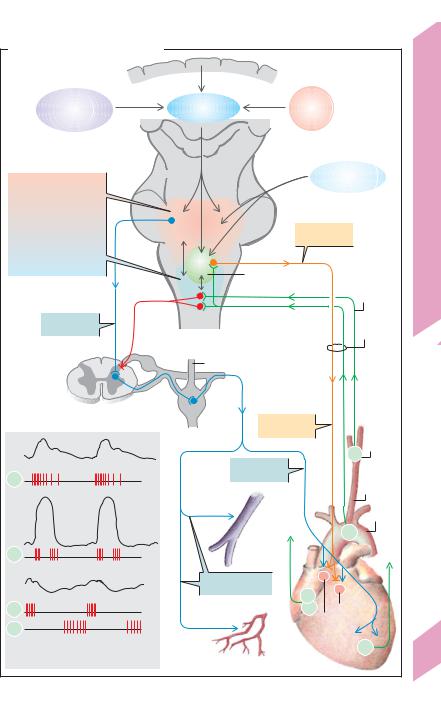
During exposure to extremely low temperatures, the cold-induced vasoconstriction of cutaneous vessels is periodically interrupted to supply the skin with blood to prevent tissue damage (Lewis response). Axoaxonal reflexes in the periphery play a role in this response, as afferent cutaneous nerve fibers transmit signals to efferent vasomotor axons. Skin reddening in response to scratching (dermatographism) is also the result of axoaxonal reflexes.
Central regulation of blood flow (!C) is the responsibility of the CNS areas in the medulla oblongata and pons. They receive information from circulatory sensors (S) or receptors (a) in the high-pressure system (barosensors or pressure sensors, SP, in the aorta and carotid artery); (b) in the low-pressure system (stretch sensors in the vena cava and atria, SA and SB); and (c) in the left ventricle (SV). The sensors measure arterial blood pressure (SP), pulse rate
(SP and SV) and filling pressure in the low pressure system (indirect measure of blood volume). The A sensors (SA) mainly react to atrial contraction, whereas the B sensors (SB) react to passive filling stretch (!C2). If the measured values differ from the set-point value, the circulatory control centers of the CNS transmit regulatory impulses through efferent nerve fibers to the heart and blood vessels (!D and p. 5 C2).
Situated laterally in the circulatory “center” is a pressor area (!C, reddish zone), the neu-
Despopoulos, Color Atlas of Physiology © 2003 Thieme |
! |
|
|
All rights reserved. Usage subject to terms and conditions of license. |
|
C. Central regulation of blood flow
1 |
Cerebral cortex |
|
Limbic |
Hypothalamus |
Tempe- |
system |
|
rature |
|
|
Respiratory |
|
|
“center” |
Pressor area |
|
|
Circulatory “center” |
|
|
in midbrain and |
|
Parasympathetic |
medulla oblongata |
|
|
|
nerve |
|
|
|
|
Depressor area |
|
|
|
Vagal nuclei |
|
IXth
cranial nerve
Sympathetic |
Vagus |
|
nervous system |
||
nerve (X) |
||
|
Inhibition |
|
|
Sympathetic trunk |
Spinal marrow
Aortic pressure
SP
Ventricular
pressure
SV
Venous pulse
SA
SB
2Afferent action potentials (AP) from circulatory sensors
|
|
2 |
|
|
|
Inhibits |
|
|
cardiac action |
Carotid |
|
|
|
|
|
|
|
|
sinus |
|
Increases |
|
SP |
|
|
Common |
|
|
cardiac action |
|
|
AP |
|
carotid |
|
|
|
||
|
|
|
artery |
|
Veins |
|
Aorta |
|
|
SP |
|
|
|
|
|
AP |
|
|
|
|
Vasoconstriction |
|
|
|
(α1 adrenoceptors) |
SA |
|
|
|
|
|
|
Arterioles |
SB |
AV node |
AP |
SA node |
||
Paintal) |
|
|
|
|
|
Heart |
|
|
(After |
|
SV |
|
|
|
|
Plate 8.15 Regulation of the Circulation II
215
Despopoulos, Color Atlas of Physiology © 2003 Thieme
All rights reserved. Usage subject to terms and conditions of license.

|
|
! |
|
|
|
|
rons of which (blue arrows) continuously |
||
|
|
transmit sympathetic nerve impulses to the |
||
|
|
heart to increase its activity (heart rate, con- |
||
|
|
duction and contractility). Their effects on ves- |
||
|
|
sels are predominantly vasoconstrictive (rest- |
||
|
|
ing tone). The pressor area is in close contact |
||
|
|
with more medial neurons (depressor area, |
||
|
|
light blue area in C). The pressor and depressor |
||
|
|
areas are connected to the dorsal nuclei of the |
||
|
|
vagus nerve (!C, green), the stimulation of |
||
|
|
which reduces the heart rate and cardiac im- |
||
|
|
pulse conduction rate (!C, orange arrows). |
||
System |
|
Homeostatic circulatory reflexes |
include |
|
|
signals along afferent nerve tracts (!D3a/b) |
|||
|
|
|||
|
|
that extend centrally from the pressosensors in |
||
Cardiovascular |
|
the aorta and carotid sinus (!C, green tracts). |
||
|
The main purpose of homeostatic control is to |
|||
|
|
|||
|
|
maintain the arterial blood pressure at a stable |
||
|
|
level. Acute increases in blood pressure |
||
|
|
heighten the rate of afferent impulses and acti- |
||
|
|
vate the depressor area. By way of the vagus |
||
8 |
|
nerve, parasympathetic neurons (!C, orange |
||
|
tract) elicit the depressor reflex response, i.e., |
|||
|
|
|||
|
|
they decrease the cardiac output (CO). In addi- |
||
|
|
tion, inhibition of sympathetic vessel innerva- |
||
|
|
tion causes the vessels to dilate, thereby reduc- |
||
|
|
|||
|
|
ing the peripheral resistance (TPR; !D4a/b). |
||
|
|
Both of these mechanisms help to lower acute |
||
|
|
increases in blood pressure. Conversely, an |
||
|
|
acute drop in blood pressure leads to activation |
||
|
|
of pressor areas, which stimulates a rise in CO |
||
|
|
and TPR as well as venous vasoconstriction |
||
|
|
(!C, blue tracts), thereby raising the blood |
||
|
|
pressure back to normal. |
|
|
|
|
Due to the fast adaptation of pressosensors |
||
|
|
(differential |
characteristics, !p. 312ff.), these |
|
|
|
regulatory measures apply to acute changes in |
||
|
|
blood pressure. Rising, for example, from a |
||
|
|
supine to a standing position results in rapid |
||
|
|
redistribution of the blood volume. Without |
||
|
|
homeostatic |
control (orthostatic |
reflex; |
!p. 204), the resulting change in venous return would lead to a sharp drop in arterial blood pressure. The circulatory centers also respond to falling PO2 or rising PCO2 in the blood (cross-links from respiratory center) to raise the blood pressure as needed.
In individuals with chronic hypertension, the input from the pressosensors is normal because they are fully adapted. Therefore, circulatory control centers
216 cannot respond to and decrease the high pressures. On the contrary, they may even help to “fix” the
blood pressure at the high levels. Chronic hypertension leads to stiffening of the carotid sinus. This may also contribute to decreasing the sensitivity of carotid pressosensors in hypertension.
A temporary increase in venous return (e.g., after an intravenous infusion) also leads to an increase in heart action (!D, right). This mechanism is known as the Bainbridge reflex. The physiological significance of this reflex is, however, not entirely clear, but it may complement the Frank–Starling mechanism (!p. 202ff.).
Hypertension
Hypertension is defined as a chronic increase in the systemic arterial blood pressure. The general criterion for diagnosis of hypertension is consistent elevation of resting blood pressure to more than 90 mmHg diastolic (!p. 206). Untreated or inadequately managed hypertension results in stress and compensatory hypertrophy of the left ventricle which can ultimately progress to left heart failure. Individuals with hypertension are also at risk for arteriosclerosis and its sequelae (myocardial infarction, stroke, renal damage, etc.). Therefore, hypertension considerably shortens the life expectancy of a large fraction of the population.
The main causes of hypertension are (a) increased extracellular fluid (ECF) volume with increased venous return and therefore increased cardiac output (volume hypertension) and (b) increased total peripheral resistance (resistance hypertension). As hypertension always leads to vascular changes resulting in increased peripheral resistance, type a hypertension eventually proceeds to type b which, regardless of how it started, ends in a vicious circle.
The ECF volume increases when more NaCl (and water) is absorbed than excreted. The usually high intake of dietary salt may therefore play a role in the development of essential hypertension (primary hypertension), the most common type of hypertension, at least in patients sensitive to salt. Volume hypertension can even occur when a relatively low salt intake can no longer be balanced. This can occur in renal insufficiency or when an adrenocortical tumor produces uncontrolled amounts of aldosterone, resulting in Na+ retention.
Other important cause of hypertension is pheochromocytoma, a tumor that secretes epinephrine and norepinephrine and therefore raises the CO and TPR. Renal hypertension can occur due to renal artery stenosis and renal disease. This results in the increased secretion of renin, which in turn raises the blood pressure via the renin–angiotensin– aldosterone (RAA) system (!p. 184).
Despopoulos, Color Atlas of Physiology © 2003 Thieme
All rights reserved. Usage subject to terms and conditions of license.

|
|
D. Circulatory reflexes |
|
|
|
Carotid and aortic |
Atrial (Bainbridge) reflex |
sinus reflex (depressant) |
(excitatory) |
|
|
IX |
X |
X |
|
4b |
4a |
3a |
3b |
3c |
4c |
|
Efferent |
Afferent |
|
Afferent |
Efferent |
SP
2b
2a SP
|
SA node |
|
|
|
|
AV node |
|
2d |
|
|
SV |
|
|
|
|
2c |
|
|
|
Vasodilatation |
|
|
Stretch |
|
|
|
|
receptors |
|
Arterial blood pressure rises |
1. |
Venous return rises |
||
Stimulus |
Atrial pressure rises |
|||
Pressosensors in: |
|
2. |
|
|
a) Aorta |
|
d) Atrial and venous |
||
b) Carotid artery |
|
Sensors |
||
|
stretch sensors |
|||
|
|
|||
c) Left ventricle |
|
|
|
|
a) Glossopharyngeal nerve (IXth nerve) |
3. |
c) Vagus nerve (Xth nerve) |
||
b) Vagus nerve (Xth nerve) |
Afferent path |
|||
|
||||
a) Stimulation of |
|
|
|
|
parasympathetic system |
4. |
c) Stimulation of |
||
|
b) Inhibition of |
Efferent path |
sympathetic system |
|
|
sympathetic system |
|||
Bradycardia |
Vasodilatation |
|
Tachycardia, |
|
|
5. |
|||
|
|
Response |
myocardial contractility |
|
Cardiac output |
Peripheral resistance |
|
increases |
|
|
|
|||
decreases |
|
|
||
decreases |
|
|
||
|
|
|
||
Arterial blood pressure decreases |
6. |
Cardiac output rises |
||
Goal achieved |
||||
|
|
|
||
Plate 8.16 Regulation of the Circulation III
217
Despopoulos, Color Atlas of Physiology © 2003 Thieme
All rights reserved. Usage subject to terms and conditions of license.