Добавил:
Опубликованный материал нарушает ваши авторские права? Сообщите нам.
Вуз: Предмет: Файл:

книги студ / color atlas of physiology 5th ed[1]. (a. despopoulos et al, thieme 2003)

.pdf
Скачиваний:
124
Добавлен:
03.09.2020
Размер:
31.56 Mб
Скачать

Reabsorption of Organic Substances


The filtered load of a substance is the product


of its plasma concentration and GFR. Since the


GFR is high (ca. 180 L/day), enormous quanti-


ties of substances enter the primary urine each


day (e.g., 160 g/day of D-glucose).


Fractional excretion (FE, !p. 154) of D-glu-

Balance

cose is very low (FE !0.4%). This virtually

complete reabsorption is achieved by second-


ary active transport (Na+-glucose symport) at

Water

the luminal cell membrane (!B and p. 29 B1).

About 95% of this activity occurs in the proxi-


mal tubule. If the plasma glucose conc. exceeds

and

10–15 mmol/L, as in diabetes mellitus (nor-

mally 5 mmol/L), glucosuria develops, and uri-

Salt,

nary glucose conc. rises (!A). Glucose reab-

sorption therefore exhibits saturation kinetics

Kidneys,

(Michaelis-Menten kinetics; !p. 28). The

above example illustrates prerenal glucosuria.


Renal glucosuria can also occur when one of

7

the tubular glucose carriers is defective.

Low-affinity carriers in the luminal cell membrane of


the pars convoluta (sodium-glucose transporter type


2 = SGLT2) and high-affinity carriers (SGLT1) in the


pars recta are responsible for D-glucose reabsorp-


tion. The co-transport of D-glucose and Na+ occurs in


each case, namely at a ratio of 1 : 1 with SGLT2 and


1 : 2 with SGLT1. The energy required for this form of


secondary active glucose transport is supplied by the


electrochemical Na+ gradient directed towards the


cell interior. Because of the co-transport of two Na+


ions, the gradient for SGLT1 is twice as large as that


for SGLT2. A uniporter (GLUT2 = glucose transporter


type 2) on the blood side facilitates the passive trans-


port of accumulated intracellular glucose out of the


cell (facilitated diffusion, !p. 22). D-galactose also


makes use of the SGLT1 carrier, while D-fructose is


passively absorbed by tubule cells (GLUT5).


The plasma contains over 25 amino acids, and


about 70 g of amino acids are filtered each day.


Like D-glucose, most L-amino acids are reab-


sorbed at proximal tubule cells by Na+-coupled


secondary active transport (!B and p. 29 B3).


At least 7 different amino acid transporters are


in the proximal tubule, and the specificities of


some overlap. Jmax and KM (!p. 28) and, there-


fore, saturability and reabsorption capacities


vary according to the type of amino acid and


carrier involved. Fractional excretion of most

158

amino acids ! to 1% (ranging from 0.1% for L-

valine to 6% for L-histidine).

Increased urinary excretion of amino acids (hyperaminoaciduria) can occur. Prerenal hyperaminoaciduria occurs when plasma amino acid concentrations are elevated (and reabsorption becomes saturated, as in A), whereas renal hyperaminoaciduria occurs due to deficient transport. Such a dysfunction may be specific (e.g., in cystinuria, where only L-cystine, L- arginine and L-lysine are hyperexcreted) or unspecific (e.g., in Fanconi’s syndrome, where not only amino acids but also glucose, phosphate, bicarbonate etc. are hyperexcreted).

Certain substances (lactate, sulfate, phosphate, dicarboxylates, etc.) are also reabsorbed at the proximal tubule by way of Na+ symport, whereas urea is subject to passive back diffusion (!p. 166). Urate and oxalate are both reabsorbed and secreted (!p. 160), with the predominant process being reabsorption for urate (FE !0.1) and secretion for oxalate (FE !1). If the urinary conc. of these poorly soluble substances rises above normal, they will start to precipitate (increasing the risk of urinary calculus formation). Likewise, the excessive urinary excretion of cystine can lead to cystine calculi.

Oligopeptides such as glutathione and angiotensin II are broken down so quickly by luminal peptidases in the brush border that they can be reabsorbed as free amino acids (!C1). Dipeptides resistant to luminal hydrolysis (e.g., carnosine) must be reabsorbed as intact molecules. A symport carrier (PepT2) driven by the inwardly directed H+ gradient (!p. 174) transports the molecules into the cells tertiary active H+ symport; !p. 26, 29 B4). The dipeptides are then hydrolyzed within the cell (!C2). The PepT2 carrier is also used by certain drugs and toxins.

Proteins. Although albumin has a low sieving coefficient of 0.0003 (!p. 154, 2400 mg/ day are filtered at a plasma conc. of 45 g/L (180 L/day · 45 g/L · 0.0003 = 2400 mg/day). Only 2 to 35 mg of albumin are excreted each day (FE !1%). In the proximal tubule, albumin, lysozyme, α1-microglobulin, "2-microglobulin and other proteins are reabsorbed by receptormediated endocytosis (!p. 28) and are “digested” by lysosomes (!D). Since this type of reabsorption is nearly saturated at normal filtered loads of proteins, an elevated plasma protein conc. or increased protein sieving coefficient will lead to proteinuria.

25-OH-cholecalciferol, which is bound to DBP (vitamin D-binding protein) in plasma and glomerular filtrate, is reabsorbed in combination with DBP by re- ceptor-mediated endocytosis (!p. 292).

Despopoulos, Color Atlas of Physiology © 2003 Thieme

All rights reserved. Usage subject to terms and conditions of license.

A. Reabsorption of glucose and amino acids

(mmol/min)

3


Filtered


glucose


Maximum


Reabsorbed


reabsorption


2

rate


glucose


amount/time


s

u ria

Excreted


Glucose

1


l u

c

o


glucose


G


(GFR=120 mL/min)


0


0

10

20


30

40


Plasma glucose


Normal

concentration (mmol/L)


C. Reabsorption of oligopeptides


Dipeptides


Oligopeptides


1


Extracellular


peptidases


a


Reabsorbed as


free amino acids


H+

Intracellular


peptidases


2


b


Lumen

Cell

Blood

a

Na+ symport carrier


b

H+ symport carrier


B. Reabsorption of organic substances


Lumen

Cell

Blood


Na+


Substances


x

b


Organic

x

a


x


ATP


Na+


of


Na+


Reabsorption


Brush border


Basolateral


membrane


membrane


Glucose,

a

Luminal

7.6


amino acids


Na+ symport


(several systems),


Plate

x

phosphate,


Passive


lactate,

b


sulfate,

carrier transport


dicarboxylates


D. Reabsorption of proteins by endocytosis


Albumin and


other proteins


Receptor


recycling


Receptor


Endosome


Primary


lysosome


Secondary


H+


lysosome


Amino


acids

159


Lumen

Proximal tubule cell

Blood


Despopoulos, Color Atlas of Physiology © 2003 Thieme

All rights reserved. Usage subject to terms and conditions of license.

Excretion of Organic Substances


Food provides necessary nutrients, but also


contains inert and harmful substances. The


body can usually sort out these substances al-


ready at the time of intake, either based on


their smell or taste or, if already eaten, with the


help of specific digestive enzymes and intesti-

Balance

nal absorptive mechanisms (e.g., D-glucose

and L-amino acids are absorbed, but L-glucose


and D-amino acids are not). Similar distinc-

Water

tions are made in hepatic excretion (! bile !

stools): useful bile salts are almost completely


reabsorbed from the gut by way of specific car-

and

riers, while waste products such as bilirubin

are mainly eliminated in the feces. Likewise,

Salt,

the kidney reabsorbs hardly any useless or

harmful substances (including end-products

Kidneys,

such as creatinine). Valuable substances (e.g.,

D-glucose and L-amino acids), on the other


hand, are reabsorbed via specific transporters

7

and thus spared from excretion (!p. 158).

The liver and kidney are also able to modify


endogenous waste products and foreign com-


pounds (xenobiotics) so that they are ”detox-


ified” if toxic and made ready for rapid elimi-


nation. In unchanged form or after the enzy-


matic addition of an OH or COOH group, the


substances then combine with glucuronic


acid, sulfate, acetate or glutathione to form


conjugates. The conjugated substances are


then secreted into the bile and proximal tubule


lumen (with or without further metabolic


processing).


Tubular Secretion


The proximal tubule utilizes active transport


mechanisms to secrete numerous waste prod-


ucts and xenobiotics. This is done by way of


carriers for organic anions (OA) and organic


cations (OC+). The secretion of these sub-


stances makes it possible to raise their clear-


ance level above that of inulin and, therefore,


to raise their fraction excretion (FE) above 1.0 =


100% (!p. 152) in order to eliminate them


more effectively (!A; compare red and blue


curves). Secretion is carrier-mediated and is


therefore subject to saturation kinetics. Unlike


reabsorbed substances such as D-glucose

160

(!p. 159 A), the fractional excretion (FE) of or-

ganic anions and cations decreases when their


plasma concentrations rise (!A; PAH secre-

tion curve reaches plateau, and slope of PAH excretion curve decreases). Some organic anions (e.g., urate and oxalate) and cations (e.g., choline) are both secreted and reabsorbed (bidirectional transport), which results in net reabsorption (urate, choline) or net secretion (oxalate).

The secreted organic anions (OA) include indicators such as PAH (p-aminohippurate; !p. 150) and phenol red; endogenous substances such as oxalate, urate, hippurate; drugs such as penicillin G, barbiturates, and numerous diuretics (!p. 172); and conjugated substances (see above) containing glucuronate, sulfate or glutathione. Because of its high affinity for the transport system, probenecid is a potent inhibitor of OAsecretion.

The active step of OAsecretion (!B) occurs across the basolateral membrane of proximal tubule cells and accumulates organic anions in the cell whereby the inside-negative membrane potential has to be overcome. The membrane has a broad specificity carrier (OAT1 = organic anion transporter type 1) that transports OAfrom the blood into the tubule cells in exchange for a dicarboxylate, such as succinate2– or α-ketoglutarate2 ; !B1). The latter substance arises from the glutamine metabolism of the cell (!p. 177 D2); the human Na+-dicarboxylate transporter hNADC-1 also conveys dicarboxylates (in combination with 3 Na+) into the cell by secondary active transport (!B2). The transport of OAis therefore called tertiary active transport. The efflux of OAinto the lumen is passive (facilitated diffusion; !B3). An ATP-dependent conjugate pump (MRP2 = multi-drug resistance protein type 2) in the luminal membrane is also used for secretion of amphiphilic conjugates, such as glutathione-linked lipophilic toxins (!B4).

The organic cations (OC+) secreted include endogenous substances (epinephrine, choline, histamine, serotonin, etc.) and drugs (atropine, quinidine, morphine, etc.).

The active step of OC+ secretion occurs across the luminal membrane of proximal tubule cells (luminal accumulation occurs after overcoming the negative membrane potential inside the cell). The membrane contains (a) direct ATP-driven carriers for organic cations (mdr1; primary active OC+ transport; !C1) and (b) a multispecific OC+/H+ antiporter (tertiary active transport; !C2). The OC+ diffuse passively from the blood into the cell by way of a polyspecific organic cation transporter (OCT; !C3).

Despopoulos, Color Atlas of Physiology © 2003 Thieme

All rights reserved. Usage subject to terms and conditions of license.

A. Secretion and excretion of p-aminohippurate (PAH)

(mmol/min)

1.00


Excreted PAH


0.75


amount/time


Secreted PAH


0.50


Saturation


PAH


0.25


Filtered PAH


0

1

2

3

4

5


PAH concentration in plasma (free PAH) (mmol/L)


B. Secretion of organic anions (OA)

Lumen

Proximal tubule cell


Blood


OA


Probenecid


3

OAT1


1


OA


α-ketoglutarate2–


PPAH,


OA


hNaDC1

phenol red,


2

hippurate,


ATP


3Na+


urate,

4


MRP2


penicillin,


barbiturates,


Amphiphilic


diuretics,

OA


conjugates


conjugates,

Excretion


etc.


C. Secretion of organic cations (OC+)

Lumen

Proximal tubule cell


Blood

OC+


OC+


OCT

3

Epinephrine,

OC+

2


choline,


histamine,


H+

OC

+


serotonin,


atropine,


mdr1


quinine,


1


cimetidine,


morphine,


ATP


OC+


etc.

Excretion


Plate 7.7 Excretion of Organic Substances

161

Despopoulos, Color Atlas of Physiology © 2003 Thieme

All rights reserved. Usage subject to terms and conditions of license.

7 Kidneys, Salt, and Water Balance

162

Reabsorption of Na+ and Cl

About 99% of the filtered load of Na+ is reabsorbed (ca. 27 000 mmol/day), i.e., the fractional excretion of Na+ (FENa) is about 1%. The precise value of FENa needed (range 0.5 to 5%) is regulated by aldosterone, atriopeptin (ANF) and other hormones (!p. 170).

Sites of Na+ reabsorption. The reabsorption of Na+ occurs in all parts of the renal tubule and collecting duct. About 65% of the filtered Na+ is reabsorbed in the proximal tubule, while the luminal Na + conc. remains constant (!p. 166). Another 25% is reabsorbed in the loop of Henle, where luminal Na+ conc. drops sharply; !p. 157 D, columns 2 and 3). The distal convoluted tubule and collecting duct also reabsorb Na+. The latter serves as the site of hormonal fine adjustment of Na+ excretion.

Mechanisms of Na+ reabsorption. Na+-K+- ATPase pumps Na+ ions out of the cell while

conveying K+ ions into the

cell (!A and

p. 156), thereby producing a

chemical Na+

gradient (!A2). Back diffusion of K+ (!A3) also leads to the development of a membrane potential (!A4). Both combined result in a high electrochemical Na+ gradient that provides the driving “force” for passive Na+ influx, the features of which vary in the individual nephron segments (!B).

In the proximal tubule, Na+ ions diffuse passively from the tubule lumen into the cells via

(a) the electroneutral Na+/H+ exchanger type 3 (NHE3), an Na+/H+-antiport carrier for electroneutral exchange of Na+ for H+ (!B1, p. 29 B4 and p. 174) and (b) various Na+ symport carriers for reabsorption of D-glucose etc. (!B1 and p. 158). Since most of these symport carriers are electrogenic, the luminal cell membrane is depolarized, and an early proximal lumen-negative transepithelial potential (LNTP) develops.

In the thick ascending limb (TAL) of the loop of Henle (!B6), Na+ is reabsorbed via the bumetanide-sensitive co-transporter BSC, a

Na+-K+-2Cl symport carrier (!p. 172). Although BSC is primarily electroneutral, the absorbed K+ recirculate back to the lumen through K+ channels. This hyperpolarizes the luminal membrane, resulting in the development of a lumen-positive transepithelial potential (LPTP).

In the distal convoluted tubule, DCT (!B8), Na+ is reabsorbed via the thiazide-sensitive cotransporter TSC, an electroneutral Na+-Cl- symport carrier (!p. 172).

In principal cells of the connecting tubule and collecting duct (!B9), Na+ exits the lumen via Na+ channels activated by aldosterone and antidiuretic hormone (ADH) and inhibited by prostaglandins and ANF (!p. 170).

Since these four passive Na+ transport steps in the luminal membrane are serially connected to active Na+ transport in the basolateral membrane (Na+-K+-ATPase), the associated transepithelial Na+ reabsorption is also active. This makes up about 1/3 of the Na+ reabsorption in the proximal tubule, and 1 ATP molecule is

consumed for each 3 Na+ ions absorbed (!p. 26). The other 2/3 of proximal sodium reabsorption is passive and paracellular.

Two driving “forces” are responsible for this: (1) the LPTP in the mid and late proximal tubule (!B5) and in the loop of Henle (!B7) drives Na+ and other cations onto the blood side of the epithelium. (2) Solvent drag (!p. 24): When water is reabsorbed, solutes for which the reflection coefficient !1 (including Na+) are “dragged along” due to friction forces (like a piece of wood drifts with flowing water). Since driving forces (1) and (2) are indirect products of Na+-K+-ATPase, the energy balance rises to about 9 Na+ per ATP molecule in the proximal tubule (and to about 5 Na+ per ATP molecule in the rest of the kidney).

On the basolateral side, Na+ ions exit the proximal tubule cell via Na+-K+-ATPase and an Na+-3 HCO3symport carrier (!p. 174). In the latter case, Na+ exits the cell via tertiary active transport as secondary active secretion of H+ (on the opposite cell side) results in intracellular accumulation of HCO3.

The fractional Cl- excretion (FECl) ranges from 0.5% to 5%. About 50% of all Clreabsorption occurs in the proximal tubule. The early proximal LNTP drives Clthrough paracellular spaces out of the lumen (!B3). The reabsorption of Cllags behind that of Na+ and H2O, so the luminal Clconc. rises. As a result, Clstarts to diffuse down its chemical gradient paracellularly along the mid and late proximal tubule (!B4), thereby producing a LPTP (reversal of potential, !B5). At the TAL and the DCT, Clenters the cells by secondary active transport and exits passively through ADH-ac- tivated basolateral Clchannels (!B6, 8).

Despopoulos, Color Atlas of Physiology © 2003 Thieme

All rights reserved. Usage subject to terms and conditions of license.

A. Electrochemical Na+ gradient


Lumen

K+

Blood


Na+


145


Na+


ATP


mmol/L

Na+


15


2

mmol/L


+

+

K+


[Na+] = Chemical driving force for Na+

Na -K -ATPase


Cl

1


+ and

Primary active Na+


+


and K+ transport

K+

K+

Na


Na


Na+

of


–70 mV

–70 mV


Reabsorption


3


4

–70 mV


Back diffusion of K+


Membrane potential


creates membrane potential

= electrical driving force for Na+

B. Na+ and Clreabsorption


Thick ascending limb of Henle’s loop


Early proximal


6

Na+-2Cl-K+ symport

7.8

Lumen

Electroneutral

Blood


2Cl


Plate

Na+/H+ exchanger


1


Na+


K+


K+


BSC


H+


Na+


2

Glucose0 etc.


Na+


Recirculation of K+


Electrogenic Na+ symport


LPTP


7


K+


LNTP


Na+


Reabsorption of cations Ca2+


3


Cl


Mg2+


Clreabsorption


Distal convoluted tubule


8

Na+-Cl symport


Mid/late proximal


Na+


Cl


Filtrate: 100%


H2O


Late proximal: 35%


Early distal: 10%


Na+


[Cl]


Final urine: 0.5–5%


TSC


K+


Cl


Cl


4 Clreabsorption


Collecting duct (principal cells)


9 Na+ transported via channels


LPTP


K+


Na+

Aldosterone,


Na+


5 Reabsorption of cations

Ca2+

ADH


Prostaglandins,


K+


Mg

2+

ANP


163


SD7.8

Despopoulos, Color Atlas of Physiology © 2003 Thieme

All rights reserved. Usage subject to terms and conditions of license.


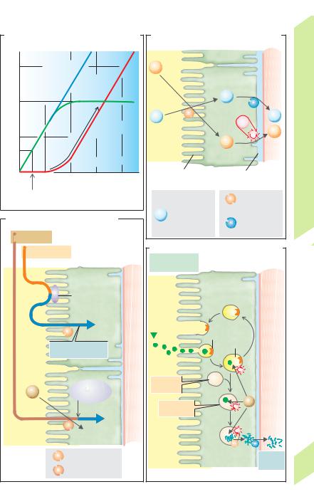
Reabsorption of Water, Formation of

pertonic towards the papillae (see below) and


if the vasa recta are permeable to water. Part of


Concentrated Urine


the water diffuses by osmosis from the de-


The glomeruli filter around 180 L of plasma

scending vasa recta to the ascending ones,


water each day (= GFR; !p. 152). By compari-

thereby “bypassing” the inner medulla (!A4).


.


Due to the extraction of water, the concentra-


son, the normal urine output (VU) is relatively


small (0.5 to 2 L/day). Normal fluctuations are

tion of all other blood components increases as


.


the blood approaches the papilla. The plasma


called antidiuresis (low VU) and diuresis (high


.


osmolality in the vasa recta is therefore con-

Balance

VU; !p. 172). Urine output above the range of

normal is called polyuria. Below normal output

tinuously adjusted to the osmolality of the sur-


is defined as oliguria (!0.5 L/day) or anuria

rounding interstitium, which rises towards the

Water

(!0.1 L/day). The osmolality (!p. 377) of

papilla. The hematocrit in the vasa recta also

plasma and glomerular filtrate is

about

rises. Conversely, substances entering the


290 mOsm/kg H2O (= Posm); that of the final

blood in the renal medulla diffuse from the as-

and

urine (Uosm) ranges from 50 (hypotonic urine in

cending to the descending vasa recta, provided

extreme water diuresis) to about 1200 mOsm/

the walls of both vessels are permeable to

Salt,

kg H2O (hypertonic urine in maximally con-

them (e.g., urea; !C). The countercurrent ex-

centrated urine). Since water diuresis permits

change in the vasa recta permits the necessary

Kidneys,

the excretion of large volumes of H2O without

supply of blood to the renal medulla without

the simultaneous loss of NaCl and other sol-

significantly altering the high osmolality of the


utes, this is known as “free water excretion”, or

renal medulla and hence impairing the urine

7

“free water clearance” (CH2O). This allows the

concentration capacity of the kidney.

kidney to normalize decreases in plasma

In a countercurrent multiplier such as the


osmolality, for example (!p. 170). The CH2O

loop of Henle, a concentration gradient be-


represents to the volume of water that could be

tween the two limbs is maintained by the ex-


theoretically extracted in order for the urine to

penditure of energy (!A5). The countercur-


reach the same osmolality as the plasma:

rent

flow amplifies the relatively small


.

[7.11]

gradient at all points between the limbs (local


CH2O " VU (1–[Uosm/Posm]).


Countercurrent Systems


gradient of about 200 mOsm/kg H2O) to a rela-


tively large gradient along the limb of the loop


A simple exchange system (!A1) can consist of

(about 1000 mOsm/kg H2O). The longer the


two tubes in which parallel streams of water flow, one

loop and the higher the one-step gradient, the


cold (0 #C) and one hot (100 #C). Due to the exchange

steeper the multiplied gradient. In addition, it


of heat between them, the water leaving the ends of

is inversely proportional to (the square of) the


both tubes will be about 50 #C, that is, the initially


flow rate in the loop.


steep temperature gradient of 100 #C will be offset.


In countercurrent exchange of heat (!A2), the

Reabsorption of Water


fluid within the tubes flows in opposite directions.


Approximately 65% of the GFR is reabsorbed at


Since a temperature gradient is present in all parts of


the tube, heat is exchanged along the entire length.

the proximal convoluted tubule, PCT (!B and


Molecules can also be exchanged, provided the wall

p. 157 D). The driving “force” for this is the re-


of the tube is permeable to them and that a concen-

absorption of solutes, especially Na+ and Cl.


tration gradient exists for the substance.


This slightly dilutes the urine in the tubule, but


If the countercurrent exchange of heat occurs in a


H2O immediately follows this small osmotic


hairpin-shaped loop, the bend of which is in contact


gradient because the PCT is “leaky” (!p. 154).


with an environment with a temperature different


from that inside the tube (ice, !A3), the fluid exit-

The reabsorption of water can occur by a para-


ing the loop will be only slightly colder than that

cellular route (through leaky tight junctions)


entering it, because heat always passes from the

or transcellular route, i.e., through water chan-


warmer limb of the loop to the colder limb.


nels (aquaporin type 1 = AQP1) in the two cell


Countercurrent exchange of water in the vasa

membranes. The urine in PCT therefore re-

164

recta of the renal medulla (!A6 and p. 150) oc-

mains

(virtually) isotonic. Oncotic pressure

curs if the medulla becomes increasingly hy-

(!p. 378) in the peritubular capillaries pro-


!

Despopoulos, Color Atlas of Physiology © 2003 Thieme

All rights reserved. Usage subject to terms and conditions of license.

A. Countercurrent systems


100°


10°


20°

30°


Exchange of heat

50°

60°


70°

80°


50°


50°


90°

100°


1 Simple exchange system

2 Countercurrent exchange


Heat


H2O


37°

35°


600

600

600


600

27°

25°


600

800


17°

15°


800

800

800


800

1000


1000

1000

1000


1000


1200


mosm/kgH2O 1200


Eis


Eis


3 Countercurrent exchange (heat)

4 Countercurrent exchange (water)

in loop


in loop (e.g. vasa recta)


H2O


Watertight


Cortex


400

400

400

200

400


NaCl


NaCl


600

600

600

400

600


NaCl


NaCl


800

800

800

600

800

Medulla


NaCl


NaCl


1000

1000

1000

800

1000


1200


Vasa


1200


recta


Collecting duct


Henle’s loop

5 Countercurrent multiplier


6 Countercurrent systems in renal medulla

(Henle’s loop)


Plate 7.9 Water Reabsorption, Concentration of Urine I

165

Despopoulos, Color Atlas of Physiology © 2003 Thieme

All rights reserved. Usage subject to terms and conditions of license.


!


vides an additional driving force for water re-


absorption. The more water filtered at the glo-


merulus, the higher this oncotic pressure.


Thus, the reabsorption of water at the proxi-


mal tubule is, to a certain extent, adjusted in


accordance with the GFR (glomerulotubular


balance).


Because the descending limb of the loop of

Balance

Henle has aquaporins (AQP1) that make it per-

meable to water, the urine in it is largely in


osmotic balance with the hypertonic inter-

Water

stitium, the content of which becomes increas-

ingly hypertonic as it approaches the papillae


(!A5). The urine therefore becomes increas-

and

ingly concentrated as it flows in this direction.

In the thin descending limb, which is only

Salt,

sparingly permeable to salt, this increases the

conc. of Na+ and Cl. Most water drawn into the

Kidneys,

interstitium is carried off by the vasa recta

water, Na+ and Clpassively diffuses (thin


(!B). Since the thin and thick ascending limbs


of the loop of Henle are largely impermeable to

7

limb) and is actively transported (thick limb)


out into the interstitium (!B). Since water


cannot escape, the urine leaving the loop of


Henle is hypotonic.


Active reabsorption of Na+ and Clfrom the


thick ascending limb of the loop of Henle


(TAL; !p. 162) creates a local gradient (ca.


200 mOsm/kg H2O; !A5) at all points be-


tween the TAL on the one side and the de-


scending limb and the medullary interstitium


on the other. Since the high osmolality of fluid


in the medullary interstice is the reason why


water is extracted from the collecting duct (see


below), active NaCl transport is the ATP-con-


suming “motor” for the kidney’s urine-concen-


trating mechanism and is up-regulated by sus-


tained stimulation of ADH secretion.

Along the course of the distal convoluted tubule and, at the latest, at the connecting tubule, which contains aquaporins and ADH receptors of type V2 (explained below), the fluid in the tubule will again become isotonic (in osmotic balance with the isotonic interstice of the renal cortex) if ADH is present (!p. 168), i.e., when antidiuresis occurs. Although Na+ and Clare still reabsorbed here (!p. 162), the osmolality does not change significantly because H2O is reabsorbed (ca. 5% of the GFR) into the interstitial space due to osmotic forces and

166urea increasingly determines the osmolality of the tubular fluid.

Final adjustment of the excreted urine volume occurs in the collecting duct. In the presence of antidiuretic hormone, ADH (which binds to basolateral V2 receptors, named after vasopressin, the synonym for ADH), aquaporins (AQP2) in the (otherwise water-impermeable) luminal membrane of principal cells are used to extract enough water from the urine passing through the increasingly hypertonic renal medulla. Thereby, the Uosm rises about four

times higher than the Posm (Uosm/Posm !4), corresponding to maximum antidiuresis. The ab-

sence of ADH results in water diuresis, where

Uosm/Posm can drop to !0.3. The Uosm can even fall below the osmolality at the end of TAL,

since reabsorption of Na+ and Clis continued in the distal convoluted tubule and collecting duct (!p. 162) but water can hardly follow.

Urea also plays an important role in the formation of concentrated urine. A protein-rich diet leads to increased urea production, thus increased the urine-concentrating capacity of the kidney. About 50% of the filtered urea leaves the proximal tubule by diffusion (!C). Since the ascending limb of the loop of Henle, the distal convoluted tubule, and the cortical and outer medullary sections of the collecting duct are only sparingly permeable to urea, its conc. increases downstream in these parts of the nephron (!C). ADH can (via V2 receptors) introduce urea carriers (urea transporter type 1, UT1) in the luminal membrane, thereby making the inner medullary collecting duct permeable to urea. Urea now diffuses back into the interstitium (where urea is responsible for half of the high osmolality there) via UT1 and is then transported by UT2 carriers back into the descending limb of the loop of Henle, comprising the recirculation of urea (!C). The non-re-

absorbed fraction of urea is excreted: FEurea ! 40%. Urea excretion increases in water diuresis

and decreases in antidiuresis, presumably due to up-regulation of the UT2 carrier.

Urine concentration disorders primarily occur due to (a) excessive medullary blood flow (washing out Na+, Cland urea); (b) osmotic diuresis; (c) loop diuretics (!p. 172); (d) deficient secretion or effectiveness of ADH, as seen in central or peripheral diabetes insipidus, respectively.

Despopoulos, Color Atlas of Physiology © 2003 Thieme

All rights reserved. Usage subject to terms and conditions of license.

B. Water reabsorption and excretion


O


2

Cortex

mosm/kg H


290

Outer medulla

600

Inner medulla

1200

GFR

=

100%

H2O follows NaCl, etc.

65%

220

Rest:

35%

High osmolality of interstitium:

H2O outflow 10%

Watertight

Plasma

Water follows NaCl

water


Rest: 25%

NaCl

permeabilitycontrol OADHto 2 ADH Hsubject

High H2O permeability

in antidiuresis

0.5% Maximum

antidiuresis

FEH2O

Maximum

5% and water diuresis more

Plate 7.10 Water Reabsorption, Concentration of Urine II

C. Urea in the kidney


100%

Urea


100%


Urea


concentration


(mmol/L)

5


Impermeable to urea


Passive


reabsorption


Rest:

Circulates via countercurrent exchange in vasa recta


Urea permeability subject to ADH control


50%

500


Recirculation of urea


40%


FEurea

167

Despopoulos, Color Atlas of Physiology © 2003 Thieme

All rights reserved. Usage subject to terms and conditions of license.

Соседние файлы в папке книги студ