

книги студ / color atlas of physiology 5th ed[1]. (a. despopoulos et al, thieme 2003)
.pdf8 Cardiovascular System
218
Circulatory Shock
Shock is characterized by acute or subacute progressive generalized failure of the circulatory system with disruption of the microcirculation and failure to maintain adequate blood flow to vital organs. In most cases, the cardiac output (CO) is insufficient due to a variety of reasons, which are explained below.
Hypovolemic shock is characterized by reduced central venous pressure and reduced venous return, resulting in an inadequate stroke volume (Frank– Starling mechanism). The blood volume can be reduced due to bleeding (hemorrhagic shock) or any other conditions associated with the external loss of fluids from the gastrointestinal tract (e.g., severe vomiting, chronic diarrhea), the kidneys (e.g., in diabetes mellitus, diabetes insipidus, high-dose diuretic treatment) or the skin (burns, profuse sweating without fluid intake). An internal loss of blood can also occur, e.g., due to bleeding into soft tissues, into the mediastinum or into the pleural and abdominal space.
Cardiogenic shock: Acute heart failure can be caused by acute myocardial infarction, acute decompensation of heart failure or impairment of cardiac filling, e.g. in pericardial tamponade. The central venous pressure is higher than in hypovolemic shock.
Shock can occur due to hormonal causes, such as adrenocortical insufficiency, diabetic coma or insulin overdose (hypoglycemic shock).
Vasogenic shock: Reduced cardiac output can also be due to peripheral vasodilatation (absence of pallor) and a resultant drop of venous return. This occurs in Gram-positive septicemia (septic shock), anaphylactic shock, an immediate hypersensitivity reaction (food or drug allergy, insect bite/sting) in which vasoactive substances (e.g., histamines) are released.
Symptoms. Hypovolemic and cardiovascular shock are characterized by decreased blood pressure (weak pulse) increased heart rate, pallor with cold sweats (not observed with shock caused by vasodilatation), reduced urinary output (oliguria) and extreme thirst.
Shock index. The ratio of pulse rate (beats/min) to systolic blood pressure (mmHg), or shock index, provides a rough estimate of the extent of volume loss. An index of up to 0.5 indicates normal or !10% blood loss; up to 1.0 = !20–30% blood loss and impending shock; up to 1.5 = "30–50% blood loss and manifest shock..
Most of the symptoms described reflect the counterregulatory measures taken by the
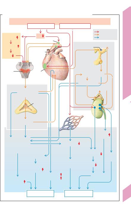
body during the non-progressive phase of shock in order to ward off progressive shock (!A). Rapid-acting mechanisms for raising the blood pressure and slower-acting mechanisms to compensate for volume losses both play a role.
Blood pressure compensation (!A left): A drop in blood pressure increases sympathetic tonus (!A1 and p. 214). Arterial vasoconstriction (absent in shock due to vasodilatation) shunts the reduced cardiac output from the skin (pallor), abdominal organs and kidneys (oliguria) to vital organs such as the coronary arteries and brain. This is known as centralization of blood flow (!A2). Sympathetic constriction of venous capacitance vessels (which raises ventricular filling), tachycardia and positive inotropism increase the diminished cardiac output to a limited extent.
Compensation for volume deficits (!A, right): When shock is imminent, the resultant drop in blood pressure and peripheral vasoconstriction lead to a reduction of capillary filtration pressure, allowing interstitial fluid to enter the bloodstream. Atrial stretch sensors detect the decrease in ECF volume (reduced atrial filling) and transmit signals to stop the atria from secreting atriopeptin (= ANP) and to start the secretion of antidiuretic hormone (ADH) from the posterior lobe of the pituitary (Gauer–Henry reflex; !p. 170). ADH induces vasoconstriction (V1 receptors) and fluid retention (V2 receptors). The drop in renal blood pressure triggers an increase in renin secretion and activation of the renin–angiotensin–al- dosterone (RAA) system (!p. 184). If these measures are successful in warding off the impending shock, the lost red blood cells are later replaced (via increased renal erythropoietin secretion, !p. 88) and the plasma protein concentration is normalized by increased hepatic synthesis.
Manifest (or progressive) shock will develop if these homeostatic compensation mechanisms are unable to prevent impending shock and the patient does not receive medical treatment (infusion, etc.). Severe hypotension (!90 mmHg systolic or !60 mmHg mean blood pressure) can persist for extended periods, even in spite of volume replacement. The resulting development of hypoxia leads to organ damage and multiple organ failure, ultimately culminating in irreversible shock and death.
Despopoulos, Color Atlas of Physiology © 2003 Thieme
All rights reserved. Usage subject to terms and conditions of license.
A. Compensation mechanisms for impending hypovolemic shock
Acute heart failure |
Hormonal causes |
|
Volume deficit |
|
||
|
Blood pressure |
|
Blood volume |
|
|
|
|
decreases |
|
decreases |
|
|
|
|
|
|
|
|
Stimulus |
|
Anaerobic |
PO2 |
|
|
|
Signal |
|
glycolysis |
|
|
|
Compensation |
||
|
Chemosensors |
|
|
|
|
|
Lactic acid |
|
|
Hypo- |
|
Thirst |
|
Presso- |
|
thalamus |
|
|
||
|
|
|
|
|||
|
|
|
|
|
|
|
|
sensors |
|
|
|
|
|
pH |
|
|
|
3 |
|
|
|
|
|
|
|
|
|
Medullary |
|
|
|
|
|
|
centers |
Volume |
Heart |
Renin |
ADH |
|
|
|
|
|||||
|
sensors |
|
|
|||
|
|
|
|
|
|
|
|
|
|
Angiotensin II |
|
||
1 |
|
|
|
|
Aldosterone |
|
Stimulation of |
|
|
4 |
|
|
|
sympathetic system |
|
|
|
|
||
Adrenal |
|
Adrenal |
|
|
||
medulla |
|
|
|
|||
|
cortex |
|
|
|||
|
|
|
|
|
||
|
|
|
Kidney |
|
|
|
Epinephrine |
|
|
Capillaries |
|
|
|
|
|
|
|
|
||
|
|
|
|
|
Re- |
|
|
|
|
|
|
absorption |
|
Arterial |
|
|
|
|
of water |
|
|
|
|
|
|
|
|
vasoconstriction |
|
|
Capillary |
|
|
|
mainly in kidneys, |
|
|
pressure |
|
|
|
stomach, gut |
|
|
|
|
|
|
and skin |
|
|
|
Renal |
|
|
Venous |
|
|
|
|||
|
blood flow |
Na+ |
||||
vasoconstriction |
|
|||||
|
|
|
|
|
retention |
|
2 |
|
H2O influx |
GFR |
|
||
Venous |
into capillaries |
|
||||
|
|
|
|
|
||
|
return |
|
|
|
|
|
|
Heart rate |
|
|
|
Oliguria |
|
Peripheral |
Myocardial |
|
|
|
|
|
|
|
|
|
|
||
contractility |
|
|
|
|
|
|
resistance |
|
|
|
|
|
|
|
|
|
|
|
|
|
Blood pressure rises |
|
Blood volume rises |
|
|
||
Despopoulos, Color Atlas of Physiology © 2003 Thieme
All rights reserved. Usage subject to terms and conditions of license.
Plate 8.17 Circulatory Shock
219

8 Cardiovascular System
220
Fetal and Neonatal Circulation
Placenta. The maternal placenta acts as the “gut” (absorption of nutrients), “kidneys” (removal of waste products) and “lungs” of the fetus (uptake of O2 and elimination of CO2). Although the fetal O2-hemoglobin dissociation curve is shifted to the left compared to that of adults (!p. 129 C), only 60% (0.6) of placental hemoglobin is saturated with O2 (!A).
Fetal blood is distributed according to need. Inactive or hardly active organs receive little blood. The fetal cardiac output (from both ventricles) is about 0.2 L/min per kg body weight. The fetal heart rate rises from an initial 65 min–1 (week 5) to 130–160 min–1 in later weeks. Approx. 50% of the blood ejected from the heart flows through the placenta, the other half supplies the body (35%) and lungs (15%) of the fetus. This is supplied by the left and right heart, which function essentially in parallel. The serial connection of the systemic circulation to the pulmonary circuit (as in adults) is not fully necessary in the fetus.
Fetal circulation. The blood flows through the fetal body as follows (!A): After being arterialized in the placenta, the blood passes into the fetus via the umbilical vein and part of it travels through the ductus venosus (Arantii), thereby bypassing the liver. When entering the inferior vena cava, the blood mixes with venous blood from the lower half of the body. Guided by special folds in the vena cava, the mixed blood passes directly from right atrium to the left atrium through an opening in the atrial septum (foramen ovale). From the left atrium, it then proceeds to the left ventricle. While in the right atrium, the blood mingles with venous blood from the superior vena cava (only slight mixing), which is received by the right ventricle. Only about one-third of this blood reaches the lungs (due to high flow resistance since the lungs are not yet expanded, and due to hypoxic vasoconstriction, !C and p. 122). The other two-thirds of the blood travels through the ductus arteriosus (Botalli) to the aorta (right-to-left shunt). Due to the low peripheral resistance (placenta), the blood pressure in the aorta is relatively low—only about 65 mmHg towards the end of pregnancy.
The arteries of the head and upper body are supplied with partly arterialized blood from the left ventricle (!A). This is important since brain tissue is susceptible to hypoxia. The remaining blood leaves the aorta and mixes with venous blood from the ductus arteriosus. As a result, the blood supplied to the lower half of the body has a relatively low O2 concentration (O2 saturation = 0.3; !A). The majority of this blood returns via the umbilical arteries to the placenta, where it is oxygenated again.
Circulation during birth. The exchange of O2, nutrients, and waste materials through the placenta stops abruptly during birth. This leads to a rise in blood PCO2, triggering chemosensors (!p. 132) that induce a strong breathing reflex. The resultant inspiratory movement causes negative pressure (suction) in the thoracic cavity, which removes the blood from the placenta and umbilical vein (placental transfusion) and expands the lungs. The unfolding of the lungs and the rise in alveolar PO2 reduces the resistance in the pulmonary circulation, and the blood flow increases while the pressure decreases (!B1, 2). Meanwhile, the resistance in the systemic circulation increases due to occlusion or clamping of the umbilical cord. This changes the direction of blood flow in the ductus arteriosus, resulting in a left-to-right shunt. The pulmonary circulation therefore receives aortic blood for a few days after birth. The right atrial filling volume decreases due to the lack of placental blood, while that of the left atrium increases due to the increased pulmonary blood flow. Due to the resultant pressure gradient from the left to right atrium and to a decrease in vasodilatory prostaglandins, the foramen ovale usually closes within about 2 weeks after birth. The ductus arteriosus and ductus venosus also close, and the systemic and pulmonary circulation now form serial circuits.
Shunts occur when the foramen ovale or ductus arteriosus remains open, placing a strain on the heart. In patent foramen ovale (atrial septum defect), the blood flows from left atrium !right atrium (left-to-right shunt)
!right ventricle (volume overload) !lungs
!left atrium. In patent ductus arteriosus, the blood flows from aorta !pulmonary artery (= left-to-right shunt) !lungs (pressure overload) !aorta.
Despopoulos, Color Atlas of Physiology © 2003 Thieme
All rights reserved. Usage subject to terms and conditions of license.

A. Fetal circulation |
|
|
|
|
|
|
Upper body |
|
|
O2 |
|
0.37 |
|
|
|
|
|
|
|
O2 saturation |
|
78 |
Lungs |
Circulation |
(fully saturated=1.0) |
|
|||
|
|
(not yet |
||
(mL/min) |
|
156 |
expanded) |
|
|
|
|||
|
|
13 |
||
Approx. blood |
|
|
||
|
|
|
||
flow/kg |
0.16 |
|
|
|
|
|
Neonatal |
||
body weight |
|
|
||
|
Ductus arteriosus |
|
||
|
78 |
Pulmonary |
||
|
Pulmonary artery |
|||
|
Foramen |
|
vein |
|
|
|
|
||
|
ovale |
|
Aorta |
|
|
104 |
and |
||
|
|
|
||
0.40 |
|
|
|
|
|
|
|
Fetal |
|
|
|
|
0.30 |
|
182 |
|
|
|
|
|
0.37 |
|
8.18 |
|
|
|
|
||
|
|
169 |
182 |
|
|
Ductus venosus |
|
||
|
|
|
Plate |
|
0.36 |
|
0.6 |
|
|
|
|
|
||
78 |
|
130 |
|
|
|
|
|
|
|
|
|
130 |
|
|
|
|
|
52 |
|
|
|
|
Lower body |
|
|
Portal vein |
|
|
|
|
Umbilical cord |
|
|
|
|
Umbilical arteries |
Umbilical vein |
|
|
|
|
Placenta |
|
|
|
|
|
|
|
|
|
|
C. Hypoxic vasoconstriction |
|
B. Pulmonary circulation before |
|
|
|
|
and after birth |
|
|
|
in fetus |
Pulmonary artery: |
3 |
|
|
|
|
|
|
|
resistancevascularPulmonary |
(mmHg·min·mL |
|
|
|
|
|
|
1 Blood flow |
2 |
|
|
|
|
|
|
|
1.0 |
|
|
|
|
|
||
|
|
|
|
|
|
|
|
) |
|
|
|
|
|
|||
|
|
|
|
|
|
|
|
|
|
|
|
|
|
|||
(L/min) |
1 |
|
|
|
|
|
|
|
|
–1 |
|
|
|
|
|
|
|
|
|
|
|
|
|
|
|
0.8 |
|
|
|
|
|
||
|
|
|
|
|
|
|
|
|
|
|
|
|
|
|
|
|
|
0 |
|
|
|
|
|
|
|
|
|
0.6 |
|
|
|
|
|
|
75 |
|
|
|
|
|
|
|
|
|
|
|
|
|
|
|
|
|
|
|
|
|
|
|
|
|
|
|
|
|
|
|
|
2 Systolic |
50 |
|
|
|
|
|
|
|
|
|
0.4 |
|
|
|
|
|
pressure in |
|
|
|
|
|
|
|
|
|
|
|
|
|
|
|
|
|
|
|
|
|
|
|
|
|
|
|
|
|
|
|
|
|
pulmonary |
25 |
|
|
|
|
|
|
|
|
|
0.2 |
|
|
|
|
|
artery |
|
|
|
|
|
|
|
|
|
|
|
|
|
|
|
|
0 |
|
|
|
|
|
|
|
|
|
|
|
|
|
|
|
|
(mmHg) |
20 |
28 |
36 |
1 |
2 |
3 |
4 |
|
|
00 |
5 |
10 |
15 |
20 |
25 |
|
|
|
|
|
|
|
|
|
|
|
|
|
Week of |
Weeks |
O2 pressure in pulmonary artery |
|
|
|
gestation |
after birth |
|
(mmHg) |
221 |
(After Rudolph) |
Birth |
|
(After Levine) |
(Measured in lamb fetus) |
|
|
|
|
|||
Despopoulos, Color Atlas of Physiology © 2003 Thieme
All rights reserved. Usage subject to terms and conditions of license.

9
222
Thermal Balance and Thermoregulation
Thermal Balance
The body temperature of humans remains relatively constant despite changes in the environmental temperature. This homeothermy applies only to the core temperature (!37 !C) of the body. The extremities and skin (“shell”) exhibit poikilothermy, i.e., their temperature varies to some extent with environmental temperature. In order to maintain a constant core temperature, the body must balance the amount of heat it produces and absorbs with the amount it loses; this is thermoregulation
(!p. 224).
Heat production. The amount of heat produced is determined by energy metabolism (! p. 228). At rest, approximately 56% of total heat production occurs in the internal organs and about 18% in the muscles and skin (!A2, top). During physical exercise, heat production increases several-fold and the percentage of heat produced by muscular work can rise to as much as 90% (!A2, bottom). To keep warm, the body may have to generate additional voluntary (limb movement) and involuntary (shivering) muscle contractions. Newborns also have tissue known as brown fat, which enables them to produce additional heat without shivering (!p. 225). Cold stimulates a reflex pathway resulting in norepinephrine release (!3-adrenergic receptors) in fatty tissues, which in turn stimulates (1) lipolysis and (2) the expression of lipoprotein lipase (LPL) and thermogenin. LPL increases the supply of free fatty acids ( !p. 254). Thermogenin localized in the inner mitochondrial membrane is an uncoupling protein that functions as an H+ uniporter (UCP1, !p. 230). It short-circuits the H+ gradient across the inner mitochondrial membrane (!p. 17/B2), thereby uncoupling the (heat-producing) respiratory chain of ATP production.
Heat produced in the body is absorbed by the bloodstream and conveyed to the body surface. In order for this internal flow of heat to occur, the temperature of the body surface must be lower than that of the body interior. The blood supply to the skin is the chief determinant of heat transport to the skin (!p. 224).
Heat loss occurs by the physical processes of radiation, conduction, convection, and evaporation (!B).
1.Radiation (!B1, C). The amount of heat lost by radiation from the skin is chiefly determined by the temperature of the radiator (fourth power of its absolute temperature). Heat net–radiates from the body surface to objects or individuals when they are cooler than the skin, and net–radiates to the body from objects (sun) that are warmer than the skin. Heat radiates from the body into the environment when no radiating object is present (night sky). Heat radiation does not require the aid of any vehicle and is hardly affected by the air temperature (air itself is a poor radiator). Therefore, the body loses heat to a cold wall (despite warm air in between) and absorbs radiation from the sun or an infrared radiator without air (space) or cold air, respectively, in between.
2.Conduction and convection (!B2, C). These processes involve the transfer of heat from the skin to cooler air or a cooler object (e.g. sitting on rock) in contact with the body (conduction). The amount of heat lost by conduction to air increases greatly when the warmed air moves away from the body by natural convection (heated air rises) or forced convection (wind).
3.Evaporation (!B3, C). The first two mechanisms alone are unable to maintain adequate temperature homeostasis at high environmental temperatures or during strenuous physical activity. Evaporation is the means by which the body copes with the additional heat. The water lost by evaporation reaches the skin surface by diffusion (insensible perspiration) and by neuron-activated sweat glands (!B3, pp. 73ff. and 225 D). About 2428 kJ (580 kcal) of heat are lost for each liter of water evaporating and thereby cooling the skin. At temperatures above 36 !C or so, heat loss occurs by evaporation only (!C, right). At even higher environmental temperatures, heat is absorbed by radiation and conduction/convection. The body must lose larger amounts of heat by evaporation to make up for this. The surrounding air must be relatively dry in order for heat loss by evaporation to occur. Humid air retards evaporation. When the air is extremely humid (e.g., in a tropical rain forest), the average person cannot tolerate temperatures above 33 !C, even under resting conditions.
Despopoulos, Color Atlas of Physiology © 2003 Thieme
All rights reserved. Usage subject to terms and conditions of license.

A. Relative contribution of organs to body weight and heat production
1 |
|
2 |
|
|
Percentage of body weight |
10% |
16% |
||
(=100%) |
|
Percentage of |
|
|
2% |
Brain |
heat production |
18% |
|
(=100%) |
|
|||
|
|
|
|
|
|
|
|
|
56% |
8% |
Other |
a |
|
|
|
|
At rest |
|
|
34% |
Thoracic and |
|
1% |
|
|
1% |
|
||
|
abdominal |
|
8% |
|
56% |
organs |
|
|
|
|
|
|
|
|
|
Skin and |
|
|
|
|
muscles |
|
|
|
At rest |
|
b |
|
90% |
|
|
|
||
|
|
During |
|
|
|
|
physical exercise |
|
|
B. Mechanisms of heat loss |
|
|
|
|
1 Radiation |
2 Conduction and convection |
3 Evaporation |
||
Heat rays |
|
Skin |
Loss of heat |
|
Convection |
|
H2O |
by |
|
|
|
|||
50°C |
|
|
|
evaporation |
|
Diffusion |
|
||
|
Heat |
|
||
|
|
|
|
|
|
conduction |
|
|
|
|
20°C |
|
|
|
|
|
H2O |
|
|
|
|
|
Sweat |
|
|
|
|
glands |
|
C.Heat production at different environmental temperatures (unclothed, at rest)
Room temperature 


Total heat loss
63J· m–2 · s–1 =100% |
|
38J· m–2 · s–1 |
=100% |
30°C |
43 J· m–2 |
· s–1 |
=100% |
36°C |
|
|
|
||||||
By way of: |
|
Distribution: |
|
Distribution: |
|
|||
13% |
20°C |
27% |
|
|
|
|
100% |
|
Evaporation |
|
|
|
|
|
|
|
|
|
|
|
|
|
|
|
|
|
26% |
|
27% |
|
|
0% |
|
|
|
Conduction |
|
|
|
|
|
|
|
|
|
|
|
|
|
|
|
|
|
and convection |
|
|
|
|
|
|
|
|
61% |
|
46% |
|
|
0% |
|
|
|
Radiation |
|
|
|
|
|
|
|
|
Despopoulos, Color Atlas of Physiology © 2003 Thieme
All rights reserved. Usage subject to terms and conditions of license.
Plate 9.1 Thermal Balance
223

9 Thermal Balance and Thermoregulation
224
Thermoregulation
Thermoregulation maintains the core temperature (!A) at a constant set point
(!37 !C) despite fluctuations in heat absorption, production, and loss (!p. 222). The core temperature exhibits circadian variation. It fluctuates by about 0.6 !C and is lowest around 3 a.m., and highest around 6 p.m. (!p. 381 C). The set point changes are controlled by an intrinsic biological clock (!p. 334). Extended set-point fluctuations happen during the menstrual cycle (!p. 299/A3) and fever.
The control center for body temperature and central thermosensors are located in the hypothalamus (!p. 330). Additional thermosensors are located in the spinal cord and skin (!p. 314). The control center compares the actual core temperature with the set-point value and initiates measures to counteract any deviations (!D and p. 4f.).
When the core temperature rises above the set point (e.g., during exercise), the body increases the internal heat flow (!p. 222) by dilating the blood vessels of the skin. Moreover, arteriovenous anastomoses open in the periphery, especially in the fingers. The blood volume transported per unit time then not only conveys more heat, but also reduces the countercurrent exchange of heat between the arteries and their accompanying veins (!B). In addition, venous return in the extremities is re-routed from the deep, accompanying veins to the superficial veins. Sweat secretion also increases. The evaporation of sweat cools the skin, thereby creating the core/skin temperature gradient needed for the internal heat flow. Central warm sensors emit the signals that activate the sweat glands. (In this case, the thermosensors of the skin do not detect warmth because their environment is cooler than the core temperature). The efferent nerve fibers to the sweat glands are cholinergic fibers of the sympathetic nervous system (! D).
Acclimatization to high environmental temperatures (e.g., in the tropics) is a slow process that often takes years. Characteristically, the sweat secretion rate rises, the salt content of the sweat decreases, and thirst and thus H2O intake increase.
When the core temperature falls below set point, the body checks heat loss by constricting the blood vessels in the shell (!A, left) and increases heat production by generating voluntary and involuntary (shivering) muscle activity (! D). Although infants can quickly become hypothermic because of their high surface/volume ratio, their brown fat allows them to produce additional heat (non-shivering thermogenesis; !p. 222). Upon exposure to low ambient temperatures, these three mechanisms are activated by the cold receptors of the skin (!p. 314) before the core temperature falls.
The range of ambient temperatures between the sweating and shivering thresholds is known as the thermoneutral zone. It lies between ca. 27 !C and 32 !C in the nearly unclothed test subject. The only thermoregulatory measure necessary within this range is variation of blood flow to the skin. The narrow range of this zone shows the thermoregulatory importance of behavior. It involves choosing the appropriate clothing, seeking shade, heating or cooling our dwellings, etc. Behavioral adaptation is the chief factor in survival at extreme ambient temperatures (!C).
The thermoneutral zone is subjectively perceived as the comfort zone. 95% of all subjects wearing normal office attire and performing normal office activities perceive an indoor climate with the following conditions to be comfortable: ambient and radiant (wall) temperature !23 !C, wind velocity " 0.1 m/s, and relative humidity !50%. A resting, unclothed subject feels comfortable at about 28 !C and ca. 31 !C to 36 !C in water depending on the thickness of subcutaneous fat (heat isolator).
Fever. Exogenous (e.g., bacteria) and endogenous pyrogens (various interleukins and other cytokines from macrophages) can cause the set-point temperature to rise above normal. This is triggered by prostaglandin PGE2 in the hypothalamus. In the initial phase of fever, the core temperature (although at its normal level) is too low compared to the elevated set-point. This results in shivering to raise the core temperature. As the fever decreases, i.e. the set-point returns toward the normal temperature, the core temperature is now too warm compared to the normalized set-point, resulting in vasodilatation and sweating to lower the core temperature again.
Despopoulos, Color Atlas of Physiology © 2003 Thieme
All rights reserved. Usage subject to terms and conditions of license.

A. Temperature zones of the body |
C. Environmental temperature and |
|
|||||||||
|
|
|
|
|
|
temperature control |
|
|
|
||
|
|
|
|
|
|
Environmental |
°C |
Thermo- |
|
||
|
|
Core |
|
|
temperature |
600 |
regulation |
|
|||
|
|
|
|
|
|
|
|
||||
|
|
temperature |
|
|
|
|
|
|
|
||
|
|
|
|
|
|
Lunar |
400 |
|
|
|
|
|
|
|
37°C |
|
|
|
|
|
|
||
|
|
|
36°C |
|
|
day |
200 |
|
|
|
|
|
|
|
|
|
|
|
|
|
|||
|
|
|
|
|
|
|
|
|
|
||
|
|
|
34°C |
|
|
|
|
Behavioral |
Thermoregulation |
||
|
|
|
32°C |
|
|
|
80 |
adaptation |
|||
|
|
|
|
|
|
|
only |
||||
|
|
|
31°C |
|
|
|
|
||||
|
|
|
|
|
|
60 |
|
|
|||
|
|
|
28°C |
|
|
|
|
|
|||
|
|
|
|
|
Tropics |
|
|
|
|||
20°C |
Room temperature |
35°C |
40 Core temp- |
Sweating |
|||||||
|
|||||||||||
|
|
|
|
|
(After Aschoff) |
|
erature Thermoneutral |
||||
|
|
|
|
|
|
|
|||||
B. Arteriovenous exchange of heat |
|
20 |
|
|
|||||||
|
|
|
|
||||||||
|
|
|
|
|
|
|
|
Shivering, etc. |
|||
Artery |
|
Vein |
|
|
|
|
0 |
9.2 |
|||
|
|
|
|
Arctic/Antarctic |
|
|
|||||
|
|
|
|
|
|
|
|||||
37 36 |
|
37 36.5 |
|
|
|
||||||
Exchange |
region |
|
|
|
|||||||
|
Behavioral |
Plate |
|||||||||
|
|
|
|
|
|
|
|||||
|
|
|
of heat |
|
|
|
–100 |
||||
|
|
|
|
|
|
adaptation |
|||||
35 34 |
|
36.5 34 |
|
||||||||
|
Lunar |
|
|
only |
|||||||
|
|
|
|
||||||||
Narrowed |
|
|
Widened |
|
|
night |
–200 |
|
|
|
|
vessel |
|
|
vessel |
36 |
35.5 |
|
|
|
|
|
|
33 |
32 |
|
|
–273 |
|
|
|
||||
|
|
|
|
|
|
|
|
|
|
||
32 |
|
31 |
35. |
|
35 |
Absolute |
|
|
|
|
|
|
|
zero |
|
|
|
|
|||||
|
31 |
|
Capillaries |
|
35 |
|
|
|
|
|
|
|
low |
Blood flow |
high |
|
|
|
(After Hardy) |
|
|||
|
|
|
|
|
|
|
|
|
|
||
D. Neural factors affecting thermoregulation
|
|
|
Central |
|
|
|
|
|
Peripheral |
|
|
|||||||
|
|
|
|
|
|
|
|
thermosensors |
|
|
||||||||
|
|
thermosensors |
|
|
|
|
|
|
|
|||||||||
|
|
|
|
|
|
|
|
|
|
|
|
|
|
|
||||
|
|
|
|
|
|
Hypothalamus |
|
|
|
|
|
|
||||||
|
|
|
|
|
|
|
|
|
|
|
|
|||||||
|
|
|
Sympathetic nervous system |
|
|
|
|
|
Somatic nervous system |
|
|
|||||||
|
|
|
|
|
|
|
|
|
|
|
|
|
|
|
|
|
|
|
cholinergic |
|
α1-adrenergic |
|
|
|
|
β3-adrenergic |
|
|
cholinergic |
|
|
||||||
|
|
|
|
|
|
|
|
|
|
|
|
|
|
|
|
|
|
|
|
Sweat |
|
|
|
Blood |
|
|
Brown fat |
|
|
Skeletal |
|
|
|||||
|
|
|
|
vessels |
|
|
|
|
|
|
|
|
muscle |
|
|
|||
|
glands |
|
|
|
|
|
|
|
|
|
|
|
|
|
||||
|
|
|
|
|
|
|
|
|
|
|
|
|
|
|
|
|
||
|
Heat loss |
|
|
Internal flow of heat |
|
|
Non-shivering |
|
|
Heat production |
225 |
|||||||
|
by evaporation |
|
|
|
|
thermogenesis |
|
|
by shivering |
|||||||||
|
|
|
|
|
(core |
skin) |
|
|
|
|
(in infants) |
|
|
|
|
|
|
|
|
|
|
|
|
|
|
|
|
|
|
|
|
|
|
|
|
|
|
Despopoulos, Color Atlas of Physiology © 2003 Thieme
All rights reserved. Usage subject to terms and conditions of license.

10 Nutrition and Digestion
Nutrition
An adequate diet must meet the body’s energy requirements and provide a minimum of carbohydrates, proteins (incl. all essential amino acids) and fats (incl. essential fatty acids). Minerals (incl. trace elements), vitamins, and sufficient quantities of water are also essential. To ensure a normal passage time, especially through the colon, the diet must also provide a sufficient amount of roughage (indigestible plant fibers—cellulose, lignin, etc.).
The total energy expenditure (TEE) or total metabolic rate consists of (1) the basal metabolic rate (BMR), (2) the activity energy costs, and the (3) diet-induced thermogenesis (DIT;
!p. 228, 231 A). TEE equals BMR when measured (a) in the morning (b) 20 h after the last meal, (3) resting, reclining, (4) at normal body temp., and (5) at a comfortable ambient temp. (!p. 224). The BMR varies according to sex, age, body size and weight. The BMR for a young adult is ca. 7300 kJ/day (!1740 kcal/ day; see p. 374 for units) in men, and ca. 20% lower in women. During physical activity, TEE increases by the following factors: 1.2-fold for sitting quietly, 3.2-fold for normal walking, and 8-fold for forestry work. Top athletes can perform as much as 1600 W (= J/s) for two hours (e.g., in a marathon) but their daily TEE is much lower. TEE also increases at various degrees of injury (1.6-fold for sepsis, 2.1-fold for burns). 1!C of fever increases TEE 1.13-fold.
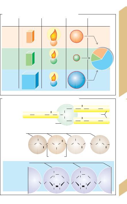
Protein, fats and carbohydrates are the three basic energy substances (!B).
An adequate intake of protein is needed to maintain a proper nitrogen balance, i.e., balance of dietary intake and excretory output of nitrogen. The minimum requirement for protein is 0.5 g/kg BW per day ( functional minimum). About half of dietary protein should be animal protein (meat, fish, milk and eggs) to ensure an adequate supply of essential amino acids such as histidine, isoleucine, leucine, lysine, methionine, phenylalanine, threonine, tryptophan and valine (children also require arginine). The content of most vegetable proteins is only about 50% of animal protein.
226 Carbohydrates (starch, sugar, glycogen) and fats (animal and vegetable fats and oils) pro-
vide the largest portion of the energy requirement. They are basically interchangeable sources of energy. The energy contribution of carbohydrates can fall to about 10% (normally 60%) before metabolic disturbances occur.
Fat is not essential provided the intake of fat-soluble vitamins (vitamins E, D, K and A) and essential fatty acids (linoleic acid) is sufficient. About 25–30% of dietary energy is supplied by fat (one-third of which is supplied as essential fatty acids; !A), although the proportion rises according to energy requirements (e.g., about 40% during heavy physical work). Western diets contain generally too much energy (more fats than carbohydrates!) considering the generally low level of physical activity of the Western lifestyle. Alcohol also
contains superfluous energy (ca. |
30 kJ/g = |
||
7.2 kcal/g). The excessive |
intake |
of |
dietary |
energy leads to weight |
gain and |
obesity |
|
(!p. 230). |
|
|
|
An adequate intake of minerals (inorganic compounds), especially calcium (800 mg/day; !p. 290ff.), iron (10–20 mg/day; !p. 90) and iodine (0.15 mg/day; !p. 288), is essential for proper body function. Many trace elements (As, F, Cu, Si, V, Sn, Ni, Se, Mn, Mo, Cr, Co) are also essential. The normal diet provides sufficient quantities of them, but excessive intake has toxic effects.
Vitamins (A, B1, B2, B6, B12, C, D2, D3, E, H (biotin), K1, K2, folic acid, niacinamide, pan-
tothenic acid) are compounds that play a vital role in metabolism (usually function as coenzymes). However, the body cannot produce (or sufficient quantities of) them. A deficiency of vitamins (hypovitaminosis) can lead to specific conditions such as night blindness (vit. A), scurvy (vit. C), rickets (vit. D = calciferol; !p. 292), anemia (vit. B12 = cobalamin; folic acid; !p. 90), and coagulation disorders (vit. K; !p. 104). An excessive intake of certain vitamins like vitamin A and D, on the other hand, can be toxic (hypervitaminosis).
Despopoulos, Color Atlas of Physiology © 2003 Thieme
All rights reserved. Usage subject to terms and conditions of license.
A. Energy content of foodstuffs and energy requirement
|
Requirement |
Physiological |
Energy content |
% of energy |
|
(g/day) |
fuel value (kJ/g) |
(kJ/day) |
requirement |
|
|
38.9 |
|
|
Fats |
65 |
|
2500 |
|
|
|
1g |
|
|
|
|
17.2 |
|
25% |
Proteins |
70 |
1g |
1200 |
12% |
|
|
|
63% |
|
|
|
|
|
|
|
|
17.2 |
|
|
Carbo- |
370 |
|
6300 |
|
hydrates |
|
|
||
1g |
|
|
||
|
|
|
|
*Values for an adult male weighing 70 kg, during light physical activity
B.Chemical structure of fats, proteins and carbohydrates
|
|
|
|
|
|
|
|
|
|
|
O |
|
|
|
|
|
|
|
|
|
O |
|
|
H2C |
O |
C |
|
(CH2)n |
|
CH3 |
|
|
|
|
|
|
|
|
|
|
|
|
|
|
|
|
|
Fats |
CH3 |
|
(CH2)n |
C |
|
O |
CH |
|
O |
|
Fatty acids |
|
|||
(e.g., triacyl- |
|
|
|
|
|
||||||||||
glycerol) |
|
|
|
|
|
|
|
H2C |
O |
C |
|
(CH2)n |
|
CH3 |
|
|
|
|
|
|
Glycerol |
|
|
|
|||||||
|
|
|
|
|
|
|
|
|
|
|
|
|
|
||
|
|
|
|
|
|
|
|
|
|
|
|
|
|
|
|
Amino acids |
|
|
|
|
|
|
|
|
|
|
|
|
|
|
|
|
|
|
R1 |
|
H |
|
|
O |
|
R3 |
|
|
|
Rn |
|
|
|
|
|
|
|
|
|
|
|
etc. |
|
|
|||
Proteins |
|
|
C |
|
|
|
|
|
C |
|
C |
|
|||
|
|
C |
N |
|
C |
C |
N |
C |
|
N |
C O– |
||||
H3N+ |
|
|
|
|
|
||||||||||
|
|
|
|
|
|
H |
|
|
|
|
|
||||
|
|
|
|
O |
|
|
R2 |
|
|
O |
|
H |
|
O |
|
|
|
|
|
|
|
|
|
|
|
|
|||||
|
|
|
|
|
|
|
|
|
|
|
|
|
|
|
|
|
|
|
|
|
|
|
|
|
|||||||
|
|
Amino |
|
Peptide bonds |
|
|
|
Carboxyl terminus |
|||||||
|
|
terminus |
|
|
|
|
|
|
|
|
|
|
|
||
Monosaccharides |
|
|
|
|
|
|
|
|
|
|
|
|
|
|
|
(e.g., glucose) |
|
|
|
|
|
|
|
|
|
|
|
|
|
|
|
|
|
|
|
|
H2COH |
|
|
H2COH |
|
|
|
|
|||
Carbohydrates |
|
|
|
|
|
C |
|
O |
|
|
C |
O |
|
|
etc. |
|
|
|
|
|
H |
|
|
|
|
H |
|
|
|
||
(e.g., amylose) |
|
|
|
|
CH |
|
|
CH |
CH |
|
CH |
(ca. 250x) |
|||
|
|
|
|
O |
|
OH H |
O |
|
OH H |
O |
|
|
|||
|
|
|
|
|
C |
|
C |
|
C |
C |
|
|
|||
|
|
|
|
|
|
|
|
|
|
|
|
||||
|
|
|
|
|
|
H |
|
OH |
|
|
H |
OH |
|
|
|
Plate 10.1 Nutrition
227
Despopoulos, Color Atlas of Physiology © 2003 Thieme
All rights reserved. Usage subject to terms and conditions of license.