

книги студ / color atlas of physiology 5th ed[1]. (a. despopoulos et al, thieme 2003)
.pdf1 Fundamentals and Cell Physiology
8
The Cell
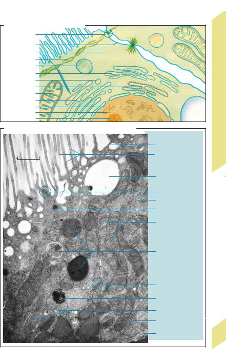
The cell is the smallest functional unit of a living organism. In other words, a cell (and no smaller unit) is able to perform essential vital functions such as metabolism, growth, movement, reproduction, and hereditary transmission (W. Roux) (!p. 4). Growth, reproduction, and hereditary transmission can be achieved by cell division.
Cell components: All cells consist of a cell membrane, cytosol or cytoplasm (ca. 50 vol.%), and membrane-bound subcellular structures known as organelles (!A, B). The organelles of eukaryotic cells are highly specialized. For instance, the genetic material of the cell is concentrated in the cell nucleus, whereas “digestive” enzymes are located in the lysosomes. Oxidative ATP production takes place in the mitochondria.
The cell nucleus contains a liquid known as karyolymph, a nucleolus, and chromatin. Chromatin contains deoxyribonucleic acids (DNA), the carriers of genetic information. Two strands of DNA forming a double helix (up to 7 cm in length) are twisted and folded to form chromosomes 10 µm in length. Humans normally have 46 chromosomes, consisting of 22 autosomal pairs and the chromosomes that determine the sex (XX in females, XY in males). DNA is made up of a strand of three-part molecules called nucleotides, each of which consists of a pentose (deoxyribose) molecule, a phosphate group, and a base. Each sugar molecule of the monotonic sugar–phosphate backbone of the strands (...deoxyribose – phosphate–deoxyribose...) is attached to one of four different bases. The sequence of bases represents the genetic code for each of the roughly 100 000 different proteins that a cell produces during its lifetime (gene expression). In a DNA double helix, each base in one strand of DNA is bonded to its complementary base in the other strand according to the rule: adenine
(A) with thymine (T) and guanine (G) with cytosine (C). The base sequence of one strand of the double helix (!E) is always a “mirror image” of the opposite strand. Therefore, one strand can be used as a template for making a new complementary strand, the information content of which is identical to that of the orig-
inal. In cell division, this process is the means by which duplication of genetic information (replication) is achieved.
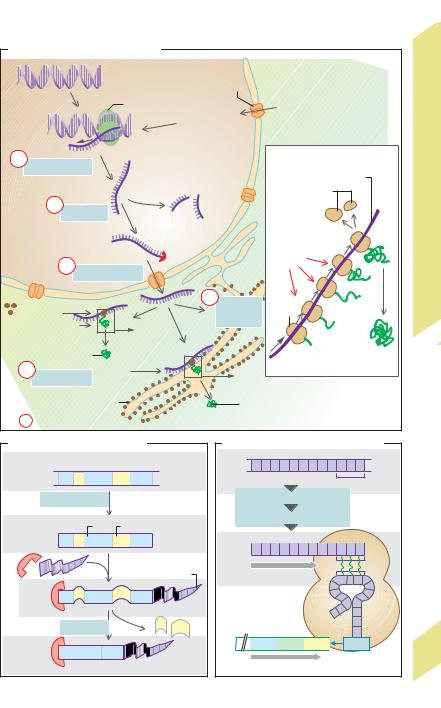
Messenger RNA (mRNA) is responsible for code transmission, that is, passage of coding sequences from DNA in the nucleus (base sequence) for protein synthesis in the cytosol (amino acid sequence) (!C1). mRNA is formed in the nucleus and differs from DNA in that it consists of only a single strand and that it contains ribose instead of deoxyribose, and uracil (U) instead of thymine. In DNA, each amino acid (e.g., glutamate, !E) needed for synthesis of a given protein is coded by a set of three adjacent bases called a codon or triplet (C–T–C in the case of glutamate). In order to transcribe the DNA triplet, mRNA must form a complementary codon (e.g., G–A–G for glutamate). The relatively small transfer RNA (tRNA) molecule is responsible for reading the codon in the ribosomes (!C2). tRNA contains a complementary codon called the anticodon for this purpose. The anticodon for glutamate is C–U–C (!E).
RNA synthesis in the nucleus is controlled by RNA polymerases (types I–III). Their effect on DNA is normally blocked by a repressor protein. Phosphorylation of the polymerase occurs if the repressor is eliminated (de-repres- sion) and the general transcription factors attach to the so-called promoter sequence of the DNA molecule (T–A–T–A in the case of polymerase II). Once activated, it separates the two strands of DNA at a particular site so that the code on one of the strands can be read and transcribed to form mRNA (transcription, !C1a, D). The heterogeneous nuclear RNA (hnRNA) molecules synthesized by the polymerase have a characteristic “cap” at their 5! end and a polyadenine “tail” (A–A–A–...) at the 3! end (!D). Once synthesized, they are immediately “enveloped” in a protein coat, yielding heterogeneous nuclear ribonucleoprotein (hnRNP) particles. The primary RNA or premRNA of hnRNA contains both coding sequences (exons) and non-coding sequences (introns). The exons code for amino acid sequences of the proteins to be synthesized, whereas the introns are not involved in the coding process. Introns may contain 100 to 10 000 nucleotides; they are removed from the
!
Despopoulos, Color Atlas of Physiology © 2003 Thieme
All rights reserved. Usage subject to terms and conditions of license.
A. Cell organelles (epithelial cell)
Tight junction
Cell membrane |
|
|
|
Cytosol |
|
|
|
Cytoskeleton |
|
|
|
Lysosome |
|
|
|
Smooth ER |
|
|
|
Golgi vesicle |
|
|
|
Rough ER |
|
|
|
Mitochondrion |
|
|
|
Golgi complex |
|
|
|
Nucleus |
|
|
|
Chromatin |
Vacuole |
I |
|
Nucleolus |
Cell |
||
|
|||
|
|
||
B. Cell structure (epithelial cell) in electron micrograph |
|
The |
|
|
|
||
|
Cell membrane |
1.4 |
|
|
Brush border |
Plate |
|
1 m |
|
||
|
|
||
|
Vacuole |
|
|
|
Tight junction |
|
|
|
Free ribosomes |
|
|
|
Cell border |
|
|
|
Mitochondria |
|
|
|
Lysosomes |
|
|
|
Rough |
|
|
|
endoplasmic |
|
|
|
reticulum |
|
|
|
Autophagosome |
|
|
|
Golgi complex |
|
|
|
Basal labyrinth |
|
|
|
(with cell membranes) |
|
|
|
Basal membrane |
9 |
|
|
|
||
|
Photo: W. Pfaller |
|
Despopoulos, Color Atlas of Physiology © 2003 Thieme
All rights reserved. Usage subject to terms and conditions of license.

1 Fundamentals and Cell Physiology
10
!
primary mRNA strand by splicing (!C1b, D) and then degraded. The introns, themselves, contain the information on the exact splicing site. Splicing is ATP-dependent and requires the interaction of a number of proteins within a ribonucleoprotein complex called the spliceosome. Introns usually make up the lion’s share of pre-mRNA molecules. For example, they make up 95% of the nucleotide chain of coagulation factor VIII, which contains 25 introns. mRNA can also be modified (e.g., through methylation) during the course of posttranscriptional modification.
RNA now exits the nucleus through nuclear pores (around 4000 per nucleus) and enters the cytosol (!C1c). Nuclear pores are high-molecular-weight protein complexes (125 MDa) located within the nuclear envelope. They allow large molecules such as transcription factors, RNA polymerases or cytoplasmic steroid hormone receptors to pass into the nucleus, nuclear molecules such as mRNA and tRNA to pass out of the nucleus, and other molecules such as ribosomal proteins to travel both ways. The (ATP-dependent) passage of a molecule in either direction cannot occur without the help of a specific signal that guides the molecule into the pore. The abovementioned 5! cap is responsible for the exit of mRNA from the nucleus, and one or two specific sequences of a few (mostly cationic) amino acids are required as the signal for the entry of proteins into the nucleus. These sequences form part of the peptide chain of such nuclear proteins and probably create a peptide loop on the protein’s surface. In the case of the cytoplasmic receptor for glucocorticoids (!p. 278), the nuclear localization signal is masked by a chaperone protein (heat shock protein 90, hsp90) in the absence of the glucocorticoid, and is released only after the hormone binds, thereby freeing hsp90 from the receptor. The “activated” receptor then reaches the cell nucleus, where it binds to specific DNA sequences and controls specific genes.
The nuclear envelope consists of two membranes (= two phospholipid bilayers) that merge at the nuclear pores. The two membranes consist of different materials. The external membrane is continuous with the mem-
brane of the endoplasmic reticulum (ER), which is described below (!F).
The mRNA exported from the nucleus travels to the ribosomes (!C1), which either float freely in the cytosol or are bound to the cytosolic side of the endoplasmic reticulum, as described below. Each ribosome is made up of dozens of proteins associated with a number of structural RNA molecules called ribosomal RNA (rRNA). The two subunits of the ribosome are first transcribed from numerous rRNA genes in the nucleolus, then separately exit the cell nucleus through the nuclear pores. Assembled together to form a ribosome, they now comprise the biochemical “machinery” for protein synthesis (translation) (!C2). Synthesis of a peptide chain also requires the presence of specific tRNA molecules (at least one for each of the 21 proteinogenous amino acids). In this case, the target amino acid is bound to the C–C–A end of the tRNA molecule (same in all tRNAs), and the corresponding anticodon that recognizes the mRNA codon is located at the other end (!E). Each ribosome has two tRNA binding sites: one for the last incorporated amino acid and another for the one beside it (not shown in E). Protein synthesis begins when the start codon is read and ends once the stop codon has been reached. The ribosome then breaks down into its two subunits and releases the mRNA (!C2). Ribosomes can add approximately 10–20 amino acids per second. However, since an mRNA strand is usually translated simultaneously by many ribosomes (polyribosomes or polysomes) at different sites, a protein is synthesized much faster than its mRNA. In the bone marrow, for example, a total of around 5 !1014 hemoglobin copies containing 574 amino acids each are produced per second.
The endoplasmic reticulum (ER, !C, F) plays a central role in the synthesis of proteins and lipids; it also serves as an intracellular Ca2+ store (! p. 17 A). The ER consists of a net-like system of interconnected branched channels and flat cavities bounded by a membrane. The enclosed spaces (cisterns) make up around 10% of the cell volume, and the membrane comprises up to 70% of the membrane mass of a cell. Ribosomes can attach to the cytosolic surface of parts of the ER, forming a rough endo-
!
Despopoulos, Color Atlas of Physiology © 2003 Thieme
All rights reserved. Usage subject to terms and conditions of license.
C. Transcription and translation |
|
|
|
|
|
|
||||
|
|
|
|
Genomic DNA |
Nucleus |
Cytoplasm |
|
|
|
|
|
|
|
|
|
|
|
|
|
||
|
|
|
|
|
Nuclear pore |
|
|
|
|
|
|
|
|
|
RNA polymerase |
|
|
|
|
|
|
|
|
|
|
|
Transcription |
|
|
|
|
|
|
|
|
|
|
factors and |
|
|
|
|
|
|
|
|
|
RNA |
signal |
|
|
|
|
|
|
|
5’ |
|
|
|
|
|
|
|
|
a |
|
|
|
|
2 Translation in ribosomes |
|
||||
Transcription |
Primary |
|
|
|||||||
|
|
|
|
RNA |
|
Ribosome |
mRNA |
|
||
|
|
|
|
|
|
|
|
|
||
|
|
|
|
|
|
subunits |
|
3’ |
|
|
|
b |
Splicing |
|
|
|
|
|
end |
|
|
|
|
|
|
|
|
Stop |
II |
|||
|
|
|
|
|
|
|
|
|
||
|
|
|
|
|
|
|
|
|
Cell |
|
|
|
|
|
mRNA |
tRNA |
|
|
|
||
|
|
|
|
|
|
|
The |
|||
|
|
|
|
amino |
|
|
|
|||
|
|
|
|
|
|
|
|
|
||
|
|
c |
|
|
|
acids |
|
|
|
|
|
|
mRNA export |
|
|
|
|
|
|||
|
|
|
|
|
|
|
1.5 |
|||
|
|
|
|
|
|
|
|
|
|
|
|
|
|
|
|
d |
|
Growing |
|
Plate |
|
Ribosomes |
|
|
|
mRNA |
Ribosome |
|
||||
|
|
|
breakdown |
peptide chain |
|
|||||
tRNA amino acids |
|
|
|
|
|
|
||||
|
|
|
|
|
|
|
|
|||
1 |
Cytosolic |
|
|
Start |
|
|
Finished |
|
||
|
|
|
|
|
|
|||||
|
e |
protein |
|
|
5’ end |
|
peptide chain |
|
||
|
|
|
tRNA |
|
|
|
||||
|
|
|
|
|
|
|
|
|||
|
Translation |
amino acids |
|
|
|
|
|
|
||
|
|
|
Ribosomes |
|
Membrane-bound |
|
|
|
|
|
|
Control |
|
|
|
|
and export proteins |
|
|
|
|
|
|
|
|
|
(cf. Plate F.) |
|
|
|
|
|
|
|
|
|
|
|
|
|
|
|
|
D. Transcription and splicing |
|
|
|
E. Protein coding in DNA and RNA |
|||||||||
|
Coding for amino acid no. ... |
|
|
|
|
3’ |
|
|
5’ |
|
|||
Genomic |
1–15 |
16–44 |
45–67 |
|
|
DNA |
T A A A A T G C T C T C |
|
|||||
|
|
|
|
|
|
|
|
|
|
|
Codogen |
|
|
DNA |
|
|
|
|
|
|
|
|
|
|
|
|
|
|
Transcription |
|
|
|
|
Transcription and Splicing |
|
||||||
Primary |
5’ |
|
Exon |
Intron |
3’ |
|
|
|
Export from nucleus |
|
|
||
end |
|
end |
|
|
|
|
|
|
|
||||
RNA |
|
|
|
|
|
|
|
|
5’ |
|
|
3’ |
|
(hnRNA) |
|
|
|
|
|
|
|
|
|
|
|
||
|
|
|
|
|
|
|
|
mRNA |
A U U U U A C G A G A G |
|
|||
|
|
|
|
|
|
|
|
|
|
|
|
Codon |
|
|
|
|
|
3’-poly-A tail |
|
|
Reading direction |
|
Anti- |
||||
|
|
|
|
|
|
C U C |
codon |
||||||
|
|
|
|
|
|
|
|
|
|||||
5’ |
|
|
|
|
A |
A |
A A A |
|
|
|
|
|
Ribosome |
cap |
|
|
|
|
|
|
|
|
|
|
tRNAGlu |
C |
|
|
|
|
|
|
|
|
|
|
|
|
|||
|
|
Splicing |
|
|
Introns |
Protein |
|
|
5’ |
C |
|
||
|
|
|
|
|
|
|
A |
|
|||||
|
|
|
|
|
|
|
|
|
|
|
|
|
|
mRNA |
|
|
|
A A |
A A |
A |
|
NH2 |
Ile |
Leu |
Arg |
Glu |
11 |
|
|
|
|
|
|
|
|
|
|
||||
|
|
|
|
|
|
|
|
|
|
|
|
||
|
1 |
15 |
44 |
67 |
|
|
|
|
Growth of peptide chain |
|
|||
|
|
|
|
|
|
|
|
|
|
||||
Despopoulos, Color Atlas of Physiology © 2003 Thieme
All rights reserved. Usage subject to terms and conditions of license.

1 Fundamentals and Cell Physiology
12
!
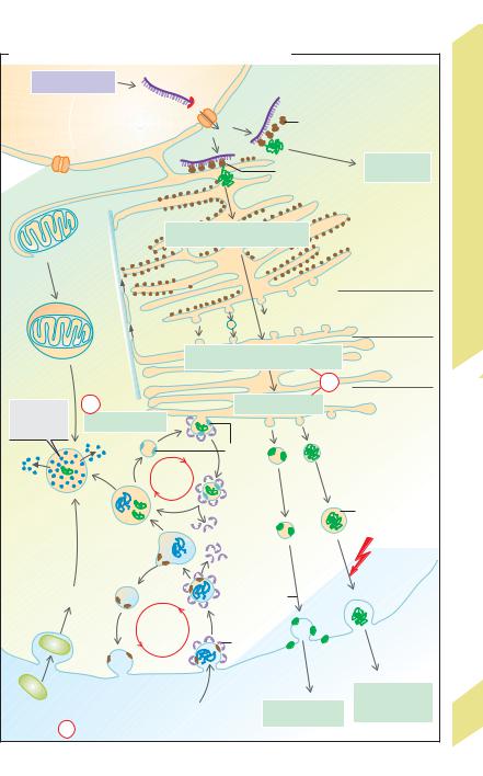
plasmic reticulum (RER). These ribosomes synthesize export proteins as well as transmembrane proteins (!G) for the plasma membrane, endoplasmic reticulum, Golgi apparatus, lysosomes, etc. The start of protein synthesis (at the amino end) by such ribosomes (still unattached) induces a signal sequence to which a signal recognition particle (SRP) in the cytosol attaches. As a result, (a) synthesis is temporarily halted and (b) the ribosome (mediated by the SRP and a SRP receptor) attaches to a ribosome receptor on the ER membrane. After that, synthesis continues. In export protein synthesis, a translocator protein conveys the peptide chain to the cisternal space once synthesis is completed. Synthesis of membrane proteins is interrupted several times (depending on the number of membrane-spanning domains (!G2) by translocator protein closure, and the corresponding (hydrophobic) peptide sequence is pushed into the phospholipid membrane. The smooth endoplasmic reticulum (SER) contains no ribosomes and is the production site of lipids (e.g., for lipoproteins, !p. 254 ff.) and other substances. The ER membrane containing the synthesized membrane proteins or export proteins forms vesicles which are transported to the Golgi apparatus.
The Golgi complex or Golgi apparatus (!F) has sequentially linked functional compartments for further processing of products from the endoplasmic reticulum. It consists of a cis- Golgi network (entry side facing the ER), stacked flattened cisternae (Golgi stacks) and a trans-Golgi network (sorting and distribution). Functions of the Golgi complex:
polysaccharide synthesis;
protein processing (posttranslational modification), e.g., glycosylation of membrane proteins on certain amino acids (in part in the ER) that are later borne as glycocalyces on the external cell surface (see below) and γ-carboxy- lation of glutamate residues (!p. 102 );
phosphorylation of sugars of glycoproteins (e.g., to mannose-6-phosphate, as described below);
“packaging” of proteins meant for export into secretory vesicles (secretory granules), the contents of which are exocytosed into the extracellular space; see p. 246, for example.
Hence, the Golgi apparatus represents a central modification, sorting and distribution center for proteins and lipids received from the endoplasmic reticulum.
Regulation of gene expression takes place on the level of transcription (!C1a), RNA modification (!C1b), mRNA export (!C1c), RNA degradation (!C1d), translation (!C1e), modification and sorting (!F,f), and protein degradation (!F,g).
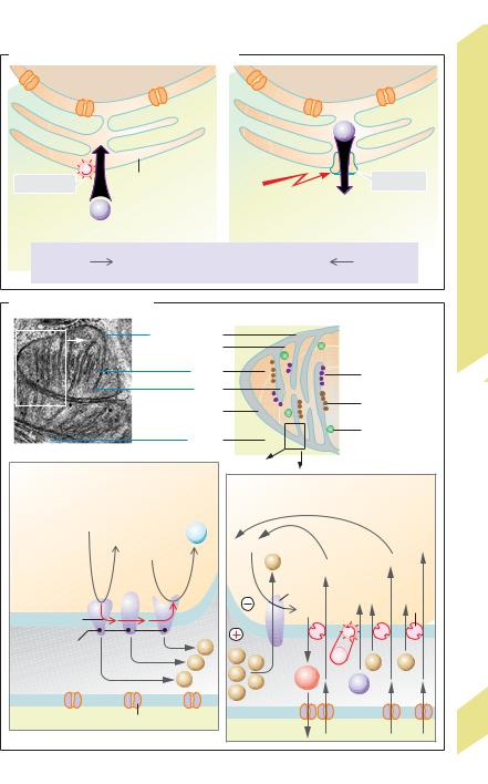
The mitochondria (!A, B; p. 17 B) are the site of oxidation of carbohydrates and lipids to CO2 and H2O and associated O2 expenditure. The Krebs cycle (citric acid cycle), respiratory chain and related ATP synthesis also occur in mitochondria. Cells intensely active in metabolic and transport activities are rich in mito- chondria—e.g., hepatocytes, intestinal cells, and renal epithelial cells. Mitochondria are enclosed in a double membrane consisting of a smooth outer membrane and an inner membrane. The latter is deeply infolded, forming a series of projections (cristae); it also has important transport functions (!p. 17 B). Mitochondria probably evolved as a result of symbiosis between aerobic bacteria and anaerobic cells (symbiosis hypothesis). The mitochondrial DNA (mtDNA) of bacterial origin and the double membrane of mitochondria are relicts of their ancient history. Mitochondria also contain ribosomes which synthesize all proteins encoded by mtDNA.
Lysosomes are vesicles (!F) that arise from the ER (via the Golgi apparatus) and are involved in the intracellular digestion of macromolecules. These are taken up into the cell either by endocytosis (e.g., uptake of albumin into the renal tubules; !p. 158) or by phagocytosis (e.g., uptake of bacteria by macrophages; !p. 94 ff.). They may also originate from the degradation of a cell’s own organelles (autophagia, e.g., of mitochondria) delivered inside autophagosomes (!B, F). A portion of the endocytosed membrane material recycles (e.g., receptor recycling in receptor-mediated endocytosis; !p. 28). Early and late endosomes are intermediate stages in this vesicular transport. Late endosomes and lysosomes contain acidic hydrolases (proteases, nucleases, lipases, glycosidases, phosphatases, etc., that are active only under acidic conditions). The
!
Despopoulos, Color Atlas of Physiology © 2003 Thieme
All rights reserved. Usage subject to terms and conditions of license.
F. Protein synthesis, sorting, recycling, and breakdown |
|
|
|||
Transcription |
Nucleus |
Cytosol |
|
|
|
|
|
|
|
||
|
|
mRNA |
|
|
|
|
|
|
Free |
|
|
|
|
|
ribosomes |
|
|
|
|
|
ER-bound |
Cytosolic |
|
|
|
|
proteins |
|
|
|
|
|
ribosomes |
|
|
|
|
Protein and lipid synthesis |
|
III |
|
|
|
|
|
|
|
Mitochondrion |
|
|
Endoplasmatic |
Cell |
|
|
|
|
|
||
|
|
|
|
reticulum (ER) |
The |
|
|
|
|
|
|
|
|
|
|
cis-Golgi |
1.6 |
|
|
|
|
Plate |
|
|
|
|
|
network |
|
|
|
|
|
|
|
Auto- |
Micro- |
Protein and lipid modification |
Golgi stacks |
|
|
phagosome |
|
|
|
|
|
|
tubule |
|
f |
|
|
|
|
|
|
|
|
Breakdown |
g |
|
Sorting |
trans-Golgi |
|
|
|
|
network |
|
|
of macro- |
Protein breakdown |
|
|
|
|
molecules |
|
|
|
|
|
|
|
M6P |
|
|
|
|
|
receptor |
|
|
|
|
|
Recycling |
|
|
|
Lysosome |
|
|
|
|
|
|
Late |
|
|
Secretory |
|
|
|
|
vesicle |
|
|
|
endosome |
|
|
|
|
|
|
|
|
|
|
|
Early |
|
|
Signal |
|
|
|
|
|
|
|
|
endosome |
|
|
|
|
|
|
|
Protein |
Cytosol |
|
Phagocytosis |
|
|
|
||
|
inclusion in |
|
|
||
|
|
|
Extra- |
|
|
|
|
cell membrane |
|
||
|
|
Recycling |
|
cellular |
|
|
|
|
space |
|
|
|
|
of receptors |
|
|
|
|
|
|
|
|
|
|
|
|
Clathrin |
|
|
|
|
|
Exocytose |
|
|
|
|
Endocytosis |
|
Controlled |
|
Bacterium |
|
|
Constitutive |
protein |
|
|
|
secretion |
13 |
||
|
|
|
secretion |
||
|
|
|
|
||
|
Control |
|
|
|
|
Despopoulos, Color Atlas of Physiology © 2003 Thieme
All rights reserved. Usage subject to terms and conditions of license.

1 Fundamentals and Cell Physiology
14
!
membrane contains an H+-ATPase that creates an acidic (pH 5) interior environment within the lysosomes and assorted transport proteins that (a) release the products of digestion (e.g., amino acids) into the cytoplasm and (b) ensure charge compensation during H+ uptake (Cl– channels). These enzymes and transport proteins are delivered in primary lysosomes from the Golgi apparatus. Mannose-6-phosphate (M6 P) serves as the “label” for this process; it binds to M6 P receptors in the Golgi membrane which, as in the case of receptor-mediated endocytosis (!p. 28 ), cluster in the membrane with the help of a clathrin framework. In the acidic environment of the lysosomes, the enzymes and transport proteins are separated from the receptor, and M6 P is dephosphorylated. The M6 P receptor returns to the Golgi apparatus (recycling, !F). The M6 P receptor no longer recognizes the dephosphorylated proteins, which prevents them from returning to the Golgi apparatus.
Peroxisomes are microbodies containing enzymes (imported via a signal sequence) that permit the oxidation of certain organic molecules (R-H2), such as amino acids and
fatty acids: R-H2 + O2 !R + H2O2. The peroxisomes also contain catalase, which transforms
2 H2O2 into O2 + H2O and oxidizes toxins, such as alcohol and other substances.
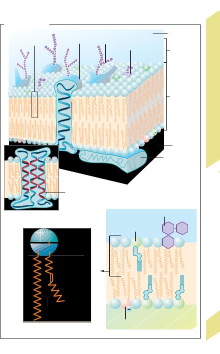
Whereas the membrane of organelles is responsible for intracellular compartmentalization, the main job of the cell membrane (!G) is to separate the cell interior from the extracellular space (!p. 2). The cell membrane is a phospholipid bilayer (!G1) that may be either smooth or deeply infolded, like the brush border or the basal labyrinth (!B). Depending on the cell type, the cell membrane contains variable amounts of phospholipids, cholesterol, and glycolipids (e.g., cerebrosides). The phospholipids mainly consist of phosphatidylcholine (!G3), phosphatidylserine, phosphatidylethanolamine, and sphingomyelin. The hydrophobic components of the membrane face each other, whereas the hydrophilic components face the watery surroundings, that is, the extracellular fluid or cytosol (!G4). The lipid composition of the two layers of the membrane differs greatly. Glycolipids are present only in the external layer, as described
below. Cholesterol (present in both layers) reduces both the fluidity of the membrane and its permeability to polar substances. Within the two-dimensionally fluid phospholipid membrane are proteins that make up 25% (myelin membrane) to 75% (inner mitochondrial membrane) of the membrane mass, depending on the membrane type. Many of them span the entire lipid bilayer once (!G1) or several times (!G2) (transmembrane proteins), thereby serving as ion channels, carrier proteins, hormone receptors, etc. The proteins are anchored by their lipophilic amino acid residues, or attached to already anchored proteins. Some proteins can move about freely within the membrane, whereas others, like the anion exchanger of red cells, are anchored to the cytoskeleton. The cell surface is largely covered by the glycocalyx, which consists of sugar moieties of glycoproteins and glycolipids in the cell membrane (!G1,4) and of the extracellular matrix. The glycocalyx mediates cell– cell interactions (surface recognition, cell docking, etc.). For example, components of the glycocalyx of neutrophils dock onto endothelial membrane proteins, called selectins (!p. 94).
The cytoskeleton allows the cell to maintain and change its shape (during cell division, etc.), make selective movements (migration, cilia), and conduct intracellular transport activities (vesicle, mitosis). It contains actin filaments as well as microtubules and intermediate filaments (e.g., vimentin and desmin filaments, neurofilaments, keratin filaments) that extend from the centrosome.
Despopoulos, Color Atlas of Physiology © 2003 Thieme
All rights reserved. Usage subject to terms and conditions of license.
G. Cell membrane |
|
|
|
|
|
Integral |
|
Extracellular |
|
|
|
|
|
|
Lipid molecule |
membrane protein |
Glycoprotein |
|
|
|
|
|
|
|
|
|
Glycolipid |
Glycocalyx |
|
|
|
|
|
|
|
|
|
Lipid |
|
|
|
|
bilayer |
|
|
|
|
(ca. 5 nm) |
The Cell IV |
|
|
|
|
|
|
|
|
Cytosol |
1.7 |
|
|
|
Plate |
|
|
|
|
Peripheral |
|
|
|
|
|
|
|
|
|
membrane |
|
|
|
|
protein |
|
|
|
1 Membrane constituents |
|
|
|
Lipophilic amino |
|
|
|
|
acid residues |
|
|
|
2 Multiple membrane- |
|
|
Glycolipid |
|
spanning integral protein |
|
|
|
|
|
|
Cholesterol |
|
|
Choline |
Polar |
|
|
|
|
head group |
|
|
|
Glycerol |
(hydrophilic) |
|
|
|
|
|
|
|
|
Double |
|
|
|
|
bond |
|
|
|
|
Fatty acids |
|
|
|
|
(hydrophobic) |
|
|
|
|
3 Phospholipid (phosphatidylcholine) |
Phosphatidylserine |
|
15 |
|
4 Membrane lipids |
|
|||
|
|
|
|
|
Despopoulos, Color Atlas of Physiology © 2003 Thieme
All rights reserved. Usage subject to terms and conditions of license.

Transport In, Through and Between
Cells
|
The lipophilic cell membrane protects the cell |
||
|
interior from the extracellular fluid, which has |
||
|
a completely different composition (!p. 2). |
||
|
This is imperative for the creation and main- |
||
Physiology |
tenance of a cell’s internal |
environment by |
|
means of metabolic energy expenditure. Chan- |
|||
nels (pores), carriers, ion pumps (!p. 26ff.) |
|||
|
|||
|
and the process of cytosis (!p. 28) allow |
||
|
transmembrane transport of selected sub- |
||
Cell |
stances. This includes the import and export of |
||
metabolic substrates and metabolites and the |
|||
|
|||
and |
selective transport of ions used to create or |
||
modify the cell potential (!p. 32), which plays |
|||
Fundamentals |
an essential role in excitability of nerve and |
||
|
|||
|
muscle cells. In addition, the effects of sub- |
||
|
stances that readily penetrate the cell mem- |
||
|
brane in most cases (e.g., water and CO2) can be |
||
|
mitigated by selectively transporting certain |
||
1 |
other substances. This allows the cell to com- |
||
pensate for undesirable changes in the cell |
|||
|
|||
|
volume or pH of the cell interior. |
||
|
Intracellular Transport |
|
|
|
The cell interior is divided into different com- |
||
|
partments by the organelle membranes. In |
||
|
some cases, very broad intracellular spaces |
||
|
must be crossed during transport. For this pur- |
||
|
pose, a variety of specific intracellular trans- |
||
|
port mechanisms exist, for example: |
||
|
Nuclear pores in the nuclear envelope pro- |
||
|
vide the channels for RNA export out of the nu- |
||
|
cleus and protein import into it (!p. 11 C); |
||
|
Protein transport from the rough endo- |
||
|
plasmic reticulum to the |
Golgi complex |
|
|
(!p. 13 F); |
|
|
|
Axonal transport in the nerve fibers, in |
|
|
which distances of up to 1 meter can be |
|
|
crossed (!p. 42). These transport processes |
|
|
mainly take place along the filaments of the |
|
|
cytoskeleton. Example: while expending ATP, |
|
|
the microtubules set dynein-bound vesicles in |
|
|
motion in the one direction, and kinesin- |
|
|
bound vesicles in the other (!p. 13 F). |
|
|
Intracellular Transmembrane Transport |
|
|
Main sites: |
|
16 |
Lysosomes: Uptake of H+ ions from the cyto- |
|
sol and release of metabolites such as amino |
||
|
||
|
acids into the cytosol (!p. 12); |
Endoplasmic reticulum (ER): In addition to a translocator protein (!p. 10), the ER has two other proteins that transport Ca2+ (!A). Ca2+ can be pumped from the cytosol into the ER by a Ca2+-ATPase called SERCA (sarcoplasmic endoplasmic reticulum Ca2+-transporting ATPase). The resulting Ca2+ stores can be released into the cytosol via a Ca2+ channel (ryanodine receptor, RyR) in response to a triggering signal (!p. 36).
Mitochondria: The outer membrane contains large pores called porins that render it permeable to small molecules (!5 kDa), and the inner membrane has high concentrations of specific carriers and enzymes (!B). Enzyme complexes of the respiratory chain transfer electrons (e–) from high to low energy levels, thereby pumping H+ ions from the matrix space into the intermembrane space (!B1), resulting in the formation of an H+ ion gradient directed into the matrix. This not only drives ATP synthetase (ATP production; !B2), but also promotes the inflow of pyruvate – and
anorganic phosphate, Pi– (symport; !B2b,c and p. 28). Ca2+ ions that regulate Ca2+-sensi- tive mitochondrial enzymes in muscle tissue can be pumped into the matrix space with ATP expenditure (!B2), thereby allowing the mitochondria to form a sort of Ca2+ buffer space for protection against dangerously high concentrations of Ca2+ in the cytosol. The insidenegative membrane potential (caused by H+ release) drives the uptake of ADP3 – in exchange for ATP4 – (potential-driven transport; !B2a and p. 22).
Transport between Adjacent Cells
In the body, transport between adjacent cells occurs either via diffusion through the extracellular space (e.g., paracrine hormone effects) or through channel-like connecting structures (connexons) located within a so-called gap junction or nexus (!C). A connexon is a hemichannel formed by six connexin molecules (!C2). One connexon docks with another connexon on an adjacent cell, thereby forming a common channel through which substances with molecular masses of up to around 1 kDa can pass. Since this applies not only for ions such as Ca2+, but also for a number of organic substances such as ATP, these types of cells are
!
Despopoulos, Color Atlas of Physiology © 2003 Thieme
All rights reserved. Usage subject to terms and conditions of license.
A. Ca2+ transport through the ER membrane |
|
|
|
|
|
|
|
|
|
|
|
||||||
|
|
|
|
|
|
|
|
|
|
|
|
|
|
|
|
|
I |
1 |
Nucleus |
|
|
|
2 |
|
|
|
|
|
|
|
|
|
|
Cells |
|
|
|
|
|
|
|
|
|
|
|
|
|
|
|
|
|||
|
|
|
|
|
|
|
|
|
|
|
|
|
|
|
|
|
|
|
|
|
Cytosol |
|
|
|
|
|
|
|
|
|
|
and Between |
|||
|
|
|
|
|
|
|
|
|
|
|
|
Ca2+ |
|
|
|
||
|
|
|
|
|
|
|
|
|
|
|
|
|
|
|
|
|
|
Ca2+-ATPase |
|
Endoplasmic |
|
|
Signal |
|
|
|
|
|
|
Ca |
2+ |
channel |
In, Through |
||
|
reticulum (ER) |
|
|
|
|
|
|
|
|
|
|||||||
|
|
|
|
|
|
(depolarization, |
|
|
Discharge |
|
|
|
|||||
Ca2+ |
|
|
|
|
hormon, etc.) |
|
|
|
|
|
|
||||||
|
|
|
|
|
|
|
|
|
|
|
|
|
|||||
Storage |
|
|
|
|
|
|
|
|
|
|
|
|
|
|
|
||
(10–5) |
10–8 mol/l |
Ca |
2+ |
Cytosolic |
|
|
|
10–5 |
|
(10–8)mol/l |
Transport |
||||||
|
|
|
|
concentration |
|
|
|
|
|
|
|
|
|
|
|||
|
|
|
|
|
|
|
|
|
|
|
|
|
|
|
|
|
|
B. Mitochondrial transport |
|
|
|
|
|
|
|
|
|
|
|
|
|
|
1.8 |
||
|
|
|
|
|
|
|
|
|
|
|
|
|
|
|
|
|
|
|
|
Outer membrane |
|
|
|
|
|
|
|
|
|
|
Plate |
||||
|
|
Inner membrane |
|
|
|
|
|
|
|
|
|
|
|||||
|
|
|
|
|
|
|
|
|
|
|
|
|
|||||
|
|
|
|
|
Matrix |
|
|
|
|
|
|
ATP synthetase |
|
||||
|
|
|
|
|
|
|
|
|
|
|
|
|
|
||||
|
|
|
|
|
Crista |
|
|
|
|
|
|
|
|
|
|
|
|
|
|
|
|
|
Inter- |
|
|
|
|
|
|
Ribosomes |
|
||||
|
|
|
membranous |
|
|
|
|
|
|
|
|
|
|
|
|||
|
|
|
|
|
space |
|
|
|
|
|
|
Granules |
|
||||
|
|
|
|
Cytosol |
|
|
|
|
|
|
|
||||||
|
|
|
|
|
|
|
|
|
|
|
|
|
|
|
|||
1 |
|
|
|
|
|
2 |
|
|
|
|
|
|
|
|
|
|
|
Formation of H+ gradient |
|
|
|
|
|
|
|
|
|
|
|
|
|
|
|
||
|
|
|
|
|
|
H+ gradient driving ATP synthesis and carriers |
|
||||||||||
NADH + H+ |
|
|
H2O |
|
|
|
|
|
|
|
|
|
Pyruvate– |
|
|||
|
NAD+ |
|
|
|
|
|
|
|
|
|
|
|
|
||||
|
|
2H+ + ½O2 |
|
|
Pi + ADP3– |
ATP synthetase |
|
|
|
|
|
|
etc. |
|
|||
|
|
|
|
H+ |
|
|
|
|
|
Pi |
|
||||||
|
|
|
|
|
|
|
ADP3– |
|
|
|
|||||||
|
|
|
|
|
|
|
|
|
|
|
|
|
|||||
Matrix |
|
|
|
|
|
|
|
|
|
|
|
|
Carrier |
|
|||
|
|
|
|
|
|
|
|
|
|
|
|
|
|
||||
e– |
|
|
|
|
|
|
|
|
ATP4– |
|
|
|
|
|
|
||
|
|
|
|
|
|
|
|
|
|
|
|
|
|
|
|
|
|
|
|
|
|
|
|
|
|
|
|
|
a |
|
b |
|
c |
|
|
Enzyme complexes |
|
|
|
|
H+ |
|
|
|
|
|
|
|
|
|
|
|
|
of respiratory chain |
|
|
|
H+ |
H+ |
|
|
|
|
|
ATP |
H+ |
|
|
H+ |
|
|
|
|
|
|
H+ |
|
|
|
|
|
|
|
|
|
||||
Intermembranous |
|
|
H |
+ |
|
H+ |
|
ATP |
4– |
|
|
|
|
|
|
||
space |
|
|
|
|
H+ |
|
|
|
Ca2+ |
|
|
|
|
||||
|
|
|
|
|
|
H+ |
|
|
|
|
|
|
|
|
|
|
|
Cytoplasm |
|
Porins |
|
|
|
|
|
|
|
|
|
|
|
|
|
|
17 |
|
|
|
|
|
|
|
|
|
|
|
|
|
|
|
|
||
Despopoulos, Color Atlas of Physiology © 2003 Thieme
All rights reserved. Usage subject to terms and conditions of license.