

Учебники / Diagnosis in Otorhinolaryngology Onerci 2009
.pdf2.7 Nasal Polyposis
2.7
Nasal Polyposis
Polyps are one of the most frequent causes of nasal obstruction. Polyps may be isolated or diffuse. The behavior of nasal polyps depends on the type of granulocytes. Eosinophils play a significant role in the classification of polyps. Polyps with significant eosinophilia behave differently from those with neutrophils. The majority of diffuse nasal polyposis is eosinophilic. Analgesic intolerance and asthma might accompany diffuse eosinophilic nasal polyposis in the later decades of a patient’s life. Isolated polyps can originate from an anatomic structure such as the ethmoid bulla or uncinate process. If polypsoriginatefromthemucosainsidethesinus,theyarenamed according to the sinus. If the polyp originates from the maxillarysinusitiscalledanantrochoanalpolyp,andifitoriginates from the sphenoid sinus it is called a sphenochoanal polyp.
Nasal polyps are unusual in children. If a child presents withnasalpolyposis,thepossibilityofdiseasessuchascystic fibrosis or primary ciliary dyskinesia should be eliminated. If the nasal polyp is unilateral, nasal encephaloceles should be ruled out with MR imaging.
In diffuse nasal polyposis patients, the presence of asthma or analgesic intolerance should always be questioned. Due to thedangerofintolerancewhichcandeveloplaterintheirlife, NSAIDs should not be prescribed to patients with diffuse eosinophilic nasal polyposis.
In adults, unilateral polyps should always raise the clinical suspicion of malignancy.
Fig. 2.7.1 Isolated polyp originating from the right middle meatus and extending anterior to the middle turbinate
Fig. 2.7.2 Polyp originating from the uncinate process
Table 2.7.1 Classification of nasal polyposis
Polyps
Inflammatory
Choanal/ isolated
Eosinophilic
Additional criteria
Acetylsalicylic acid intolerance
Asthma/COPD
Allergy
Associated diseases
Cystic fibrosis
Immune insufficiency (acquired/congenital)
Primary ciliary dyskinesia
Vasculitis, granulomatosis
Fig. 2.7.3 Right sphenochoanal polyp originating from the mucosa inside the sphenoid sinus, filling the sphenoethmoidal recess, and extending to the choana
87
A E R
O N
E S
H T
O R
AT
K C E N D N A

88 |
Chapter 2 |
Nose |
|
a |
a |
b
b
Fig. 2.7.4 (a) Antrochoanal polyp has three parts. The cystic portion is the part originating in the maxillary sinus. If this part is not removed, the polyp recurs. The neck is the portion of the polyp passing through the ostium. The main polyp mass is the part filling the nasal passage and causing obstruction. Although antrochoanal polyps are very often unilateral, they may cause bilateral nasal obstruction by extending and occluding the other choana. If the cystic part is not removed, the polyp recurs. (b) Coronal CT scan of antrochoanal polyp; the left
maxillary sinus is opaque and the maxillary sinus ostium is widened. c This widening of the maxillary sinus ostium is almost diagnostic for antrochoanal polyp
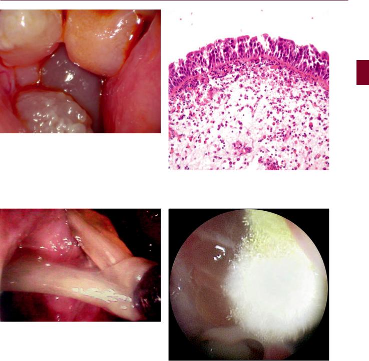
Fig. 2.7.5 (a) Diffuse eosinophilic nasal polyposis; the nasal passage is completely obstructed. Since the whole ethmoidal mucosa is polypoid there is no originating point. (b) Coronal CT scan shows that all sinuses are opaque. (c) Removal of the polyps
2.7 Nasal Polyposis
Fig. 2.7.6 Diffuse eosinophilic nasal polyposis completely obstructing the nasal passage
Fig. 2.7.7 In eosinophilic diffuse nasal polyposis, the mucus is thick, viscid, and yellow-green. It contains many eosinophils. It is referred to as allergic mucin
Fig. 2.7.8 Diffuse nasal polyposis. Diffuse eosinophilia is seen on histological examination (H&E)
Fig. 2.7.9 Fungal ball in sphenoid sinus
89
A E R
O N
E S
H T
O R
AT
K C E N D N A

90 |
Chapter 2 |
Nose |
|
a |
b |
Fig. 2.7.10 Kartagener syndrome. (a) Dextrocardia on chest X-ray; (b) diffuse bronchiectasia on axial CT scan
Fig. 2.7.11 Nasal bone expansion due to extensive diffuse nasal polyposis in the younger patient. Rhinoplasty is needed to restore the appearance
Fig. 2.7.12 Diffuse nasal polyposis completely filling the nasal passages. On the left side, nasal polyps protrude from the nostril. The patient did not agree to the operation because of her fear of anesthesia
Table 2.7.2 Treatment evidence and recommendations for postoperative treatment in adults with nasal polypsa
Therapy |
Level |
Grade of |
Relevance |
|
|
recommendation |
|
Oral antibiotics: short term < 2 weeks |
No data |
D |
Immediately postoperative, if pus was |
|
|
|
seen during operation |
Oral antibiotics: long term > 12 weeks |
Ib |
A |
Yes |
Topical steroids after Functional endoscopic |
Ib (two studies |
B |
Yes |
sinus surgery (FESS) |
one +, one −) |
|
|
Topical steroids after polypectomy |
Ib |
A |
Yes |
Oral steroids |
No data |
D |
Yes |
Nasal douche |
No data |
D |
Yes |
After Table 13.6 of European Position Paper in Rhinosinusitis and Nasal Polyposis, Suppl 20, 2007. Reproduced with permission of Rhinology aSome of these studies also included patients with Chronic Rhinosinusitis (CRS) without nasal polyps

2.7 Nasal Polyposis
2 symptoms: one of which should be nasal obstruction or discoloured discharge
+/- frontal pain, headache +/- smell disturbance
ENT examination including endoscopy (size of polyps) consider CT scan
consider diagnosis and treatment of co-morbidities eg. ASA
|
mild |
|
|
|
moderate |
|
|
|
|
|
severe |
|
||||||
|
VAS 0-3 |
|
|
|
VAS >3-7 |
|
|
|
|
|
VAS >7-10 |
|
||||||
|
|
|
|
|
|
|
|
|
|
|
|
|
|
|
|
|
|
|
|
|
|
|
|
|
|
|
|
|
|
|
|
|
|
|
|
|
|
|
|
|
|
|
|
|
|
|
|
|
|
|
|
|
|
|
|
|
|
topical steroids |
|
|
|
topical steroids |
|
|
|
|
oral steroids |
|
|||||||
|
(spray) |
|
|
|
|
(drops) |
|
|
|
|
|
|
||||||
|
|
|
|
|
|
|
|
|
|
(short course) |
|
|||||||
|
|
|
|
|
|
|
|
|
|
|
|
|
|
|
|
|
||
|
|
|
|
|
|
|
|
|
|
|
|
|
|
|
topical steroids) |
|
||
|
|
|
|
|
|
|
|
|
|
|
|
|
|
|
|
|
|
|
|
|
|
|
|
|
|
|
|
|
|
|
|
|
|
|
|
||
|
|
|
review after 3 months |
|
|
|
|
|
|
|
|
|
||||||
|
|
|
|
|
|
|
|
|
|
|
|
|
|
|
|
review after 1 month |
||
|
|
|
|
|
|
|
|
|
|
|
|
|
|
|||||
|
|
|
|
|
|
|
|
|
|
|
|
|
|
|
|
|
|
|
|
improvement |
|
|
|
no improvement |
|
|
|
|
|
|
|
||||||
|
|
|
|
|
|
|
|
|
|
|
|
|
|
|
|
|
|
|
|
|
|
|
|
|
|
|
|
|
|
|
|
|
|
|
|
|
|
|
|
|
|
|
|
|
|
|
|
|
|
|
|
|
|
improvement |
|
|
|
continue with |
|
|
|
|
|
|
|
|
|
||||||||
|
|
|
|
|
|
|
|
|
|
|
|
|
|
|
|
|||
|
topical steroids |
|
|
|
|
|
|
|
|
|
|
|
|
|
|
|
||
|
|
|
|
|
|
|
|
|
|
|
|
|
|
|
|
|
|
|
|
|
|
|
|
|
|
|
|
|
|
|
|
|
|
|
|
|
|
|
|
|
|
|
|
|
|
|
|
|
|
|
|
|
|
|
|
|
|
|
|
|
|
|
|
|
|
|
|
|
|
follow up |
|
||||
|
review every 6 months |
|
|
|
|
|
|
|
|
|
|
|||||||
|
|
|
|
|
|
|
|
|
|
douching |
|
|||||||
|
|
|
|
|
|
|
|
|
|
|
|
|
|
|||||
|
|
|
|
|
|
|
|
|
|
|
topical ± oral steroids |
|
||||||
|
|
|
|
|
|
|
|
|
|
|
± long term antibiotics |
|
||||||
|
|
|
|
|
|
|
|
|
|
|
|
|
|
|
|
|
|
|
consider other diagnosis unilateral symptoms
bleeding crusting cacosmia
orbital symptoms: peri-orbital oedema displaced globe
double or reduced vision ophthalmoplegia
severe frontal headache frontal swelling
signs of meningitis or focal neurological signs
urgent investigation and intervention
no improvement
CT scan
surgery
Table. 2.7.3 Treatment scheme for ENT specialists for adults with nasal polyps. After Fig. 13.5 of European Position Paper in Rhinosinusitis and Nasal Polyposis, Suppl 20, 2007. Reproduced with permission of Rhinology
91
A E R
O N
E S
H T
O R
AT
K C E N D N A

92 |
Chapter 2 Nose |
2.8
Nasal Obstruction
a
b |
Fig. 2.8.2 Septal deviation to the right |
Fig. 2.8.3 Nasal obstruction due to alar insufficiency, which is the result of overexcision of the lateral crura of the lower lateral cartilages during rhinoplasty. The depressions on both sides lateral to the nasal tip are due to overresection of the alar cartilages
c
Fig. 2.8.1 (a) Right inferior turbinate hypertrophy. (b) Nodular type left inferior turbinate hypertrophy. (c) Posterior part of the inferior turbinate is polypoid causing nasal obstruction
Fig. 2.8.4 Bilateral concha bullosa. No surgery is necessary for asymptomatic cases

a |
b |
c |
d |
e |
f |
2.8 Nasal Obstruction 93
A E R
O N
E S
H T
O R
AT
K C E N D N A
Fig. 2.8.5 Choanal atresia is a congenital abnormality. A bony plate or a membrane obstructs the posterior nares. Unilateral atresia may not cause symptoms. However, bilateral choanal atresia presents an emergency situation since the newborn is totally dependent on the nasal airway for breathing. During feeding, the newborn becomes cyanotic. The diagnosis is made by the inability to pass a soft catheter perinasally, or demonstrating the atretic plate after instillation of radiopaque dye. CT scan shows the atretic plate. The atretic plate can be seen on endoscopic examination. As soon as the diagnosis is made, a transnasal airway should be established. Blindly perforating
the bony plate or the membrane should be avoided because of the narrow nasopharynx and unsatisfactory results. Endoscopic transnasal surgery of choanal atresia gives better results. (a) Mucoid discharge in the nasal cavity. (b) Endoscopic view of atretic plate from inside the nose. (c) Right-sided unilateral choanal atresia; view from the nasopharynx. (d) Axial CT scan shows right-sided unilateral choanal atresia. Note the narrow nasal cavity on the atresia side due to thickened pterygoid plates. (e) Mucosal flaps have been elevated and the bony plate is drilled. (f) Complete opening of the atresia; the flaps are placed in position, the nasopharynx is seen

94 |
Chapter 2 |
Nose |
|
a |
b |
c |
d |
e |
f |
Fig. 2.8.6 Nasal encephaloceles are rare lesions. The brain and meninges herniate through a defect generally in the lamina cribrosa. Encephaloceles are bluish, pulsatile, compressible masses. CT and MR imaging is necessary. Treatment comprises surgical removal of the encephalocele and closure of the defect. (a, b) Nasal view of
encephaloceles. (c) On the coronal CT scan, the defect at the ethmoid roof is visible. (d, e) Coronal and sagittal MR images of the encephalocele herniating through the cranial defect into the nasal cavity.
(f) Encephalocele after resection. (g) Closure of the defect with temporalis fascia

g
Fig. 2.8.6 (Continued)
2.8 Nasal Obstruction
Table 2.8.1 Nasal obstructions
Rhinitis (acute, chronic)
Mechanical factors
Nasopharyngeal diseases (Thornwald cyst, adenoid vegetation)
Turbinate pathologies
Middle turbinate pathologies (paradox middle turbinate, concha bullosa)
Inferior turbinate hypertrophy
Anatomic abnormalities
Septal deviation, septal abscess
Alar collapse
Nasal valve insufficiency
Choanal atresia
Foreign bodies
Nasal masses
Nasal polyps
Encephalocele
Benign tumors
Malignant tumors
Fig. 2.8.7 Empty nose. Although bilateral functional endoscopic sinus surgery and total inferior turbinate resection were performed in a previous operation, the patient felt that his nose was still obstructed
95
A E R
O N
E S
H T
O R
AT
K C E N D N A

96 |
Chapter 2 |
Nose |
|
a |
b |
c
Fig. 2.8.8 Rhinolith. A rhinolith is a large foreign body with deposits of Ca and Mg around a nidus. On examination there is a unilateral mass that is hard on palpation. Radiologic examination helps to make the diagnosis. (a) Waters view; an opaque foreign body lateral to the
middle turbinate. (b) Coronal paranasal sinus CT showing opaque foreign material inferior and lateral to the inferior turbinate. (c) The rhinolith after removal