

Bioregenerative Engineering Principles and Applications - Shu Q. Liu
..pdf606 CARDIAC REGENERATIVE ENGINEERING
mitogenic cellular activities such as cell proliferation, adhesion, and migration. PDGFRα can also interact with integrins, including integrin αV and integrin 3, to mediate synergistically the transduction of signals from extracellular matrix proteins and thus augment mitogenic cellular processes.
PLATELET-DERIVED GROWTH FACTOR RECEPTOR β [14.20]. Platelet-derived growth factor receptor β (PDGFRβ), also known as PDGFR1, is a receptor protein tyrosine kinase of 1106 amino acids with molecular weight 124 kDa. The receptor is composed of an extracellular region, a transmembrane region, and a cytoplasmic region. The extracellular region contains several domains, including two Ig-like domains and an Ig constant 2 domain. The cytoplasmic region contains a protein tyrosine kinase domain. The gene of PDGFRβ is localized to chromosome 5 at locus 5q21–q32. PDGFRβ is expressed in the lung, kidney, blood vessels, bone, and prostate gland.
The extracellular region of PDGFR β can interact with primarily the PDGF B chain, inducing dimerization of the receptors and autophosphorylation of the receptor cytoplasmic domains. Phosphorylated PDGFR β can in turn activate a number of intracellular signaling molecules, including SH2 domain-containing protein tyrosine phosphatase (SHP)2, Raf1, focal adhesion kinase, growth factor receptor-bound protein 2 (Grb2), Grb4, and the Ras protein. These signaling molecules further induce activation of mitogenic processes such as cell proliferation and migration. PDGFRβ can also interact with integrins, including integrin αV and β3. This interaction synergistically enhances mitogenic activities induced by extracellular ligands and matrix components.
VASCULAR ENDOTHELIAL GROWTH FACTOR A [14.21]. Vascular endothelial growth factor (VEGF) A, also known as the vascular permeability factor, is a 189 amino acid protein of molecular weight approximately 22 kDa. This growth factor is encoded by the VEGF A gene, which contains 8 exons. Alternative splicing of the VEGF A gene may give rise to different sizes of the VEGF protein. The missing of the exon 6-encoded residues results in the formation of a VEGF variant of 165 amino acids, whereas the missing of exons 6 and 7 results in the formation of a VEGF variant of 121 amino acids. All these forms possess the normal biological activity of the growth factor. This growth factor may exist in the form of homodimer with molecular weight 45 kDa.
The VEGF gene is localized to chromosome 6 at locus 6p12. VEGF A is expressed in a number of tissues, including the heart, blood vessels, kidney, adrenal gland, lung, liver, stomach, pancreas, uterus, retina, and skin. VEGF A can interact with VEGF receptor 1 (VEGFR1) and VEGFR2. Such interaction activates these receptors, leading to mitogenic cellular responses such as cell proliferation, differentiation, and migration. In particular, VEGF A is involved in the regulation of endothelial cell differentiation and angiogenesis, or the formation of blood vessels on the basis of an established vascular network. Another mitogenic factor, angiopoietin 2, acts synergistically with VEGF A in the regulation of angiogenesis. Under conditions with reduced oxygen and nutrient supply, VEGF plays a critical role for the survival of vascular endothelial cells and the formation of blood vessels, which is an adaptive process in response to hypoxia and nutrient depletion.
VASCULAR ENDOTHELIAL GROWTH FACTOR B [14.22]. Vascular endothelial growth factor B (VEGF B), also known as vascular endothelial growth factor-related factor (VRF), is a 22 kDa protein, which exists in two different isoforms: one with 186 amino acids and
CARDIAC DISORDERS |
607 |
the other with 167 amino acids. The two VEGF B isoforms differ at their carboxyl ends due to a shift in the open reading frame. Both VEGF B isoforms possess similar function. VEGF B exhibits strong homology to VEGF A. The VEGF B gene is localized to chromosome 11 at locus 11q13. The human VEGF B gene shares about 88% amino acid sequence identity with the mouse VEGF B gene. VEGF B is expressed in the heart, skeletal muscle, pancreas, and prostate gland. VEGF B can bind to VEGF receptor (VEGFR) 1 to induce phosphorylation of the receptor protein tyrosine kinase, leading to activation of intracellular mitogenic signaling cascades, involving Grb2, Ras, mitogen-activated protein kinases, and mitogenic cellular activities, such as cell proliferation, migration, and angiogenesis. The deficiency of VEGF B in transgenic mice is associated with abnormal vascular structure and impaired recovery from cardiac injury.
VASCULAR ENDOTHELIAL GROWTH FACTOR RECEPTOR 1 [14.23]. Vascular endothelial growth factor 1 (VEGF1) is a protein tyrosine kinase-containing transmembrane receptor. It is also known as vascular permeability factor receptor, FLT, and FLT1. The length of the receptor is 1338 amino acids and the molecular weight is approximately 151 kDa. The receptor is composed of five immunoglobulin-like domains and two immunoglobulin constant 2 domains in the extracellular region, a transmembrane region, and a cytoplasmic region, which contains a protein tyrosine kinase domain. VEGFR1 is encoded by the oncogene flt1, which belongs to the src oncogene family and is localized to chromosome 13 at locus 13q12. VEGFR1 is expressed primarily in vascular endothelial cells in several tissues and organs, including the bone marrow, testis, intestine, pancreas, ovary, prostate gland, and placenta. VEGFR1 can interact with VEGF A and VEGF B at the extracellular domains 2 and 3, inducing autophosphorylation of the cytoplasmic protein tyrosine kinase domain. Phosphorylated VEGFR1 activates intracellular signaling molecules, including phospholipase Cγ 2 and SHC. An important function of VEGFR1 is to regulate endothelial cell proliferation and angiogenesis. Furthermore, VEGFR1 can interact with placental growth factor (PGF), which mediates the crosstalk between VEGFR1 and VEGFR2. VEGF and PGF can form heterodimers, which stimulate the formation of VEGFR1 and VEGFR2 heterodimers. The VEGFR1/PGF combination synergistically enhances the activity of VEGFR1 and VEGFR2. A soluble form of VEGFR1 can be generated by alternative splicing of the VEGFR1 pre-mRNA and is termed sFLT1. This form of VEGFR1 can bind to VEGF with high affinity. Thus, sFLT1 sequesters VEGF and competitively inhibits the activity of VEGF.
VASCULAR ENDOTHELIAL GROWTH FACTOR RECEPTOR 2 [14.24]. Vascular endothelial growth factor receptor 2 (VEGFR2) is a transmembrane receptor with a cytoplasmic protein tyrosine kinase domain. VEGFR2 is also known as fetal liver kinase 1 (Flk1), kinase insert domain receptor (KDR), and tyrosine kinase growth factor receptor. The length of the receptor is 1356 amino acids, and the molecular weight is approximately 151 kDa. This receptor is encoded by the oncogene kdr, which is localized to the gene locus 4q12. VEGFR2 is composed of five immunoglobulin-like domains in the extracellular region, a transmembrane region, and a cytoplasmic region with a catalytic protein tyrosine kinase domain. The structure of the protein tyrosine kinase domain is similar to that of the platelet-derived growth factor receptor, colony-stimulating factor 1 receptor, fibroblast growth factor receptor, and KIT. The receptor is primarily expressed in the vascular endothelial cells and is often used as a marker for the identification of the endo-
608 CARDIAC REGENERATIVE ENGINEERING
thelial cells. VEGFR2 can interact with VEGF A. The binding of VEGF A to VEGFR2 induces autophosphorylation of the cytoplasmic protein tyrosine kinase domain, which further activates intracellular signaling molecules, including Grb2, Grb10, phospholipase Cγ2, Src-homology collagen protein (Shc), and Shc-like protein (Sck). These signaling molecules participate in the regulation of endothelial cell division and angiogenesis. VEGFR2 and associated signaling molecules also regulate the differentiation and development of vascular endothelial cells as well as vasculogenesis during the embryonic stage.
VEGFR2 is found in embryonic hematopoietic stem cells. About 0.1–0.5% of CD34+ embryonic hematopoietic stem cells co-express VEGFR2. The CD34+VEGFR2+ cell population contains pluripotent hematopoietic stem cells that can differentiate into hematopoietic progenitor cells and vascular endothelial cells. The adult bone marrow also contains CD34+VEGFR2+ cells. These cells can potentially differentiate into vascular endothelial cells. VEGFR2 is considered a marker for the identification of endothelial cell progenitor cells. Furthermore, VEGFR2+ progenitor cells contribute to the formation of blood vessels and regulate the organization of the vasculature. It is interesting to note that the growth factor VEGF stimulates the transformation of VEGFR2+ progenitor cells to endothelial cells, whereas PDGF induces the formation of the vascular smooth muscle cells.
HEPATOCYTE GROWTH FACTOR [14.25]. Hepatocyte growth factor (HGF) is a protein of 728 amino acids with molecular weight 83 kDa. HGF is also known as hepatopoeitin A, lung fibroblast-derived mitogen, and scatter factor (SF). This growth factor is composed of a heavy and light chain. The heavy chain is about 50–60-kDa in molecular weight, and the light chain is about 30–35 kDa. The two chains are linked by disulfide bonds. The HGF gene is localized to the gene locus 7q21.1 of chromosome 7. The structure of HGF is apparently different from that of other growth factors as described above. HGF is composed of an apple-like domain, four Kringle domains, and a trypsin-like serine protease domain. The apple-like domain is a fourfold repeat apple-like structure found in plasma kallikrein and coagulation factor XI. This domain mediates the binding of factor XI to platelets. The Kringle domain is a triple-loop, three-disulphide bridged structure found in several serine proteases such as prothrombin and urokinase-type plasminogen activator. This domain mediates the binding of molecules. HGF is expressed in a number of tissues, including the liver, blood vessels, brain, bone marrow, and placenta. This growth factor can interact with HGF receptor (HGFR), inducing mitogenic responses. HGF plays a critical role in regulating the development and morphogenesis of embryonic tissues and organs. The deficiency of HGF in transgenic mice is associated with incomplete liver and placental development, resulting in premature death of the animal.
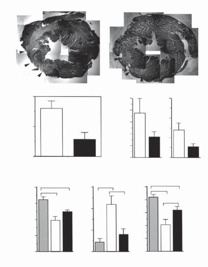
HGF exerts a protective effect on injured cardiomyocytes (Fig. 14.7). HGF is upregulated in cardiac injury and stimulates the survival and proliferation of injured cardiomyocytes. In particular, this growth factor protects injured cardiac tissue from fibrosis (note that fibrosis reduces cardiac contractility). These effects render HGF a potential therapeutic factor for the treatment of cardiac injury.
HEPATOCYTE GROWTH FACTOR RECEPTOR [14.26]. Hepatocyte growth factor receptor (HGFR), also known as MET and RCCP2, is a transmembrane receptor with a cytoplasmic protein tyrosine kinase. The length of the receptor is 1400 amino acids, and the molecular
610 CARDIAC REGENERATIVE ENGINEERING
during development) and is characterized by the presence of conserved cysteine residues. These residues form disulfide bonds and stabilize the protein structure. The PSI domain is found in plexins (proteins involved in the development of neural and epithelial tissues), semaphorins, and integrins. The IPT domain is an immunoglobulin-like structure found in plexins and certain transcription factors. HGFR is encoded by the oncogene met, which is localized to the gene locus 7q31. The protein product of the met oncogene is a dimer composed of an α subunit and a β subunit linked by disulfide bonds. The α subunit contains only an extracellular region, whereas the β subunit consists of extracellular, transmembrane, and cytoplasmic regions. The β subunit of the MET protein is the primary subunit that interacts with HGF. HGFR is expressed in the liver, brain, placenta, and skeletal muscle. The deficiency of the HGFR gene in transgenic mouse models induces liver and limb muscle defects, resulting in embryonic death of the animal. HGFR is often upregulated in human cancer cells. The expression of HGFR is enhanced during metastasis. This receptor may contribute to the metastatic properties to nontumorigenic and tumorigenic cells.
BIBLIOGRAPHY
14.1. Anatomy and Physiology
Guyton AC, Hall JE: Textbook of Medical Physiology, 11th ed, Saunders, Philadelphia, 2006.
McArdle WD, Katch FI, Katch VL: Essentials of Exercise Physiology, 3rd ed, Lippincott Williams & Wilkins, Baltimore, 2006.
Germann WJ, Stanfield CL: (with contributors Niles MJ, Cannon JG): Principles of Human Physiology, 2nd ed, Pearson Benjamin Cummings, San Francisco, 2005.
Thibodeau GA, Patton KT: Anatomy & Physiology, 5th ed, Mosby, St Louis, 2003.
Boron WF, Boulpaep EL: Medical Physiology: A Cellular and Molecular Approach, Saunders, Philadelphia, 2003.
Ganong WF: Review of Medical Physiology, 21st ed, McGraw-Hill, New York, 2003.
14.2. Pathogenesis, Pathology, and Clinical Features of Heart Failure
Schneider AS, Szanto PA: Pathology, 3rd ed, Lippincott Williams & Wilkins, Philadelphia, 2006.
McCance KL, Huether SE: Pathophysiology: The Biologic Basis for Disease in Adults & Children, 5th ed, Elsevier Mosby, St Louis, 2006.
Porth CL: Pathophysiology: Concepts of Altered Health States, 7th ed, Lippincott Williams & Wilkins, Philadelphia, 2005.
Frazier MS, Drzymkowski JW: Essentials of Human Diseases and Conditions, 3rd ed, Elsevier Saunders, St Louis, 2004.
Lloyd-Jones DM, Larson MG, Leip EP, Beiser A, D’Agostino RB et al: Lifetime risk for developing congestive heart failure: The Framingham Heart Study, Circulation 106:3068–72, 2002.
Davidson MJ, Koch WJ: Genetic manipulation of b-adrenergic signalling in heart failure, Acta Physiol Scand 173:145, 2001.
Birkeland JA, Sejersted OM, Taraldsen T, Sjaastad I: EC-coupling in normal and failing hearts,
Scand Cardiovasc J 39:13–23, 2005.
BIBLIOGRAPHY 611
Bers DM: Cardiac excitation-contraction coupling, Nature 415:198–205, 2002.
Hoshijima M: Gene therapy targeted at calcium handling as an approach to the treatment of heart failure, Pharmacol Ther 105:211–28, 2005.
Brodde OE: Beta-adrenoceptors in cardiac disease, Pharmacol Ther 60:405–30, 1993.
Rockman HA, Koch WJ, Lefkowitz RJ: Seven-transmembrane-spanning receptors and heart function, Nature 415:206–212, 2002.
Brodde OE: Beta-adrenoceptors in cardiac disease, Pharmacol Ther 60:405–30, 1993.
Wehrens XH, Marks AR: Molecular determinants of altered contractility in heart failure, Ann Med 36(Suppl 1):70–80, 2002.
Movsesian MA: cAMP-mediated signal transduction and sarcoplasmic reticulum function in heart failure, Ann NY Acad Sci 853:231–9, 1998.
Jessup M, Brozena S: Heart failure, New Engl J Med 348:2007–18, 2003.
14.3. Experimental Models of Heart Failure
Yue P et al: Post-infarction heart failure in the rat is associated with distinct alterations in cardiac myocyte molecular phenotype, J Mol Cell Cardiol 30:1615–30, 1998.
Swynghedauw B: Molecular mechanisms of myocardial remodeling Physiol Rev 79:215–62, 1999.
Gomez AM et al: Heart failure after myocardial infarction: Altered excitation-contraction coupling, Circulation 104:688–93, 2001.
Loennechen JP et al: Cardiomyocyte contractility and calcium handling partially recover after early deterioration during post-infarction failure in rat, Acta Physiol Scand 176:17–26, 2002.
Lorell BH: Transition from hypertrophy to failure, Circulation 96:3824–7, 1997.
Ito K et al: Contractile reserve and intracellular calcium regulation in mouse myocytes from normal and hypertrophied failing hearts, Circ Res 87:588–95, 2000.
Babu GJ, Periasamy M: Transgenic mouse models for cardiac dysfunction by a specific gene manipulation, Meth Mol Med 112:365–77, 2005.
Ho KKL, Anderson KM, Kannel WB, Grossman W, Levy D: Survival after the onset of congestive heart failure in Framingham Heart Study subjects, Circulation 88:107–15, 1993.
14.4. Cardiomyopathy
Hoshijima M et al: The MLP family of cytoskeletal Z disc proteins and dilated cardiomyopathy: A stress pathway model for heart failure progression, Cold Spring Harb Symp Quant Biol 67:399–408, 2002.
Pashmforoush M et al: Adult mice deficient in actinin-associated LIM-domain protein reveal a developmental pathway for right ventricular cardiomyopathy, Nat Med 7:591–7, 2001.
Zhou Q et al: Ablation of cypher, a PDZ-LIM domain Z-line protein, causes a severe form of congenital myopathy. J Cell Biol 155:605–12, 2001.
Megeney LA et al: Severe cardiomyopathy in mice lacking dystrophin and MyoD, Proc Natl Acad Sci USA 96:220–5, 1999.
Grady RM et al: Skeletal and cardiac myopathies in mice lacking utrophin and dystrophin: A model for Duchenne muscular dystrophy, Cell 90:729–38, 1997.
Hunter EG, Hughes V, White J: Cardiomyopathic hamsters, CHF 146 and CHF 147: A preliminary study, Can J Physiol Pharmacol 62:1423–8, 1984.
Durbeej M, Campbell KP: Muscular dystrophies involving the dystrophin-glycoprotein complex: An overview of current mouse models, Curr Opin Genet Dev 12:349–61, 2002.
612 CARDIAC REGENERATIVE ENGINEERING
Milner DJ et al: Disruption of muscle architecture and myocardial degeneration in mice lacking desmin, J Cell Biol 134:1255–70, 1996.
Fatkin D et al: Neonatal cardiomyopathy in mice homozygous for the Arg403Gln mutation in the alpha cardiac myosin heavy chain gene, J Clin Invest 103:147–53, 1999.
McConnell BK et al: Dilated cardiomyopathy in homozygous myosin-binding protein-C mutant mice, J Clin Invest 104:1235–44, 1999.
Hoshijima M: Models of dilated cardiomyopathy in small animals and novel positive inotropic therapies, Ann NY Acad Sci 1015:320–31, 2004.
14.5. Conventional Treatment of Cardiac Failure
Williams JF Jr, Bristow MR, Fowler MB, Francis GR, Garson A et al: Guidelines for the evaluation and management of heart failure: Report of the American College of Cardiology/American Heart Association Task Force on Practice Guidelines (Committee on Evaluation and Management of Heart Failure), J Am Coll Cardiol 26:1376–98, 1995.
Cohn JN, Franciosa JA: Vasodilator therapy of cardiac failure, New Engl J Med 297:27–31, 1977.
Packer M: The development of positive inotropic agents for chronic heart failure: How have we gone astray? J Am Coll Cardiol 22(Suppl A):119A–26A, 1993.
Cohn JN: The management of chronic heart failure, New Engl J Med 335:490–8, 1996.
Eichhorn EJ, Bristow MR: Medical therapy can improve the biological properties of the chronically failing heart: A new era in the treatment of heart failure, Circulation 94:2285–96, 1996.
Cohn JN, Archibald DG, Ziesche S, Franciosa JA, Harston WE et al: Effect of vasodilator therapy on mortality in chronic congestive heart failure: Results of a Veterans Administration Cooperative Study, New Engl J Med 314:1547–52, 1986.
The CONSENSUS Trial Study Group: Effects of enalapril on mortality in severe congestive heart failure: Results of the Cooperative North Scandinavian Enalapril Survival Study (CONSENSUS), New Engl J Med 316:1429–35, 1987.
DiBianco R, Shabetai R, Kostuk W, Moran J, Schlant RC et al: for the Milrinone Multicenter Trial Group: A comparison of oral milrinone, digoxin, and their combination in the treatment of patients with chronic heart failure, New Engl J Med 320:677–83, 1989.
The SOLVD Investigators: Effect of enalapril on survival in patients with reduced left ventricular ejection fractions and congestive heart failure, New Engl J Med 325:293–302, 1991.
Cohn JN, Johnson G, Ziesche S, Cobb F, Francis G et al: A comparison of enalapril with hydrala- zine-isosorbide dinitrate in the treatment of chronic congestive heart failure, New Engl J Med 325:303–10, 1991.
Packer M, Gheorghiade M, Young JB, Constantini PJ, Adams KF et al: for the RADIANCE Study Group: Withdrawal of digoxin from patients with chronic heart failure treated with angiotensin- converting-enzyme inhibitors, New Engl J Med 329:1–7, 1993.
Packer M, Bristow MR, Cohn JN, Colucci WS, Fowler MB et al: for the U.S. Carvedilol Heart Failure Study Group: The effect of carvedilol on morbidity and mortality in patients with chronic heart failure, New Engl J Med 334:1349–55, 1996.
The Digitalis Investigation Group: The effect of digoxin on mortality and morbidity in patients with heart failure, New Engl J Med 336:525–33, 1997.
Pfeffer MA, Braunwald E, Moye LA, Basta L, Brown EJ et al: on behalf of the SAVE Investigators: Effect of captopril on mortality and morbidity in patients with left ventricular dysfunction after myocardial infarction: Results of the Survival and Ventricular Enlargement Trial, New Engl J Med 327:669–77, 1992.
BIBLIOGRAPHY 613
The Acute Infarction Ramipril Efficacy (AIRE) Study Investigators: Effect of ramipril on mortality and morbidity of survivors of acute myocardial infarction with clinical evidence of heart failure, Lancet 342:821–8, 1993.
Køber L, Torp-Pedersen C, Carlsen JE, Bagger H, Eliasen P et al: for the Trandolapril Cardiac Evaluation (TRACE) Study Group: A clinical trial of the angiotensin-converting-enzyme inhibitor trandolapril in patients with left ventricular dysfunction after myocardial infarction, New Engl J Med 333:1670–6, 1995.
Pitt B, Segal R, Martinez FA, Meurers G, Cowley AJ et al: on behalf of the ELITE Study Investigators: Randomised trial of Losartan versus captopril in patients over 65 with heart failure (Evaluation of Losartan in the Elderly Study, ELITE), Lancet 349:747–52, 1997.
Cohn JN, Ziesche S, Smith R, Anand I, Dunkman B et al: for the Vasodilator Heart Failure Trial (VHeFT) Study Group: Effect of the calcium antagonist felodipine as supplementary vasodilatory therapy in patients with chronic heart failure treated with enalapril: VHeFT III, Circulation 96:856–63, 1997.
Elkayam U, Amin J, Mehra A, Vasquez J, Weber L et al: A prospective, randomized, double-blind, crossover study to compare the efficacy and safety of chronic nifedipine therapy with that of isosorbide dinitrate and their combination in the treatment of chronic congestive heart failure, Circulation 82:1954–61, 1990.
Hampton JR, van Veldhuisen DJ, Kleber FX, Cowley AJ, Ardia A et al: for the Second Prospective Randomised Study of Ibopamine on Mortality, and Efficacy (PRIME II) Investigators: Randomised study of effect of ibopamine on survival in patients with advanced severe heart failure, Lancet 349:971–7, 1997.
The Cardiac Arrhythmia Suppression Trial II Investigators: Effect of antiarrhythmic agent moricize on survival after myocardial infarction, New Engl J Med 327:227–33, 1992.
Levine GN, Keaney JF, Vita JA: Cholesterol reduction in cardiovascular disease: Clinical benefits and possible mechanisms, New Engl J Med 332:512–21, 1995.
Pedersen TR, Kjekshus J, Berg K, Olsson AG, Wilhelmsen L et al: for the Scandinavian Simvastatin Survival Study Group: Cholesterol lowering and the use of healthcare resources: Results of the Scandinavian Simvastatin Survival Study, Circulation 93:1796–802, 1996.
14.6. Molecular Therapy for Cardiac Failure
Williams ML, Koch WJ: Viral-based myocardial gene therapy approaches to alter cardiac function,
Annu Rev Physiol 66:49–75, 2004.
Houser SR, Margulies KB: Is depressed myocyte contractility centrally involved in heart failure? Circ Res 92:350–8, 2003.
Miyamoto MI, del Monte F, Schmidt U, DiSalvo TS, Kang ZB et al: Adenoviral gene transfer of SERCA2a improves left-ventricular function in aortic-banded rats in transition to heart failure,
Proc Natl Acad Sci USA 97:793–8, 2000.
del Monte F, Williams E, Lebeche D, Schmidt U, Rosenzweig A et al: Improvement in survival and cardiac metabolism after gene transfer of sarcoplasmic reticulum Ca2+ -ATPase in a rat model of heart failure, Circulation 104:1424–9, 2001.
Hagemann D, Xiao RP: Dual site phospholamban phosphorylation and its physiological relevance in the heart, Trends Cardiovasc Med 12:51–6, 2002.
Movsesian MA: cAMP-mediated signal transduction and sarcoplasmic reticulum function in heart failure, Ann NY Acad Sci 853:231–9, 1998.
Minamisawa S, Hoshijima M, Chu G, Ward CA, Frank K et al: Chronic phospholambansarcoplasmic reticulum calcium ATPase interaction is the critical calcium cycling defect in dilated cardiomyopathy, Cell 99:313–22, 1999.
614 CARDIAC REGENERATIVE ENGINEERING
Freeman K, Lerman I, Kranias EG, Bohlmeyer T, Bristow MR et al: Alterations in cardiac adrenergic signaling and calcium cycling differentially affect the progression of cardiomyopathy, J Clin Invest 107:967–74, 2001.
Sato Y, Kiriazis H, Yatani A, Schmidt AG, Hahn H et al: Rescue of contractile parameters and myocyte hypertrophy in calsequestrin overexpressing myocardium by phospholamban ablation, J Biol Chem 276:9392–9, 2001.
Eizema K, Fechner H, Bezstarosti K, Schneider-Rasp S, van der Laarse A et al: Adenovirus-based phospholamban antisense expression as a novel approach to improve cardiac contractile dysfunction: Comparison of a constitutive viral versus an endothelin-1-responsive cardiac promoter, Circulation 101:2193–9, 2000.
del Monte F, Harding SE, Dec GW, Gwathmey JK, Hajjar RJ: Targeting phospholamban by gene transfer in human heart failure, Circulation 105:904–7, 2002.
Hoshijima M, Ikeda Y, Iwanaga Y, Minamisawa S, Date MO et al: Chronic suppression of heartfailure progression by a pseudophosphorylated mutant of phospholamban via in vivo cardiac rAAV gene delivery, Nat Med 8:864–71, 2002.
Haghighi K, Gregory KN, Kranias EG: Sarcoplasmic reticulum Ca-ATPase-phospholamban interactions and dilated cardiomyopathy, Biochem Biophys Res Commun 322:1214–22, 2004.
Movsesian MA: cAMP-mediated signal transduction and sarcoplasmic reticulum function in heart failure, Ann NY Acad Sci 853:231–9, 1998.
Schillinger W, Janssen PM, Enami S, Henderson SA, Ross RS et al: Impaired contractile performance of cultured rabbit ventricular myocytes after adenoviral gene transfer of Na+ -Ca2+ exchanger, Circ Res 87:581–7, 2000.
Communal C, Singh K, Sawyer DB, Colucci WS: Opposing effects of B1 and B2 adrenergic receptors on cardiac myocyte apoptosis: Role of pertussis toxin-sensitive G protein, Circulation 100:2210–2, 1999.
Milano CA, Allen LF, Rockman HA, Dolber PC, McMinn TR et al: Enhanced myocardial function in transgenic mice overexpressing the β2-adrenergic receptor, Science 264:582–6, 1994.
Engelhardt S, Hein L, Wiesmann F, Lohse MJ: Progressive hypertrophy and heart failure in β1- adrenergic receptor transgenic mice, Proc Natl Acad Sci USA 96:7059–64, 1999.
Milano CA, Allen LF, Rockman HA, Dolber PC, McMinn TR et al: Enhanced myocardial function in transgenic mice overexpressing the β2-adrenergic receptor, Science 264:582–6, 1994.
Dorn GW, Tepe NM, Lorenz JN, Davis MG, Koch WJ et al: Low and high transgenic expression of β2-adrenergic receptors differentially affects cardiac hypertrophy and function in Gαq overexpressing mice, Proc Natl Acad Sci USA 96:6400–5, 1999.
Maurice JP, Hata JA, Shah AS, White DC, McDonald PH et al: Enhancement of cardiac function after adenoviral-mediated in vivo intracoronary beta-2-adrenergic receptor gene delivery, J Clin Invest 104:21–9, 1999.
Engelhardt S, Hein L, Wiesmann F, Lohse MJ: Progressive hypertrophy and heart failure in β1- adrenergic receptor transgenic mice, Proc Natl Acad Sci USA 96:7059–64, 1999.
Brodde OE: Beta-adrenoceptors in cardiac disease, Pharmacol Ther 60:405–30, 1993.
Steinberg SF: The molecular basis for distinct β-adrenergic receptor subtype actions in cardiomyocytes, Circ Res 85:1101–11, 1999.
Maurice JP, Hata JA, Shah AS, White DC, McDonald PH et al: Enhancement of cardiac function after adenoviral-mediated in vivo intracoronary β2-adrenergic receptor gene delivery, J Clin Invest 104:21–9, 1999.
Kawahira Y, Sawa Y, Nishimura M, Sakakida S, Ueda H et al: Gene transfection of β2-adrenergic receptor into the normal rat heart enhances cardiac response to β-adrenergic agonist, J Thorac Cardiovasc Surg 118:446–51, 1999.
BIBLIOGRAPHY 615
Kawahira Y, Sawa Y, Nishimura M, Sakakida S, Ueda H et al: In vivo transfer of a β2-adrenergic receptor gene into the pressure-overloaded rat heart enhances cardiac response to β-adrenergic agonist, Circulation 98:II262–7, 1998.
Shah AS, Lilly RE, Kypson AP, Tai O, Hata JA et al: Intracoronary adenovirus-mediated delivery and overexpression of the β2-adrenergic receptor in the heart: Prospects for molecular ventricular assistance, Circulation 101:408–14, 2000.
Kreusser MM, Haass M, Buss SJ, Hardt SE, Gerber SH et al: Injection of nerve growth factor into stellate ganglia improves norepinephrine reuptake into failing hearts, Hypertension 47:209–15, 2006.
14.7. Tissue Engineering Therapy for Cardiac Failure
Zimmermann WH, Didie M, Wasmeier GH, Nixdorff U, Hess A et al: Cardiac grafting of engineered heart tissue in syngenic rats, Circulation 106(Suppl 1):151–7, 2002.
14.8. Pathogenesis, Pathology, and Clinical Features of Ischemic Heart Disease
Gheorghiade M, Bonow RO: Chronic heart failure in the United States: A manifestation of coronary artery disease, Circulation 97:282–9, 1998.
Sytkowski PA, Kannel WB, D’Agostino RB: Changes in risk factors and the decline in mortality from cardiovascular disease: The Framingham Heart Study, New Engl J Med 322:1635–41, 1990.
Andrews TC, Raby K, Barry J, Naimi CL, Allred E et al: Effect of cholesterol reduction on myocardial ischemia in patients with coronary disease, Circulation 95:324–8, 1997.
Frye RL: Clinical reality of lowering total and LDL cholesterol, Circulation 95:306–7, 1997.
Mancini GBJ, Henry GC, Macaya C, O’Neill BR, Pucillo AL et al: Angiotensin-converting enzyme inhibition with quinapril improves endothelial vasomotor dysfunction in patients with coronary artery disease: The TREND (Trial on Reversing Endothelial Dysfunction) Study, Circulation 94:258–65, 1996.
Daemen MJAP, Lombardi DM, Bosman FT, Schwatz SM: Angiotensin II induces smooth muscle cell proliferation in the normal and injured rat arterial wall, Circ Res 68:450–6, 1991.
Falk E, Shah PK, Fuster V: Coronary plaque disruption, Circulation 92:657–71, 1995.
Latini R, Maggioni AP, Flather M, Sleight P, Tognoni G: ACE inhibitor use in patients with myocardial infarction: Summary of evidence from clinical trials, Circulation 92:3132–7, 1995.
Rapaport E, Gheorghiade M: Pharmacologic therapies after myocardial infarction, Am J Med 101(Suppl 4A):61S–70S, 1996.
Ambrosio G, Betocchi S, Pace L, Losi MA, Perrone-Filardi P et al: Prolonged impairment of regional contractile function after resolution of exercise-induced angina: Evidence of myocardial stunning in patients with coronary artery disease, Circulation 94:2455–64, 1996.
14.8. Molecular Regenerative Engineering for Ischemic Heart Disease
Melo LG, Pachori AS, Kong D, Gnecchi M, Wang K et al: Gene and cell-based therapies for heart disease, FASEB J 18:648–63, 2004.
Brocheriou V, Hagege AA, Oubenaissa A, Lambert M, Mallet VO et al: Cardiac functional improvement by a human Bcl-2 transgene in a mouse model of ischemia/reperfusion injury, J Gene Med 2:326–33, 2000.