

Bioregenerative Engineering Principles and Applications - Shu Q. Liu
..pdf516 NERVOUS REGENERATIVE ENGINEERING
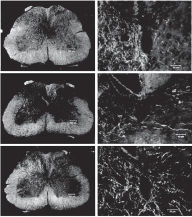
Figure 13.12. Influence of axotomy on the structure of neurons. Confocal microscopy images of facial nuclei at the intact (left) and axotomy (right) sides. D1 and D7 are days 1 and 7, respectively, after axotomy. The specimens were labeled with an antibody against mouse synaptophysin, an axonal marker. The expression level of synaptophysin is apparently reduced at the axotomy side. (Reprinted from Ikeda R, Kato F: Early and transient increase in spontaneous synaptic inputs to the rat facial motoneurons after axotomy in isolated brainstem slices of rats. Neuroscience 134:889– 99, 2005, copyright 2005, with permission from Elsevier.)
spinal cord, causing blunt injury. The degree of injury can be controlled by altering the weight and height of the impactor. Electronically controlled mechanical impactors can be developed and used to cause blunt injury based on a similar principle. In the compression model, a mechanical clip or band can be applied to an exposed spinal cord to induce circumferential compression. The degree of injury can be controlled by altering the level of compression. This model simulates spinal cord injury due to the compression of vertebral columns.
An important issue in spinal cord injury modeling is to estimate the degree of injury or whether the nerve tracts are completely severed. Axonal tracers are often used to achieve such a goal. The nerve axons are capable of conducting retrograde transport of substances toward the cell body. A tracer can be applied to a site distal to the nerve injury. The completeness of transection can be judged by observing the appearance of the tracer in the cell body. Among the three types of model described above, the contusion and compression models usually result in incomplete injury. Because of the complexity of the spinal cord, it is often difficult to estimate the degree of nerve regeneration by using these incomplete injury models.
NERVOUS DISORDERS |
517 |
(A) |
|
(B) |
(C) |
|
(D) |
(E) |
|
(F) |
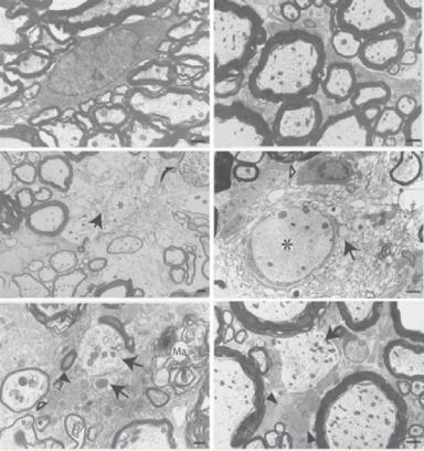
Figure 13.13. Histological micrographs showing demyelination after contusive spinal cord injury. (A,B) Normal myelin sheaths in the ventrolateral funiculus of the adult spinal cord. These sheaths surround axons. (C,D) Demyelination 1 week after contusive spinal cord injury in the ventrolateral funiculus. Many myelin sheaths degenerated and numerous intramyelinic vacuoles were seen (arrows). A macrophage was observed in close relation to degenerating myelin (open arrowhead in panel D). The axons surrounded by degenerating myelin appeared morphologically normal (asterisks). (E,F) Some demyelinated axons survived for at least 1 month after injury (arrows). (E,F) Healthy-appearing demyelinated axons were observed in the ventrolateral funiculus of spinal cord at the injury epicenter (E) and also a few millimeters away from the epicenter (panel F, arrow). Scale bars: 2.5 μm. (Reprinted with permission from Cao Q et al: J Neurosci 25:6947–57, copyright 2005.)
Peripheral Nerve Injury. Peripheral nerve injury is largely induced by trauma resulting from accidents and surgery. The peripheral nerves are capable of recovering from injury when they are not completely severed. However, when the nerves are completely severed, the distal axons are usually disintegrated into segments within several days, followed by further degradation of the nerve segments. At the same time, the myelin sheaths of the Schwann cells that enclose the axons are also degraded. The degraded axon and myelin are phagocytosed by macrophages.
The proximal neurons are able to regenerate and extend their axons and restore the lost distal axonal segment (Fig. 13.14). However, the outcome depends on the severity of the injury, especially the distance between the two ends of the severed axon. When the two
518 NERVOUS REGENERATIVE ENGINEERING
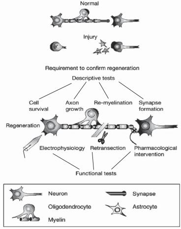
Figure 13.14. Nerve regeneration after nerve injury. A number of descriptive and functional tests have been designed and used for evaluating the outcome of nerve regeneration after nerve injury. Descriptive tests, as shown in the figure, can be used to determine the survival and integrity of the injured system, whether axonal regeneration is present, and if appropriate synaptic connections and remyelination have occurred. Functional tests, incldung electrophysiological and pharmacological intervention, can be used to assess the function and specificity of the regenerated pathway. (Reprinted by permission from Macmillan Publishers Ltd.: Horner PJ, Gage FH: Nature 407:963–70, copyright 2000.)
ends are close to each other and well aligned, nerve regeneration and reconnection can occur. In contrast, when the two ends of a severed nerve are separated and not aligned, the injured nerve segment may not be restored. The residual Schwann cells play a critical role in nerve regeneration. These cells start to enlarge and undergo mitosis when the proximal axon starts to regenerate. The Schwann cells are aligned along the injured axons, preparing for the regeneration and reconnection between the proximal and distal axons. The proximal axons that encounter the Schwann cells are able to restore the injured distal axons. However, the proximal axons that are not escorted by Schwann cells cannot reach the severed distal axons.
NERVOUS DISORDERS |
519 |
Nerve regeneration is a slow process. It usually takes about several weeks for the proximal regenerating nerves to reach the injured distal nerves. The end of each proximal nerve may form several sprouts. The sprouts that encounter the Schwann cells develop into axons and replace the severed distal nerves. The remaining sprouts are gradually disintegrated. Following the growth of the proximal axons into the Schwann cells, the regenerating axons are enclosed by newly generated myelin sheaths.
Conventional Treatment of Nerve Injury. For the injury of the central nerve system due to craniocerebral trauma, several approaches can be used for the treatment depending on the severity of the injury. For patients with concussion (transient unconsciousness), no special treatment is needed. Patients are usually given analgesics such as aspirin or acetaminophen for headache, and antidepressants for anxious depression, if any. For severe head injury, especially when patients are unconscious, several treatments should be carried out: (1) check to ensure that the airways are functional and are able to supply adequate ventilation (artificial respiration should be applied, if necessary); (2) stop bleeding, if any;
(3) restore blood pressure, if hypotension exists (note that hypertension may exist with severe head injury); (4) restore the cardiac function, if necessary; (5) measure and reduce intracranial pressure when intracranial pressure is too high; (6) supply nutrients via a nasogastric tube, if coma persists; and (7) resort to surgical intervention to stop hemorrhage, relieve intracranial pressure, repair fractured or distorted bones, and/or remove coagulated blood, if necessary. For severed peripheral nerves due to trauma, simple surgical reconnection or grafting of the injured nerves with an autologous nerve segment, if necessary, can be performed. Such an approach usually gives a satisfactory result.
Molecular Nerve Regenerative Engineering [13.3]
Strategies of Molecular Nerve Regenerative Engineering. Principal strategies for the treatment of nerve injury are to promote neuronal survival and regeneration, prevent neuronal death, protect the neurons from secondary injury, stimulate axonal adhesion, extension and reconnection, reduce fibrosis and scar formation, enhance synaptic formation. In conventional medicine, there are few available methods that can be used to effectively achieve these strategies. The recovery of injured neurons and nerve fibers is very much dependent on the self-regeneration capability. For the past decade, regenerative engineering approaches have been developed at the molecular, cellular, and tissue levels to facilitate the regeneration of injured nerves. At the molecular level, mitogenic proteins and genes can be used to promote the survival and regeneration of injured neurons and to prevent neuronal death. At the cellular level, various cell types can be used and transplanted to the injury site to assist the recovery of injured neurons. At the tissue level, various biological and mechanical devices have been constructed to serve as guidance for the regeneration of injured nerve axons. Although most of these engineering approaches have not been used in clinical therapy, experimental investigations have demonstrated potential for clinical applications.
For the central nerve system, because the function of many structures has not been completely understood and the system is difficult to access, it is often a challenge to “engineer” the brain and spinal cord. Thus, engineering treatment for injured brain and spinal cord is limited. In contrast, the peripheral nerve fibers are relatively easy to access, it is not technically difficult to manipulate these fibers. Here, the principle of molecular and cell regenerative engineering for central and peripheral nerve injuries is discussed.
520 NERVOUS REGENERATIVE ENGINEERING
Molecular regenerative engineering approaches can be established and used for facilitating the regeneration of injured neurons and nerve fibers. There are several strategies for the application of molecular engineering approaches to nerve regeneration: preventing cell death, promoting cell survival, and enhancing the differentiation of stem and progenitor cells to neurons and supporting cells. There exist several types of growth factors that are known to promote cell survival and growth, and prevent cell death. Thus, growth factors and their genes are candidate therapeutic factors for the treatment of nerve injury. At the protein level, growth factors can be applied directly to the site of nerve injury to stimulate nerve cell regeneration. At the gene level, selected growth factor genes can be manipulated to enhance the level of gene expression, resulting in an increase in the production of encoded growth factors.
There are several challenges for the application of molecular regenerative engineering approaches to nerve regeneration. These include the selection of therapeutic growth factors and/or genes, the establishment of appropriate techniques for the delivery of the therapeutic factors, and precise delivery of the therapeutic factors to the injury site. The selection of therapeutic factors and genes is dependent on the nature of therapeutic molecules, the degree of nerve injury, and the type of the injured cells. While certain types of growth factors promote nerve growth and regeneration, other types may exert an opposite effect. Fr instance, neurotrophic factors and receptors stimulate nerve regeneration, whereas epidermal growth factor receptor (EGFR) inhibits nerve regeneration. EGFR can be activated or phosphorylated in response to the stimulation of chondroitin sulfate proteoglycans and activates signaling mechanisms that suppress axonal regeneration. Other therapeutic factors and genes are discussed for different cases in the following sections.
There are several approaches for the delivery of therapeutic factors and genes to the site of nerve injury:
1.Therapeutic proteins can be delivered to the nerve system via direct injection to the injury site of the central nervous system. This method gives a local delivery of highly concentrated therapeutic factors. However, it is difficult to find the precise site of injury for injection. Furthermore, direct injection induces nerve injury.
2.Therapeutic factors and genes can be injected into the spinal cavity. This cavity is connected to the four cerebral ventricles and the arachnoid space that cover the cerebrum (see page 507). The delivered factors can reach the cranial cavity via diffusion, where they are taken up by epithelial cells and transported to the nearby nerve cells. This approach is suitable for delivering therapeutic factors to the brain and spinal cord. Compared to the direct injection method, the spinal cavity delivery is a safe and reliable method, although it may require a high dose of growth factors or genes to reach an effective concentration at the injury site.
3.Selected cell types, such as glial cells or fibroblasts, can be cultured and transfected with a therapeutic gene, rendering the cells to express a high level of a desired factor. These cells can be delivered to the site of nerve injury to release the selected therapeutic factor. Direct injection is an effective method for cell delivery.
It should be noted that the bloodstream may be considered an alternative delivery route. Therapeutic factors and genes can be injected into the blood, taken up by endothelial cells, and transported to the target tissues. However, due to the presence of the blood–brain barrier, it may be difficult to deliver therapeutic proteins and genes to the brain system with high efficiency.
NERVOUS DISORDERS |
521 |
Enhancement of Neuron Survival. There are several growth factors that can be used for the enhancement of neuronal cell survival and regeneration. These factors include brainderived neurotrophic factor (BDNF), neurotrophin (NT)3, neurotrophin 4, nerve growth factor (NGF), and glial cell-derived growth factor (GDNF) (Table 13.1). The functions of these growth factors are briefly discussed as follows.
BRAIN-DERIVED NEUROTROPHIC FACTOR [13.4]. Brain-derived neurotrophic factor is a 247 amino acid protein and a member of the nerve growth factor family. The brain-derived neurotrophic factor gene is mapped to chromosome 11 and locus 11p13 in humans. This factor shares a nucleotide sequence considerably similar to the nerve growth factor gene. The brain-derived neurotrophic factor gene is similar among different mammals. Brainderived neurotrophic factor is expressed primarily in the cortical neurons. These neurons produce the precursor of brain-derived neurotrophic factor. The precursor is released into the extracellular matrix and cleaved by several enzymes, including serine protease plasmin, matrix metalloproteinases (MMP)7 and MMP3, resulting in the formation of mature brain-derived neurotrophic factor.
Brain-derived neurotrophic factor is necessary for the survival of neurons in the central nervous system. During embryonic development, brain-derived neurotrophic factor plays a critical role in the differentiation and proliferation of neuronal cells and contributes to the development of germ cells. In response to nerve injury, the expression of the BDNF gene is often upregulated, an important mechanism for the self-repair of injured nerves (Fig. 13.15). A treatment with BDNF in cultured dorsal root ganglion explants enhances neurite outgrowth (Fig. 13.16). In neural degenerative disorders, such as Alzheimer’s and Huntington’s diseases, the expression of brain-derived neurotrophic factor is downregulated, which contributes to the development of these disorders. In transgenic model of brain-derived neurotrophic factor deficiency induced by homologous recombination or site-specific gene targeting in embryonic stem cells, sensory neurons are lost in various areas of the central nerve system. However, the motor neurons are not significantly affected by the deficiency of the brain-derived neurotrophic factor gene.
Brain-derived neurotrophic factor can be released on neuron depolarization, resulting in the activation of intracellular signaling pathways. In dopamine neurons, brain-derived neurotrophic factor stimulates the expression of dopamine D3 receptor during development and adulthood, contributing to the control of animal behavior. In serotonergic neurons, brain-derived neurotrophic factor regulates the expression of several postsynaptic serotonin receptors in the frontal cortex, hippocampus, and hypothalamus. The deficiency of brain-derived neurotrophic factor induces a reduction in the expression of these serotonin receptors, which is associated with behavioral abnormalities such as increased aggressiveness and hyperphagia. These observations suggest that brain-derived neurotrophic factor plays a critical role in the regulation of the development, function, and regeneration of dopaminergic and serotonergic neurons.
NEUROTROPHIN 3 [13.5]. Neurotrophin 3, also known as neurotrophic factor 3, is a growth factor that is similar in structure to nerve growth factor and brain-derived neurotrophic factor. All these factors are members of the nerve growth factor family. Neurotrophin 3 is composed of 258 amino acids and is expressed in the human brain and placenta. The gene of neurotrophin 3 is localized to chromosome 12 and locus 12p13. The gene sequence of neurotrophin 3 is well conserved among mammals. The primary function of

522
TABLE 13.1. Characteristics of Selected Nerve Growth-Related Factors*
|
|
|
Molecular |
|
|
|
|
Amino |
Weight |
|
|
Proteins |
Alternative Names |
Acids |
(kDa) |
Expression |
Functions |
|
|
|
|
|
|
Brain-derived |
BDNF |
247 |
28 |
Central nervous system |
Regulating the survival of neurons, stimulating |
neurotrophic |
|
|
|
(cortex, retina, and |
embryonic development |
factor |
|
|
|
spinal cord), fetal testis |
|
Ciliary |
CNTF |
200 |
23 |
Brain |
Promoting neurotransmitter synthesis and neurite |
neurotrophic |
|
|
|
|
outgrowth and regulating the survival of neurons |
factor |
|
|
|
|
and oligodendrocytes |
Glial cell line- |
Astrocyte-derived trophic |
211 |
24 |
Nervous system, kidney |
Promoting the survival and differentiation of |
derived |
factor 1 and glial cell- |
|
|
|
dopaminergic neurons and preventing apoptosis |
neurotrophic |
derived neurotrophic |
|
|
|
of motor neurons |
factor |
factor |
|
|
|
|
|
|
|
|
|
|
*Based on bibliography 13.4 and 13.8.
NERVOUS DISORDERS |
523 |
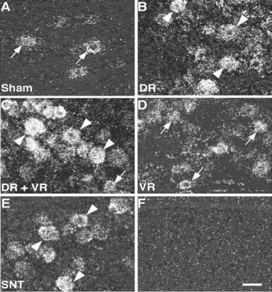
Figure 13.15. Expression of brain-derived neurotrophic factor (BDNF) in injured neurons. (A–E) Expression of BDNF mRNA in the ipsilateral L5 dorsal root ganglion (DRG) by in situ hybridization histochemistry in the sham control (A), dorsal rhizotomy only (B), dorsal rhizotomy + ventral rhizotomy (C), ventral rhizotomy only (D), and spinal nervous transection (E) groups at 7 days after surgery. Arrowheads indicate large-size sensory neurons positive for BDNF, whereas arrows indicate small-to-medium-diameter neurons. These observations show that nerve and spinal cord injury induces upregulation of the BDNF gene. (F) Hybridization with a sense probe for BDNF mRNA showed no positive signals. Scale bar: 50 μm. (Reprinted from Obata K et al: The effect of site and type of nerve injury on the expression of brain-derived neurotrophic factor in the dorsal root ganglion and on neuropathic pain behavior, Neuroscience 137:961–70, copyright 2006, with permission from Elsevier.)
neurotrophin 3 is to promote neuronal proliferation, differentiation, and survival as well as the neurite outgrowth during development.
Neurotrophin 3 can interact with the neurotrophin receptor p75 in neurons, exerting a stimulatory effect on neuronal mitogenic activities. In a rat model of spinal cord injury, the transfer of the neurotrophin 3 gene into the spinal cord stimulates the regeneration of injured axons (Fig. 13.17). Neurotrophin 3 (NT3) can also promote axonal growth of injured neurons into neural stem cell (NSC) grafts in injured spinal cord (Fig. 13.18). Furthermore, neurotrophin 3 plays a critical role, together with brain-derived neurotrophic factor, in the regulation of myelin formation by developing and regenerating Schwann
524 NERVOUS REGENERATIVE ENGINEERING
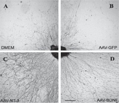
Figure 13.16. Enhancement of neurite outgrowth by neurotrophin-3 (NT3) and brain-derived neurotrophic factor (BDNF). Dorsal root ganglion explants were cultured with conditioned media collected from four groups of cells: (A) mock-infected 293 cells; (B) 293 cells transfected with adeno-associated viral vector (AAV) containing the GFP gene; (C) 293 cells transfected with AAV containing the NT3 gene; (D) 293 cells transfected with AAV containing the BDNF gene. Explant specimens were prepared and stained for neurofilaments using the RT97 antibody. In the controls (A and B), a low level of neurite outgrowth was observed. With AAV–NT3- and AAV–BDNF- conditioned medium, the rate of neurite outgrowth is significantly higher than the controls. Scale bar: 1 mm. (Reprinted from Blits B et al: Adeno-associated viral vector-mediated neurotrophin gene transfer in the injured adult rat spinal cord improves hind-limb function, Neuroscience 118:271–81, copyright 2003, with permission from Elsevier.)
cells. Neurotrophin 3 is also required for the outgrowth and innervation of sympathetic axons. In transgenic mice, the deficiency of neurotrophin 3 results in the loss of peripheral sensory and sympathetic neurons, while the motor neurons are not significantly affected. These defects are associated with the loss of peripheral muscular sensors, movement disorder, and death shortly after birth. Thus, neurotrophin 3 is a candidate factor for the treatment of nerve injury. It is interesting to note that neurotrophin 3 regulates not only the development and regeneration of the central and peripheral nervous system, but also the development of the heart. Neurotrophin 3 is required for the development of the cardiac atria and ventricles. The deficiency of neurotrophin 3 induces several major cardiac disorders, including atrial and ventricular septal defects and Fallot tetralogy, common forms of congenital cardiac defects. These disorders often result in perinatal death of transgenic mice.
|
NERVOUS DISORDERS |
525 |
A |
A' |
|
B |
B' |
C |
C' |
Figure 13.17. Enhancement of axonal growth by neurotrophin-3 (NT3) gene transfer to injured spinal cord. Rats with unilateral lesion of the corticospinal tract (CST) were transfected with Adv.EF α-NT3 or Adv.EF α-LacZ. The unlesioned corticospinal tract was labeled with BDA.
(A) Section from a sham control rat. (B) Section from an Adv.EF α-LacZ-transfected rat. (C) Section from an Adv.EF α-NT3-transfected rat. (A′–C′) Higher-magnification photomicrographs of the regions around the central canal. Note that in panel C, BDA-labeled corticospinal tract neurites can be seen arising from the intact corticospinal tract, traversing the midline, and growing into the gray matter of the lesioned side of the spinal cord. (Reprinted with permission from Zhou L et al: J Neurosci 23:1424–31, copyright 2003.)
NEUROTROPHIN 4 [13.6]. Neurotrophin 4 is a 210-amino acid neurotrophic factor (13– 14 kDa) expressed in the nervous system. It is also known as neurotrophin 5 or neurotrophin 4/5. The term neurotrophin 4 was derived based on the fact that the Xenopus neurotrophin 4 gene was used to isolate the human counterpart gene. However, the human neurotrophin 4 exhibits more diverse features and activities than the Xenopus neurotrophin 4. Thus, this growth factor was also termed neurotrophin 5. Some investigators prefer the term neurotrophin 4/5 to reflect the similarities and differences between the mammalian and Xenopus forms. The gene of neurotrophin 4 is localized to chromosome 19q13.3. Neurotrophin 4 plays a critical role in regulating the survival, proliferation, and differen-