

Bioregenerative Engineering Principles and Applications - Shu Q. Liu
..pdf426 MOLECULAR ASPECTS OF BIOREGENERATIVE ENGINEERING
selection of the gene of interest, (6) amplification of the selected gene, (7) gene sequencing and analysis, and (8) test of the function of the selected gene.
Assessing mRNA Transcription. The identification of mutant genes may start with assessing the level of gene transcription. In general, a gene defect or mutation may result in an alteration in the level of mRNA transcription. The alteration may be a complete deficiency, increase, or decrease in mRNA transcription. Since a change in the regulatory activity of gene expression may also induce an increase or decrease in mRNA transcription, an alteration in gene expression may not be indicative of the presence of a mutant gene. However, such a change may serve as a clue for identifying potential mutant genes. Genes with altered expression can be selected for further analyses, such as DNA sequencing and functional test, which provide conclusive information for identifying the mutant gene.
To assess the level of mRNA transcription or gene expression, it is necessary to prepare mRNA and measure the level of mRNA transcription. Total mRNA can be extracted from target cells, and the transcription of mRNA can be assessed by analytical approaches such as Northern blotting and gene microarray analyses. Since the gene microarray analysis provides a profile of mRNA transcription covering hundreds and thousands of genes, this method is more efficient than Northern blotting analysis. Here, the principle of gene microarray analysis is briefly discussed.
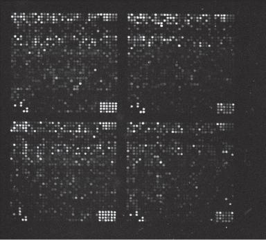
Gene microarray analysis is an approach established to detect quantitatively the transcription levels of multiple mRNAs according to the rule of complementary hybridization with prearranged DNA or cDNA (complementary DNA) samples with known sequences. In this analysis, fluorochrome-conjugated mRNA or reverse-transcribed cDNA samples, known as probes, from a specified cell type are applied to arrays of microspots coated with selected DNA samples of known sequences, inducing hybridization reactions. The fluorescent levels of the spots with mRNA-hybridized DNA can be measured and used to represent the relative level of mRNA transcription in the selected cell type (Fig. 10.3). Because the DNA samples coated on the microarrays are selected with known structure, the hybridized mRNA or cDNA samples can be identified. With the information accumulated in the gene bank for the past decades, genes involved in most known diseases have been studied, identified, and cloned. Thus, it is not difficult to establish gene microarrays that can be used for the identification of genes involved in common diseases. A major advantage for the gene microarray analysis is that a single experiment can provide an expression profile for a large number of genes.
To conduct a gene microarray analysis, it is necessary to follow several procedures, including the preparation of gene microarrays, preparation of fluorochrome-conjugated mRNA or cDNA probes, probe-DNA hybridization, detection of the relative levels of mRNA or cDNA probes hybridized to DNA targets, and data analysis. Gene microarrays can be prepared by coating denatured genomic DNA, cDNA (DNA prepared by reversetranscribing mRNA), or synthesized oligonucleotide samples to arrays of DNA-binding spots created by photolithography on a glass slide of a centimeter size. Each spot can be as small as 50–350 μm in dimension. Thus, a large number of different DNA samples can be arranged in a single slide (>5000 spots/cm2), which is also known as a gene “chip.” In practice, it is considerably easier to prepare microarrays with oligonucleotides compared to those with DNA and cDNA. Oligonucleotides can be synthesized in single-strand format. An appropriate size of oligonucleotides for gene microarray analysis should be 20–120 bases.
DNA ENGINEERING |
427 |
Figure 10.3. An image of a microarray containing >5000 genes. Each spot features a pool of identical single-stranded DNA molecules representing a single gene. The brightness of the spot is proportional to the amount of fluorescent mRNA hybridized to the DNA of the spot. The fluorescence spots can be identified by automated image analysis. The fluorescence intensity from each spot can be measured and compared to the background fluorescence. The images are further compared to images obtained from control measurements and transformed into a gene expression matrix, which can be analyzed by numerical methods. (Reprinted by permission of the Federation of the European Biochemical Society from Brazma A, Vilo J: Gene expression data analysis, FEBS Lett 480:17–24, copyright 2000.)
To detect the level of mRNA transcription from a cell sample, two types of probe can be synthesized from extracted mRNA: cDNA and RNA. cDNA probes can be synthesized by reverse transcription using extracted mRNAs as templates and the reverse transcriptase as a catalytic enzyme. Such a procedure produces single-stranded cDNA molecules. The cDNA probes can be conjugated with fluorochromes for probe identification. Alternatively, RNA probes can be synthesized from cDNA templates reverse-transcribed from mRNAs. In this preparation, T7 RNA polymerase is used to synthesize RNA molecules from double-stranded cDNA templates in the presence of fluorochrome-labeled nucleotides. The synthesized RNAs can be used as probes for gene microarray analysis.
The next step is to apply the probe, either single-stranded cDNAs or RNAs, to gene microarrays. The probing cDNA or RNA molecules hybridize with target DNA molecules via hydrogen bonds on the basis of the DNA complementary rule. Excessive and unhybridized probes can be removed by washing. The fluorescence of hybridized probes can be imaged and recorded from the microarrays by a laser scanner, and the intensity of
428 MOLECULAR ASPECTS OF BIOREGENERATIVE ENGINEERING
fluorescence can be measured and analyzed. Such intensity represents the relative level of mRNA transcription. It is important to note that positive and negative controls should be introduced to the analysis. A positive control can be a selected cDNA probe that is known to hybridize to a target DNA, whereas a negative control is a cDNA probe that does not hybridize to a given target DNA.
The profile of mRNA transcription derived from a gene microarray analysis can be analyzed and compared to the profile of mRNA transcription from a normal or specified control cell type. Thus, genes with altered transcription can be identified from the microarray analysis. Although changes in the level of mRNA transcription may not indicate a gene defect or mutation, an increased, decreased, or null gene transcription suggests a candidate gene for further investigation. Once a gene with an altered transcription level is identified, the gene can be cloned and analyzed for the identification of structural mutation. If no mutation is found, the alterations in gene expression reflect changes in the regulatory activity for gene expression.
Extracting and Digesting DNA. Once a gene with altered mRNA transcription is identified as described above, DNA can be isolated and collected for gene analyses, including gene sequencing and identification of gene mutation. To obtain DNA, a selected cell sample can be lysed (for cultured cells) or homogenized (for a tissue sample), and DNA can be extracted and purified with established methods. Since DNA is a very large molecule, it should be digested into short fragments for DNA manipulation. DNA digestion can be accomplished by using restriction enzymes, which are found in bacteria and are capable of cleaving DNA at specific sequences. Although restriction enzymes are originated from bacteria, they can cleave DNA molecules from all known species as long as the DNA contains digestion target sequences. Each DNA molecule contains a large number of restriction sites for various types of restriction enzyme. These restriction sites are randomly distributed. DNA fragments with desired lengths can be generated by selecting appropriate restriction enzymes based on the locations of restriction sites. The digestion of target DNA at specified sites is one of the most important genetic approaches used for DNA manipulation. It is impossible to clone genes without the assistance of the restriction enzymes. It should be noted that restriction enzymes are not present in mammals. Thus, DNA molecules are not digested in the body systems under physiological conditions.
Restriction enzymes can cleave DNA into fragments with two different forms of end—sticky end and blunt end—depending on the type of restriction enzyme, but not the sequence of DNA. Some restriction enzymes, such as EcoRI, EcoRII, and HindIII, can cut the two strands of a DNA molecule unevenly, generating DNA ends with uneven strand length (the 3′ end is longer than the 5′ end or vise versa). The uneven end is also known as “sticky” end, so named because two such ends can hybridize to each other based on the complementary rule. Other restriction enzymes, such as HindII, HaeIII, and SmaI, cut the two strands of a DNA molecule evenly, leaving DNA ends with even strand length. The even DNA ends are also called “blunt” ends. In most cases, the substrate sequence for a restriction enzyme is identical for the two DNA strands at a given restriction site, but oriented in antiparallel directions. Thus, a “sticky” cut can produce two identical ends at a restriction site. This is a very useful feature for the creation of recombinant DNA. A “blunt” DNA end can be remodeled into a “sticky” end by an enzymatic treatment. Once DNA fragments are prepared, these fragments can be integrated into DNA vectors and used to create recombinant DNAs.
DNA ENGINEERING |
429 |
Constructing Recombinant DNA. The extracted and digested DNA fragments need to be further processed to generate DNA structures that can be cloned, screened for the desired gene, analyzed, and tested. An effective approach for such purposes is the construction of recombinant DNA. Recombinant DNA is a complex of DNA constructed by integrating a DNA fragment selected from a donor organism into a DNA vector collected from a different organism. The process of constructing a recombinant DNA is defined as DNA recombination. The purpose of DNA recombination is to generate functional genes in vitro, so that the gene of interest can be cloned, amplified, tested, used for gene transfection, and expressed in the transfected cells and tissues.
Gene cloning is a process of gene replication, generating multiple copies of the same gene. For cloning a gene, it is necessary to prepare a gene cloning vector. A vector is a bacterial DNA structure that contains restriction sites, and can accept foreign gene inserts and replicate in living bacteria. A vector is often engineered to add multiple restriction sites for the insertion of foreign gene fragments and to add functional genes for the selection, identification, and collection of the vector. A number of vector types, such as plasmids, λ phages, and cosmids, have been established and used in gene cloning. A typical plasmid vector carrying a LacZ gene (β-galactosidase gene) is shown in Fig. 10.4. The LacZ gene is often used as a reporter gene for gene selection or gene transfection.
To make recombinant genes, a selected donor DNA or cDNA fragment and a selected vector are treated with an identical type of restriction enzyme to generate the same type of “sticky” DNA ends. The enzyme-treated DNA fragment and vector are then incubated under a desired condition to hybridize the DNA fragment with the vector. At this stage, although the donor DNA and vector are joined with hydrogen bonds, the sugar–phosphate backbone is not linked together with phosphodiester bonds. An enzyme known as DNA ligase is needed to link the backbone, completing the recombinant process.
There are potential problems for DNA recombination. One problem is that the open ends of a vector may rejoin together without the insertion of donor DNA. Such vectors should not be included in the analysis. A gene selection method is usually used to remove the vectors that do not contain the inserted gene of interest. For this method, a selection gene, such as the β-galactosidase gene, is inserted into a selected vector in a cloning region with multiple restriction sites, also known as a polylinker or a multiple cloning site. The selection mechanism is that the β-galactosidase gene can be expressed when transfected into bacterial cells, only if the β-galactosidase gene is not interrupted by another gene. The insertion of a donor DNA fragment into the polylinker interrupts the structure of the β-galactosidase gene and renders the β-galactosidase gene unfunctional. When vectors without the donor DNA fragment are transfected into bacteria, the β-galactosidase gene
amp
ori
IIIHindl HIamISmaRIEco
stPB
Linker
lac Z
Figure 10.4. Schematic representation of the pUC18 plasmid vector containing a Lac Z gene.
430 MOLECULAR ASPECTS OF BIOREGENERATIVE ENGINEERING
can be expressed. Expressed β-galactosidase can react with a reagent known as X-gal to produce a blue-colored substance, which can be visualized under an optical microscope. However, the β-galactosidase gene in the vector with the donor DNA insert cannot be expressed and no blue-colored substance is generated when reacted with X-gal. Thus, clones with inserted donor genes can be selected on the basis of the color expression.
Another problem is that dominant “blunt” ends may be generated for the donor DNA, reducing the efficiency of donor DNA integration into the cloning vectors. To resolve such a problem, the donor DNA can be treated with a specific exonuclease that degrades the DNA at only the 5′ end, thus creating uneven “sticky” ends. However, the “sticky” ends of the donor DNA may not be complementary to the ends of a vector. To resolve such a problem, the open ends of the donor DNA fragment and the vector can be modified by synthesizing a short sequence at the ends. Such a process can be induced by treating both the donor DNA and the vectors separately with terminal transferase in the presence of different yet complementary nucleotides. For instance, the donor DNA can be treated with terminal transferase in the presence of dTTP, whereas the vector can be treated with terminal transferase in the presence of dATP. The added poly-T ends of the donor DNA are thus complementary to the poly-A ends of the vector.
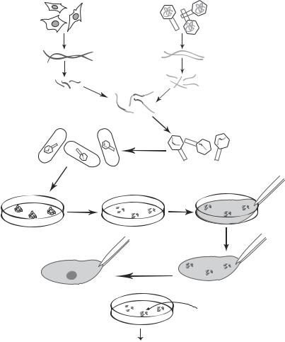
Establishing a DNA Library. To identify and isolate the gene of interest from the gene pool containing all genes from a genome, it is necessary to establish a DNA library, which is defined as a collection of all gene clones from a cell sample of interest. A gene library can be established by using the procedures as described above, namely, extracting all DNA molecules, digesting the DNA with selected restriction enzymes into fragments of appropriate lengths, and inserting the DNA fragments into gene-carrying vectors. The vectors are then transfected into Escherrichia coli to establish E. coli colonies. Each E. coli colony is supposed to contain a specific DNA fragment. A DNA library is composed a large number of DNA fragments (Fig. 10.5). It is expected that one of these fragments contains the gene of interest. Such a gene can be identified and isolated by using a specific DNA or RNA probe as discussed on page 433.
For establishing a DNA library, two issues should be taken into account: vector type used in the cloning process and the DNA source. Vectors can accommodate DNA fragments with different sizes. Thus, vectors should be selected on the basis of the DNA fragment size. Plasmids and λ-phages are suitable for small DNA fragments, whereas cosmids and yeast artificial chromosomes (YACs) are used for large DNA fragments. Various vectors give different forms of DNA library. A plasmid or cosmid library is a collection of E. coli bacteria containing the cloned genes, a λ-phage library is a collection of λ-phages, whereas a YAC library is a collection of yeasts. The choice of vectors does not affect the structure of the cloned genes.
Another factor to consider in the construction of a DNA library is the source of DNA. DNA from two sources can be used for the construction: genomic DNA and complementary DNA (cDNA). The later is DNA synthesized from mRNA by reverse transcription. There are distinct features for the two types of DNA library. Genomic DNA contains introns and exons for all genes from a genome. A genomic DNA library is suitable for investigating the regulation of gene transcription, which requires the identification and understanding of the regulatory cis elements located in the introns. In contrast, a cDNA library contains only exons that encode proteins. In addition, cDNA does not contain genes that are not transcribed into mRNA at a given time and state. Thus, a cDNA library is suitable for the identification and isolation of expressed genes. Since most pathological
|
DNA ENGINEERING |
431 |
Mammalian |
Bacteriophages |
|
cells |
|
DNA extraction |
|
Cell DNA |
Phage DNA |
|
Restriction enzyme
Ligation
E. coli culture
E. coli lysis
Identification of  the clone of interest in the culture
Recombinant DNA
Phage transfection
E. coli transfection
Transfer to filter 
|
Hybridization |
|
with a DNA |
Identification of |
probe |
|
|
probe-hybridized |
|
clone |
|
Clone of interest
Amplification of selected clone
Figure 10.5. Preparation of a gene library. Based on bibliography 10.2.
disorders involve gene expression, creating a cDNA library is a practical approach for identifying genes that contribute to pathological disorders.
Selecting a Gene of Interest from a DNA Library. A DNA library may contain thousands of gene fragments. To find and collect the gene of interest, it is necessary to screen the DNA library. There are two approaches that can be used for DNA screening: directly probing the gene of interest or probing the protein encoded by the gene integrated into the expression vector, which is designed to express proteins in cultured E. coli cells. For the direct DNA approach, it is necessary to establish a DNA probe, which is a singlestranded DNA fragment or oligodeoxynucleotide, capable of hybridizing to its complimentary strand. With the tag of an identification marker to the probe, the target gene strand can be identified and isolated. For the protein-probing approach, an antibody
432 MOLECULAR ASPECTS OF BIOREGENERATIVE ENGINEERING
specific to the protein produced by the gene of interest can be used to identify the protein, which leads to the identification of the encoding gene.
As discussed on page 426, gene selection can start from the detection of mRNA transcription in a cell sample selected from a disordered organ by gene microarray analysis. An altered transcription level (increase, decrease, or null) may help to identify a potential gene involved in the pathogenesis of a disorder. It is important to point out that a gene microarray analysis does not provide information about alterations in the gene structure. It merely demonstrates the level of mRNA transcription, suggesting potential genes for further analyses. It is the DNA library screening and probing that provide gene clones, which can be used for sequencing and functional analyses.
For direct gene probing, a key issue is how to design a DNA probe. A probe can be constructed according to a selected sequence within the gene of interest (identified by gene microarray analysis), if the gene has been identified. Usually, information for most human genes is available in the GenBank database, from which a gene sequence can be selected. A DNA probe or oligodeoxynucleotide can be synthesized based on the selected sequence. A DNA probe can also based on the structure of a protein, which may potentially contribute to a pathological disorder of interest and is identified via protein analysis. Once the identity of the protein is known (via sequencing or mass spectrometry), an oligodeoxynucleotide can be synthesized based on the amino acid sequence of the protein. It is important to note that most amino acids are coded by more than one gene codon and there may exist a number of DNA sequences for each given amino acid sequence. For instance, the amino acid tyrosine (Tyr or Y) is encoded by gene codons TAT and TAC, and serine (Ser or S) is encoded by TCT, TCC, TCA, and TCG. Thus, it may be difficult to define the exact oligonucleotide sequence on the basis of a known protein sequence. To resolve such a problem, a fragment of protein with the minimal gene codon redundancy should be used. Furthermore, all possible DNA sequences for a selected protein fragment should be synthesized and mixed together. For a peptide sequence of 5 amino acids, including Tyr, Ser, Asp (asparagines), Cys (cysteine), and His (histidine), which are encoded by 2, 4, 2, 2, and 2 gene codons, respectively, 64 oligonucleotide strands need to be synthesized. The mixture of all these oligonucleotide strands should be applied to a DNA library for hybridization. The correct oligonucleotide fragment will hybridize to the gene of interest.
In addition to DNA probes, an antibody can be used as a probe to identify the protein product of a gene, thus identifying the gene indirectly. A key procedure for this approach is to induce protein expression. Such a task can be accomplished by using an expression vector. A cDNA fragment can be inserted into a selected expression vector in frame with the gene of a bacterial protein to generate a fusion protein. When E. coli cells are transformed with the vector, the cells will produce the fusion protein that contains the protein encoded by the inserted cDNA and the bacterial protein. In the DNA library, the fusion protein is expressed within or near the gene clone. Since the structure of the bacterial protein is known, an antibody can be developed and used to screen the DNA library. Any clones marked with the antibody should contain the gene of interest. Such a clone can be excised and collected for further analyses.
Amplification of the Selected Gene. The procedures described above can only produce a small amount of recombinant DNA. To carry out further DNA analyses such as DNA sequencing and transfection, the recombinant DNA must be amplified to generate a sufficient amount of DNA. An effective approach for DNA amplification is to transfect E. coli
DNA ENGINEERING |
433 |
cells with the identified and isolated recombinant gene from the DNA library. Each recombinant gene can replicate into multiple copies once transfected into E. coli cells. These recombinant gene copies can be rapidly amplified when E. coli cells grow. After cell culture for a day or two, billions of copies of each recombinant gene can be produced. The recombinant genes can be purified and isolated. The copies of the gene are referred to as gene clones.
DNA can also be amplified by using polymerase chain reaction (PCR), a method for in vitro DNA synthesis in the presence of a DNA template, DNA polymerase, DNA primers, and deoxynucleotides (dNTPs). This method can be used to select and amplify a DNA fragment of interest from genomic DNA by using an appropriate set of primers. In practice, a template DNA/cDNA fragment or a plasmid that contains a DNA fragment of interest is incubated in a PCR buffer supplemented with a temperature-resistant DNA polymerase (e.g., Teq DNA polymerase), DNA primers (specific to each selected DNA fragment), and dNTPs. The reaction mix is subject to about 30–35 thermal cycles with three alternating temperature levels for each cycle, including 95ºC, 60ºC, and 70ºC. The temperature 95ºC is for denaturing DNA, 60ºC is for primer annealing, and 70ºC is for DNA synthesis. After the PCR reaction, a million-fold of amplification can be achieved. It is important to note that PCR is suitable for the amplification of small DNA fragments (hundreds of base pairs) before the gene is inserted into a plasmid and for the selection of a gene fragment from a plasmid. PCR is not capable of amplifying large DNA molecules, such as plasmids or cosmids.
DNA Sequencing and Analysis. Once a gene of interest is identified, cloned, and isolated, the next steps are to confirm the isolated gene, detect the sequence of the gene, and assess possible structural changes or gene mutation. A common approach used for the confirmation of the isolated gene is Southern blotting analysis, named after E. M. Southern, who initially developed the technique. To prepare for Southern blotting analysis, the isolated gene-containing vector should be amplified as described on page 432 if the samples are not sufficient, and the gene of interest should be removed from the cloning vector by digestion with restriction enzymes that cut and release precisely a selected DNA fragment. The removed DNA fragment can be fractionated by gel electrophoresis, transferred to a filter membrane, and detected with the probe used for DNA library screening or a different probe designed on the basis of the same gene. The probe-reacted band of DNA on the filter membrane is the gene of interest. The identified band can be excised from the gel and used for gene sequencing and analyses.
To determine whether altered expression of a selected gene is due to gene defect or mutation, or due to merely a change in the regulatory activity of gene transcription, it is necessary to detect the sequence of the gene and compare it with that of a control gene. The sequence of a gene can be determined by using two methods: base destruction sequencing and dideoxy sequencing. For the first method, DNA strands are labeled at the 5′ ends with 32P, the double-stranded DNA is denatured (separated), and one strand is discarded. Copies from the remaining strand are separated into four groups and used for sequencing. Each group is treated with a chemical reagent that selectively breaks one or two of the four bases. The four groups are treated with four different chemicals. For instance, group 1 is treated with a chemical that degrades the bases A and G, group 2 with a chemical for the base G, group 3 with a chemical for the base C, and group 4 with a chemical for the bases C and T. For each group, the degradation of the selected base results in the disruption of the DNA strand at all locations with the selectively degraded
434 MOLECULAR ASPECTS OF BIOREGENERATIVE ENGINEERING
base. When the degrading chemical is prepared in an appropriate concentration and reaction is carried out for an appropriate period, only a fraction of the target bases is degraded for each copy of DNA. Since the chemical reaction is completely random and a DNA strand contains a large number of the same base, DNA strands can be disrupted at different locations of the target base. Since a large number of DNA copies are present for each reaction with a selected degrading chemical, multiple DNA copies with an identical disruption location can be generated. For a group of 6 identical DNA copies, each contains 3 Gs, a treatment with a degrading chemical specific to the Gs under a well-controlled chemical concentration may induce only one disruption for each copy of DNA with an equal degradation probability for the three Gs, possibly resulting in an equal number of DNA copies disrupted at each G location. On the basis of such a principle, the four groups of single-stranded DNA samples can be disrupted randomly at distinct locations. Each group is composed of DNA fragments disrupted at a given base type with an equal disruption probability for each location of the same base. Resulting DNA fragments from each group can be run in a designated lane in gel electrophoresis. All DNA bands in each lane lost the same base at the 3′ ends, which is the target of a selected degrading chemical. The 5′ ends of the DNA fragments are tagged with 32P for the purpose of identification. By comparing electrophoretic results from all four groups, all ends of the DNA fragments can be read and analyzed, giving a sequence of the entire DNA strand.
For the dideoxy sequencing method, a single stranded DNA is prepared and used as a template for synthesizing DNA in the presence of dideoxy nucleotides (ddNTPs). A ddNTP contains a deoxyribose sugar that lacks the 3′-hydroxyl group. A ddNTP can be incorporated into a DNA chain, but DNA synthesis is terminated whenever a ddNTP is incorporated because of the lack of the 3′-hydroxyl group, which is necessary for DNA extension. In practice, single-stranded template DNA is divided into four groups, and DNA synthesis is carried out in the presence of DNA polymerase, primers labeled with a radioisotope or fluorescent marker for identification, and the four types of standard deoxynucleotides (dATP, dTTP, dCTP, and dGTP). In addition, each reaction group is mixed with a distinct type of ddNTPs. Group 1 is mixed with ddATP, group 2 with ddTTP, group 3 with ddCTP, and group 4 with ddGTP. For each reaction group, the ddNTP is mixed in such a percentage with respect to the dNTP that the ddNTP molecules are incorporated into only a fraction of growing DNA chains, while the remaining DNA chains incorporate the standard dNTPs at any given time. This gives an equal probability of ddNTP incorporation into each site of a corresponding dNTP. DNA synthesis continues when a dNTP is incorporated to the respective site, but stops when a ddNTP is incorporated. As a result, various sizes of DNA chains can be synthesized in each reaction group with all chains ending at the same ddNTP site. Synthesized DNA fragments of different sizes from each reaction group can be fractionated by gel electrophoresis in a distinct lane. Each DNA band indicates the relative location of a designated ddNTP. By comparing all four reaction groups (each reacted with a different ddNTP), the locations of all four ddNTPs can be determined. By reading along the electrophoretic lanes, the sequence of the DNA fragment can be resolved.
Testing the Function of the Selected Gene. Based on the gene sequence, gene mutation, if any, can be identified by comparing with the sequence of a respective control gene from a gene database or from a specified control cell sample. The next step is to design a physiological test and assess whether the identified gene mutation influences the physiological function of the gene. A test may be prepared with the following procedures:
DNA ENGINEERING |
435 |
(1) constructing a recombinant vector containing the gene of interest; (2) establishing a physiological testing system, which is usually cultured cells or an in vivo animal model;
(3) transferring the vector into the physiological testing system; and (4) assessing the influence of the transferred gene on the function of corresponding proteins and related cellular activities.
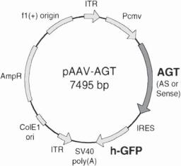
A recombinant vector with a mutant gene can be constructed as shown in Fig. 10.6. Since gene expression is a critical process for the test, it is necessary to integrate into the vector a gene promoter, which drives the gene expression. Commonly used promoters include cytomegalovirus (CMV) promoter and simian virus (SV)40 promoter, but any promoter that drives the expression of the selected gene can be used. In addition, one may integrate a reporter gene, such as a β-galactosidase gene and a green or red fluorescent protein gene, into the recombinant gene. The protein products of these reporter genes can serve as markers for the identification of the cells with positive gene transfection and expression. The constructed vector can be amplified by transforming and growing E. coli cells as described on page 432.
A functional test can be carried out in an in vitro cell culture system or an in vivo organ system. The in vitro system is easier to use and usually provides reliable information. It should be kept in mind that the endogenous wildtype gene in the testing cells may likely interfere with the transferred exogenous gene, rendering it difficult to identify the influence of the transferred exogenous gene. To resolve such a problem, a cell line with null gene mutation (complete loss of the function of a wildtype gene, also referred to as gene knockout) should be used. Such a cell line can be obtained from a transgenic animal
Figure 10.6. Schematic representation of the rat angiotensinogen (AGT) gene construct based on the adeno-associated virus (AAV)-derived plasmid. Plasmid pAAV-AGT-AS and pAAV-AGT-S were constructed by inserting a full-length cDNA (1.65 kb) of AGT into the unique Hind III site of the AAV-derived plasmid pTR-UF3 in antisense (AS) or sense (S) direction, respectively. AGT cDNA, indicated by the shaded arrow, is driven by a cytomegalovirus (CMV) early promoter (Pcmv). ITR, AAV inverted terminal repeat; IRES, polio virus type 1 internal ribosomal entry site; h-GFP, “humanized” victoria green fluorescent protein gene. Other open bars and arrows represent other basic elements of the vector. (Reprinted from Tang X et al: Am J Physiol 277:H2392–9, copyright 1999 by permission of the American Physiological Society.)