

Bioregenerative Engineering Principles and Applications - Shu Q. Liu
..pdf336 FERTILIZATION AND EARLY EMBRYONIC DEVELOPMENT
Sperm–Oocyte Fusion
After a sperm passes through the zona pellucida, it approaches and attaches to the cell membrane of the oocyte. The cell membrane of the sperm and that of the oocyte can fuse together, bringing the sperm into the oocyte cytoplasm (Fig. 7.6). Gamete membrane fusion is a process regulated by fusion proteins. While the regulatory mechanisms of fusion are not completely understood, an oocyte membrane protein, CD9 (Table 7.1), has been suggested to play a role in the mediation of gamete fusion. The knockout of the CD9 gene is associated with impaired interaction between sperm and oocyte, resulting in infertility. When CD9 mRNA is delivered into the egg with CD9 gene knockout, infertility can be reversed. In addition, a molecule found in the membrane of sperm cells, known as the Izumo immunoglobulin superfamily protein, plays a critical role in regulating sperm fusion into the oocyte. In Izumo − /− mice, the sperm cells can develop to maturity and
Granulosa cells
A B
Zona pellucida
Sperm
Nucleus
Male nucleus |
Female nucleus |
C D
Polar bodies
Centrosome
Spindle Chromosome
E
2-cell embryo
Figure 7.6. Schematic representation of sperm fusion into an oocyte. Based on bibliography 7.1.
FERTILIZATION 337
can penetrate the zona pellucida, but cannot fuse into the oocyte (Fig. 7.7). However, the exact mechanisms of Izumo action remains poorly understood.
When a sperm enters an oocyte, the oocyte immediately becomes resistant to further sperm fusion, a mechanism that prevents polyspermy. The antipolyspermy activity is controlled by a rapid shift of cell membrane potential. A sperm can fuse with an oocyte at the normal resting membrane potential (about −90 mV). Immediately after a sperm fuses into the oocyte, the normally negative resting membrane potential is reversed to a positive value (about 20 mV), induced by the opening of the sodium channels and sodium flux into the oocyte. Such a rapid change in membrane potential prevents the interaction of other sperm cells with the oocyte during the early period after a sperm fuses with the oocyte. The shift of membrane potential is a transient process that lasts only about one minute. There exists another mechanism, by which a sperm-fused oocyte prevents further sperm fusion. The oocyte cell contains a large number of cortical granules, which contain proteolytic enzymes (Fig. 7.8). The fusion of a sperm to the oocyte triggers the release of calcium, which in turn induces the exocytosis of the cortical granules, releasing the enzymes into the zona pellucida. These enzymes cleave regulatory proteins, such as zona pellucida glycoprotein 3 (ZP3; Table 7.2), which mediate sperm–oocyte interaction. Thus, sperm can no longer bind to the zona pellucida and enter the sperm-fused oocyte.
Phase |
Hoechst 33342 |
IgG
Izumo-Anti
A B
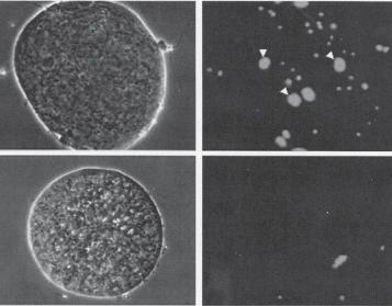
Figure 7.7. Role of the the Izumo protein in the regulation of sperm fusion to oocytes. The Izumo protein is expressed in the membrane of the sperm cell and regulates sperm fusion into eggs. (A) Izumo−/− mouse sperm cells are not able to fuse into a mouse egg compared to Izumo+/− mouse sperm cells. Note that fused sperm cells show enlarged cell nuclei. (B) Anti-human Izumo antibody (anti-hIzumo) significantly suppressed human sperm fusion into a human egg. The blue color represents cell nucei labeled with Hoechst 33342. (Reprinted by permission from Macmillan Publishers Ltd: Inoue N et al: Nature 434:234–8, copyright 2005.)

338
TABLE 7.1. Characteristics of CD9*
|
|
Amino |
|
Molecular |
|
|
|
Proteins |
Alternative Names |
Acids |
Weight (kDa) |
Expression |
|
Functions |
|
|
|
|
|
|
|
||
CD9 |
CD9 antigen, leukocyte antigen |
228 |
|
25 |
Sperm, oocyte, leukocyte, A membrane glycoprotein that interacts with |
||
|
MIC3, p24 antigen |
|
|
|
skin, intestine |
|
integrins and regulates cell adhesion, migration, |
|
|
|
|
|
|
|
and fusion |
|
|
|
|
|
|
|
|
*Based on bibliography 7.1. |
|
|
|
|
|
|
|
TABLE 7.2. Characteristics of ZP3* |
|
|
|
|
|
|
|
|
|
|
|
|
|
|
|
|
|
Amino |
Molecular |
|
|
|
|
Proteins |
Alternative Names |
Acids |
Weight (kDa) Gene Locus |
Expression |
Functions |
||
|
|
|
|
|
|
|
|
ZP3 |
Zona pellucida glycoprotein 3A, |
424 |
|
47 |
7q11.23 |
Oocyte |
A zona pellucida component that regulates |
|
sperm receptor, zona pellucida |
|
|
|
|
|
sperm–oocyte interaction and activation of |
|
protein C, zona pellucida sperm- |
|
|
|
|
fertilization processes |
|
|
binding protein 3 |
|
|
|
|
|
|
|
|
|
|
|
|
|
|
*Based on bibliography 7.1.
FERTILIZATION 339
A |
B |
C |
D |
E |
Control |
Nocodozole |
Colchicine |
Cytochalasin D |
Latrunculin A |
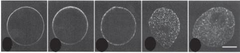
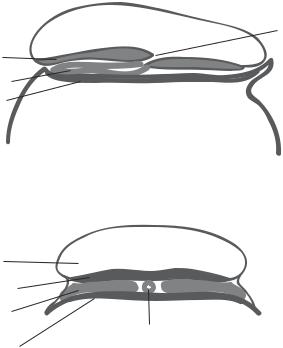
Figure 7.8. Translocation of cortical granules in oocytes. The cortical granules of oocytes are translocated to the cortical layer following germinal vesicle breakdown. This process is dependent on the organization and function of the actin filaments. A. Distribution of cortical granules in a control egg. B and C. Distribution of cortical granules in eggs treated with microtubule inhibitors nocodozole and colchicine, respectively. Note that these inhibitors did not influence the distribution of the cortical granules, suggesting that microtubules do not play a significant role in regulating the translocation of cortical granules. D and E. Distribution of cortical granules in eggs treated with actin filament inhibitors cytochalasin D and latrunculin A, respectively. Note that these inhibitors significantly influenced the distribution of the cortical granules, suggesting that actin filaments play a role in regulating the translocation of cortical granules. Reprinted from Wessel GM et al. Development 129:4315–4325, 2002 by permission of The Company of Biologists Ltd.
Activation of Embryonic Development
The fusion of a sperm cell into an oocyte triggers the activation of several important cellular activities, including DNA synthesis, restoration of mitosis, protein synthesis, and membrane synthesis. All these activities are directly or indirectly related to changes in the intracellular concentration of calcium. Since sea urchin oocytes are used extensively in the study of sperm–oocyte fusion, these cells are used here as an example. The fusion of a sperm with an oocyte, especially the binding of ZP3 to its receptor on the sperm, triggers the activation of G proteins, which further activate phospholipase C. Activated phospholipase C can cleave phosphatidylinositol biphosphate (PIP2) to form inositol triphosphate (IP3) and diacylglycerol. IP3 can act on calcium channels to induce calcium release and an increase in the level of intracellular calcium. Calcium is involved in the regulation of mitosis, DNA synthesis, protein synthesis, and membrane synthesis. Diacylglycerol can further activate protein kinase C, which is involved in the regulation of DNA and protein synthesis. These activities are essential for initiating the developmental processes of the zygote or the fertilized oocyte.
Integration of Gamete Genomes
In mammals, a sperm usually fuses with an oocyte that is arrested in the metaphase of meiosis (note that an oocyte is a developing egg before completing meiosis). The interaction of the sperm with the oocytes triggers a number of events: (1) separation of the sperm nucleus and centrioles from the flagellum, (2) degradation of the sperm mitochondria and flagellum, (3) migration of the sperm nucleus to the oocyte nucleus, (4) completion of oocyte meiosis, and (5) fusion of the sperm and oocyte genetic materials (Fig. 7.6). These
340 FERTILIZATION AND EARLY EMBRYONIC DEVELOPMENT
processes are critical to the embryonic development. Since the sperm mitochondria and microtubules are mostly degraded, the mitochondrial and microtubular systems in each individual are derived from the oocyte.
The sperm chromatids are organized and packed within the nucleus by DNA-binding proteins through disulfide bonds. When the sperm enters the oocyte cytoplasm, the oocyte is able to reduce the disulfide bonds and loose the packed chromatids. The loose sperm DNA then migrates toward the oocyte nucleus and is prepared for fusion with the oocyte DNA. At the same time, the oocyte activates its signaling pathways that stimulate the reactivation and completion of meiosis, resulting in the formation of the haploid egg nucleus (note that the oocyte has now matured to an egg). The microtubules of the egg are connected with the sperm and the egg nucleus. This process allows the migration of both nuclei toward each other. The interaction of the nuclei induces the integration of the genetic materials.
CLEAVAGE [7.2]
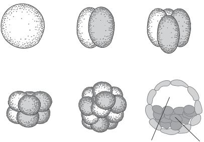
Cleavage is a process of zygote separation, which is accomplished through two events: mitotic division of the nucleus (karyokinesis) and cytoplasmic division (cytokinesis). The mitotic division takes place immediately following fertilization. Cytokinesis occurs subsequently. These events generate individual nucleated cells, known as blastomeres. The zygote is cleaved into two cells, which are subsequently cleaved into four cells, and so on, although the mammalian blastomeres may not cleave symmetrically and some cells may skip a round of cleavage (Fig. 7.9). The zygote moves along the oviduct during cleavage. The first cleavage process occurs approximately at the end portion of the oviduct. With further cleavage, the embryo moves to the uterus and is ready for implantation (Fig. 7.4).
Blastomere cleavage represents the most rapid process of cell division through the entire lifespan, including the embryonic period and adulthood. A Drosophila egg, for example, can be cleaved to generate about 50,000 blastomeres within about 12 h. The cleavage of the egg is initiated by a molecule known as mitosis-promoting factor (MPF). The fertilization process induces the activation of MPF, which stimulates the fertilized egg to enter the mitosis cycle. The mitotic process during the early cleavage undergoes only two phases: DNA synthesis (S) and mitosis (M). During the periodic cleavage cycles, the level of MPF changes cyclically: it reaches the maximal level during the M phase and reduces to the minimal level during the S phase. The periodic change in the MPF level controls the cyclically procession of cell division.
FORMATION OF THE BLASTOCYST [7.2]
Blastocyst is an embryonic structure composed of an inner cell mass of about 30 stem cells, which can differentiate into all specified cell types, and an external sac known as trophoblast, which develops into an embryo-supporting tissue called chorion (the external layer of the extraembryonic membrane and the embryonic part of the placenta) (see chapter-opening figure). Since the inner cell mass and the trophoblast serve as the origins of distinct embryonic and extraembryonic tissues, the formation of blastocyst is considered a milestone of embryogenesis.
GASTRULATION 341
Fertilized |
2-cell stage |
4-cell stage |
|
egg |
|||
|
|
8-cell stage |
32-cell stage |
Blastocyst |
Blastocoel |
Inner cell mass |
Figure 7.9. Schematic demonstration of developmental stages. Based on bibliography 7.1.
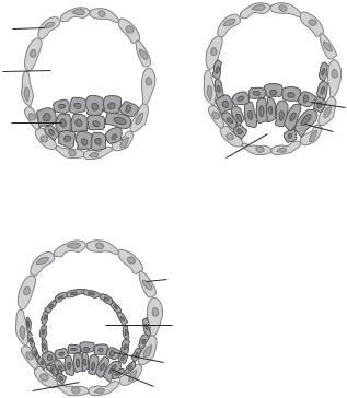
There are several key steps for the formation of the blastocyst: zygote cleavage, formation of morula, and cavitation of the morula (Fig. 7.9). Zygote cleavage has been discussed above. When cells are cleaved to a stage of 16 cells at about day 4–5, all cells are highly compacted into a solid ball-like structure. This structure is defined as a morula. With further cell divisions, the external cells of the morula transform into trophoblast cells at about day 6–7, while the internal cells transform into cells of the inner cell mass (Fig. 7.10). The inner cell mass further develops into two layers: the epiblast and the hypoblast. These layers eventually develop into embryonic and extraembryonic tissues as discussed below.
GASTRULATION [7.2]
Gastrulation is a process by which early embryonic cells are organized into three distinct layers: ectoderm, mesoderm, and endoderm (Fig. 7.11). These layers eventually develop into specified tissues and organs for the new individual (see page 346 for details). When cell cleavage generates a sufficient number of cells, the embryonic cells are organized into a blastocyst, composed of the inner cell mass and trophoblast. The inner cell mass gives rise to epiblast and hypoblast. The former develops into the embryonic epiblast and amnionic ectoderm, and the latter develops into extraembryonic endoderm. The embryonic epiblast develops into the embryonic ectoderm and primitive streak. The primitive streak gives rise to embryonic endoderm and mesoderm. The amnionic ectoderm gives rise to the amnionic sac. The hypoblast-derived extraembryonic endoderm develops into the yolk sac. The trophoblast develops into extraembryonic supporting structures.
342 FERTILIZATION AND EARLY EMBRYONIC DEVELOPMENT
7 days |
9 days |
Trophoblast |
|
Blastocoel |
|
|
Hypoblast |
Inner cell mass |
Epiblast |
|
|
|
Amniotic cavity |
11 days |
|
|
Trophoblast |
|
Yolk sac |
|
Hypoblast |
Amniotic cavity |
Epiblast |
Figure 7.10. Schematic demonstration of the formation of the inner cell mass and the epiblast and hypoblast layers. Based on bibliography 7.2.
The extraembryonic supporting structures include the chorion, amnionic sac or amnion, and yolk sac. The chorion is the outmost extraembryonic membrane developed from the trophoblast of the blastocyst. The chorionic membrane is composed of chorionic cells, a vascular system, and connective tissue. A region of the chorion gives rise to the embryonic placenta, which is composed of two types of cellular structure called cytotrophoblast and synctiotrophoblast. The cells of these structures can interact with the maternal uterine epithelial cells via adhesion molecules, which assist in the attachment and implantation of the blastocyst into the uterine wall. Furthermore, these cells produce and release enzymes that degrade the uterine tissue, facilitating the invasion of the blastocyst into the uterine wall. Such a uterine degradation process also reorganizes the uterine vascular system in favor of the transport of nutrients and oxygen from the maternal blood to the embryo and fetus. The cytotrophoblast and synctiotrophoblast eventually develop into placenta structures including chorionic villi and intervillus space. The chorionic villi contain embryonic/fetal arteries, capillaries, and veins, which absorb and transport nutrients and oxygen from the maternal placenta to the embryo/fetus.
The amnionic sac is a tough extraembryonic membrane that lines the internal surface of the chorion and, when fully developed, encloses the fetus. The amnionic sac is developed from the inner cell mass-derived epiblast. During the early embryonic stage, the amnionic sac is located between the chorionic embryonic placenta and the ectoderm. It
Anterior
Amniotic cavity
Ectoderm
Axial mesoderm
Endoderm
Median section (anterior to posterior)
Amniotic cavity
Ectoderm
Paraaxial
mesoderm
Axial mesoderm
Endoderm
Transverse section
BIBLIOGRAPHY 343
Posterior
Primitive pit and
Hensen's node
Figure 7.11. Formation of the three embryonic layers. Based on bibliography 7.2.
folds and extends gradually around the embryo, and eventually lines the chorionic membrane and encloses the embryo/fetus. The amnionic cavity is filled with a fluid called the amnionic fluid, in which the embryo/fetus resides. Together with the chorionic membrane, the amnionic membrane mediates the interaction of the embryo/fetus with the mother and protects the embryo/fetus from harmful environment. The yolk sac is a membrane structure that stores nutrients in the yolk cavity for the early development of the embryo. This structure is developed from the inner cell mass-derived hypoblast, and is located near the endoderm. The yolk cavity is relative large during the early embryonic stage, but is reduced in size and is gradually diminished during embryogenesis.
BIBLIOGRAPHY
7.1. Sperm, Egg, and Fertilization
Gilbert Scott F: Developmental Biology, 7th edn, Sinauer Associates, Sunderland, UK, 2003.
Inoue N, Isotani A, Okabe M: The immunoglobulin superfamily protein Izumo is required for sperm to fuse with eggs, Nature 434:234–8, 2005.
344 FERTILIZATION AND EARLY EMBRYONIC DEVELOPMENT
Gutierrez-Lopez MD, Ovalle S, Yanez-Mo M, Sanchez-Sanchez N, Rubinstein E et al: A functionally relevant conformational epitope on the CD9 tetraspanin depends on the association with activated beta1 integrin, J Biol Chem 278:208–18, 2003.
Zhu GZ, Miller BJ, Boucheix C, Rubinstein E, Liu CC et al: Residues SFQ (173–175) in the large extracellular loop of CD9 are required for gamete fusion, Development 129:1995–2002, 2002.
Benoit P, Gross MS, Frachet P, Frezal J, Uzan G et al: Assignment of the human CD9 gene to chromosome 12 (region p13) by use of human specific DNA probes, Hum Genet 86:268–72, 1991.
Kaji K, Oda S, Shikano T, Ohnuki T, Uematsu Y et al: The gamete fusion process is defective in eggs of Cd9-deficient mice, Nature Genet 24:279–82, 2000.
Le Naour F, Rubinstein E, Jasmin C, Prenant M, Boucheix C: Severely reduced female fertility in CD9-deficient mice, Science 287:319–21, 2000.
Miyado K, Yamada G, Yamada S, Hasuwa H, Nakamura Y et al: Requirement of CD9 on the egg plasma membrane for fertilization, Science 287:321–4, 2000.
Tachibana I, Hemler ME: Role of transmembrane 4 superfamily (TM4SF) proteins CD9 and CD81 in muscle cell fusion and myotube maintenance, J Cell Biol 146:893–904, 1999.
Waterhouse R, Ha C, Dveksler GS: Murine CD9 is the receptor for pregnancy-specific glycoprotein 17, J Exp Med 195:277–82, 2002.
Dean J: Biology of mammalian fertilization: Role of the zona pellucida, J Clin Invest 89:1055–9, 1992.
Lunsford RD, Jenkins NA, Kozak CA, Liang LF, Silan CM et al: Genomic mapping of murine Zp-2 and Zp-3, two oocyte-specific loci encoding zona pellucida proteins, Genomics 6:184–7, 1990.
van Duin M, Polman JEM, Suikerbuijk RF, Geurts van Kessel AHM, Olijve W: The human gene for the zona pellucida glycoprotein ZP3 and a second polymorphic locus are located on chromosome 7, Cytogenet Cell Genet 63:111–3, 1993.
van Duin M, Polman JEM, Verkoelen CCEH, Bunschoten H, Meyerink JH et al: Cloning and characterization of the human sperm receptor ligand ZP3: Evidence for a second polymorphic allele with a different frequency in the Caucasian and Japanese populations, Genomics 14:1064– 70, 1992.
Zhao M, Gold L, Ginsberg AM, Liang LF, Dean J: Conserved furin cleavage site not essential for secretion and integration of ZP3 into the extracellular egg coat of transgenic mice, Mol Cell Biol 22:3111–20, 2002.
7.2. Cleavage, Formation of Blastocyst, and Gastrulation
Wassarman PM: Contribution of mouse egg zona pellucida glycoproteins to gamete recognition during fertilization, J Cell Physiol 204:388–91, 2005.
Shur BD, Ensslin MA, Rodeheffer C: SED1 function during mammalian sperm-egg adhesion, Curr Opin Cell Biol 16:477–85, 2004.
Santella L, Lim D, Moccia F: Calcium and fertilization: The beginning of life, Trends Biochem Sci 29:400–8, 2004.
Shur BD, Rodeheffer C, Ensslin MA: Mammalian fertilization, Curr Biol 14:R691–2, 2004.
Klonisch T, Fowler PA, Hombach-Klonisch S: Molecular and genetic regulation of testis descent and external genitalia development, Dev Biol 270:1–18, 2004.
Dean J: Reassessing the molecular biology of sperm-egg recognition with mouse genetics, Bioessays 26:29–38, 2004.
Lenart P, Ellenberg J: Nuclear envelope dynamics in oocytes: From germinal vesicle breakdown to mitosis, Curr Opin Cell Biol 15:88–95, 2003.
BIBLIOGRAPHY 345
Evans JP, Florman HM: The state of the union: The cell biology of fertilization, Nat Cell Biol 4 (Suppl):s57–63, 2002.
Primakoff P, Myles DG: Penetration, adhesion, and fusion in mammalian sperm-egg interaction, Science 296:2183–5, 2002.
Matzuk MM, Burns KH, Viveiros MM, Eppig JJ: Intercellular communication in the mammalian ovary: oocytes carry the conversation, Science 296:2178–80, 2002.
Runft LL, Jaffe LA, Mehlmann LM: Egg activation at fertilization: Where it all begins, Dev Biol 245:237–54, 2002.
Wassarman PM, Jovine L, Litscher ES: A profile of fertilization in mammals, Nat Cell Biol 3: E59–64, 2001.
Wassarman PM: Mammalian fertilization: molecular aspects of gamete adhesion, exocytosis, and fusion, Cell 96:175–83, 1999.
Snell WJ, White JM: The molecules of mammalian fertilization, Cell 85:629–37, 1996.
Wassarman PM: Towards molecular mechanisms for gamete adhesion and fusion during mammalian fertilization, Curr Opin Cell Biol 7:658–64, 1995.
Nothias JY, Majumder S, Kaneko KJ, DePamphilis ML: Regulation of gene expression at the beginning of mammalian development, J Biol Chem 270:22077–80, 1995.
Schultz RM, Kopf GS: Molecular basis of mammalian egg activation, Curr Top Dev Biol 30:21–62, 1995.