

Bioregenerative Engineering Principles and Applications - Shu Q. Liu
..pdf436 MOLECULAR ASPECTS OF BIOREGENERATIVE ENGINEERING
model without the gene of interest. Since the function of the selected wildtype gene is completely deficient, the function of the transferred recombinant gene can be assessed by comparing to a control with the function of the corresponding wildtype gene.
The next step for testing the gene function is to transfer the gene construct into cultured cells. It is a natural property that cells can endocytose large molecules, including DNA, into the cytoplasm. A fraction of endocytosed DNA can go through the nucleus membrane and integrate into the genome. Several methods, including virus-, salt-, liposome-, recep- tor-, and electroporation-mediated gene transfer, have been established and used to facilitate gene transfection into mammalian cells. The exogenous gene can be expressed transiently, producing encoded protein. The function of the transferred gene can be tested in several aspects, including the expression and function of the encoded protein as well as the activity of the host cells. Tests can be conducted at selected times after 12 hs from gene transfer. Protein expression can be detected by Western blotting or immunoblotting, which is a sensitive method for detecting a small amount of protein. A comparison to a sample from control cells with the corresponding wildtype gene demonstrates alterations in the mutant protein in terms of molecular weight and expression level. A functional test can be designed according to the nature of the protein. If the protein of interest is an enzyme, the catalytic activity of the protein can be assessed by detecting changes in the substrate protein. In vitro tests can be carried out by using an isolated enzyme and substrates. The influence of the transferred gene on the host cells can be assessed by measuring changes in a cellular activity known to be related to the transferred gene.
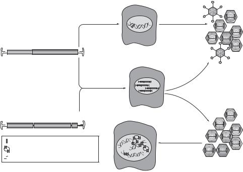
Constructing a Recombinant Therapeutic Gene. Once the pathological role of a mutant gene is identified, the next step is to design and construct a therapeutic gene that can be used to restore the physiological function of the gene (Fig. 10.7). Methods described on page 429 can be used for this purpose. Recall that there exist several types of genetic disorders, including chromosomal defects, simple inherited genetic defects, and multifactorial defects. Different strategies should be used for these types of genetic disorder. For chromosomal defects, since a large amount of DNA is involved, it is difficult to restore the structure and function of the defect chromosomes. For a simple inherited genetic defect, sine a single gene is usually involved, a corresponding wildtype gene may be isolated, cloned, and used to construct a therapeutic gene, which can be used for correcting the defect gene. For multifactorial defects, it is necessary to identify the genes involved and determine the pathogenic mechanisms of the disorder, based on which therapeutic genes can be constructed to modulate the activity of the mutant genes. For instance, the upregulation of multiple growth factor genes may contribute to the initiation and development of atherosclerosis. Growth inhibitor genes can be constructed and transferred into vascular cells to inhibit the activity of growth factors and thus to suppress atherogenesis.
Transfection of Target Cells with a Therapeutic Gene. The primary goal of molecular engineering is to correct pathological disorders due to gene mutation or changes in gene activities. An effective approach is to transfer therapeutic genes, as constructed on page 429, into target cells to restore the structure and function of the mutant or altered genes. Such an approach is referred to as gene therapy. The transferred genes, once in the cytoplasm, can be transported to the cell nucleus, integrated into the genome, and expressed to produce corresponding proteins. It is expected that the newly expressed proteins can function in place of the corresponding mutant proteins, thus reducing or suppressing

DNA ENGINEERING |
437 |
Therapeutic gene 

Plasmid vector 
Antibiotic  resistance gene
Recombinant
DNA
E. coli transfection
Selection
Amplification of recombinant gene
Figure 10.7. Construction of a therapeutic gene vector. Based on bibliography 10.2.
mutant protein-induced pathological disorders. Investigations for the past decade have provided tremendous evidence for the potential application of gene therapy to human disorders.
There are two general approaches that can be potentially applied to human gene therapy: embryonic gene transfer and somatic gene transfer. For embryonic gene transfer, a blastocyst is collected from a pregnant animal, and a therapeutic gene is injected into
438 MOLECULAR ASPECTS OF BIOREGENERATIVE ENGINEERING
the cells of the blastocyst to induce site-specific gene integration or homologous gene recombination. Embryonic cells from the blastocyst are stem cells. Once these stem cells carry the therapeutic gene, the gene can be passed to differentiated cells. Gametes may also carry the therapeutic gene and pass it to next generations. The transferred gene may function in place of the mutant gene. This is potentially an effective approach for the correction of pathological disorders due to hereditary gene defect or mutation. However, procedures for blastocyst collection induce cell injury and death. A safe, reliable, and effective technique has not been established for the treatment of hereditary human disorders. A technically similar technique has been used to modulate or remove selected genes in the embryonic stem cells of animals for the establishment of transgenic models. These models have been used extensively in scientific research.
Somatic gene transfer is an approach used for transferring therapeutic genes into the somatic cells of humans and animals after birth. Obviously, it is impossible to transfer a gene into all somatic cells in the body. Thus, this approach is used for gene transfer into selected target cells. Although mammalian cells are capable of taking up DNA, a process known as endocytosis or “naked gene transfer,” the rate of DNA taking up is very low. A number of gene transfer-mediation methods, including virus-, liposome-, receptor-, elec- troporation-, salt-mediated gene transfer, have been established to facilitate gene transfer into mammalian cells. Each of these gene transfer strategies has strengths and weaknesses. The therapeutic efficacy and effectiveness differ between these mediation methods. The viral approach usually results in a higher rate of gene transfection and longer period of gene expression than do the nonviral approaches. However, viral carriers may cause infectious disorders and gene mutation in transfected cells. In contrast, the nonviral approaches are safe and easy to carry out, and exhibit low immunogenicity. However, their efficacy is relatively low and the expression duration of the transferred gene is short compared to the viral approach.
The choice of a gene transfer approach may also be dependent on the structure, function, and accessibility of the target tissue or organ. For instance, electroporation may be applied to the skin and skeletal muscle cells for gene transfer, but not to the heart and brain, because these organs are difficult to access and electroporation causes cell injury and death. In contrast, viral gene carriers and liposome can be used for gene transfer into target cells in most types of tissues and organs. Furthermore, various cell types may possess different capabilities of accepting foreign genes, and the cell state may also influence the rate of gene transfer for a given type of gene transfer mediation approach. For instance, retroviral gene carriers can infect dividing cells, but not nondividing cells, whereas adenoviral gene carriers can infect both dividing and nondividing cells. However, the retroviral approach may induce more stable gene expression than the adenoviral approach. Thus, all possible factors should be taken into account for the choice of a gene transfer approach. Here, several common gene transfer mediating approaches are briefly discussed.
Virus-Mediated Gene Transfer. Several types of virus, including retrovirus, adenovirus, adeno-associated virus, herpes simplex virus, have been used as gene carriers for mediating gene transfer into mammalian cells. The basis for virus-mediated gene transfer is that viruses are capable of infecting mammalian cells, carrying therapeutic genes, and integrating the carried genes into the host cell genome. Viruses can be modified or genetically engineered to remove harmful components and accommodate desired therapeutic gene fragments. Once transferred into the cell, viruses are able to integrate their genome into
DNA ENGINEERING |
439 |
the host genome and use the synthetic machineries of the host cells to produce viral DNA, RNA, and proteins. Thus, viruses are natural gene transfer carries. To be used for gene transfer mediation, a viral gene carrier ought to be replication-deficient, nonimmunogenic, and nontoxic, but are able to facilitate gene transfer to the genome of a target cell. The choice of a viral carrier for gene transfer is dependent on the nature of the virus and the state of the target cells, as we will see in the following sections.
RETROVIRUS-MEDIATED GENE TRANSFER. Retroviruses belong to a family of RNA viruses. A typical retrovirus contains two parts: the viral core and the envelope. The viral core is composed of two identical RNA strands and several enzymes, including reverse transcriptase, protease, and integrase. The envelope, composed of a membrane and glycoproteins, encloses the viral core. Common retroviruses used for gene transfer include Moloney virus and lentivirus. The Moloney virus is a murine leukemia virus that causes lymphoid leukemia in mice. The lentivirus is a retroviral strain that causes maedi (a chronic pulmonary disease found in Iceland) and visna (a disease affecting the central nervous system and causing paralysis) in sheep.
Retroviruses can interact with mammalian cells through cell membrane receptors, and enter the cell through receptor mediation. In the cytoplasm, the viral RNA genome can be converted into DNA by the reverse transcriptase. The converted DNA can be integrated into the host genome and replicate together with the host DNA during cell division. Thus, genetic information of the virus can be transmitted to the genome of the host cell and carried to the next generation. Any foreign genes that are inserted into the retroviral genome can be integrated into the host genome. The virus itself can produce viral proteins based on the viral mRNA and reproduce the same type of virus.
The RNA of the retrovirus can be engineered through a packaging process, generating viral vectors. Briefly, retroviral particles can be modulated to remove a selected RNA sequence known as the packaging signal, which is responsible for packaging the RNA core into the viral envelope. When transfected into a host cell line, the viral particles can express functional genes, such as env, gag, and pol. The env and pol genes encode viral envelope proteins, whereas the gag gene encodes the reverse transcriptase. While the virus is able to convert its RNA to DNA and produce necessary proteins for the construction of the viral envelope, it cannot pack the core RNA into the envelope. At the same time, another group of viral particles is selected and engineered to remove selectively the harmful sequences without modulating the packaging signal. A therapeutic gene fragment can be inserted into the viral genome to generate a recombinant viral gene carrier. When the recombinant viral gene carrier is transfected into the same host cell line containing the empty viral envelopes, the recombinant gene carrier can be packed into the empty envelopes under the action of the packaging signal, thus generating a complete viral structure that contains the therapeutic gene without the harmful gene sequences. This engineered viral structure is harmless and nonreplicable, and can be used to for gene transfer. Retrovirus-derived gene carriers have been used in experimental models and clinical trials.
There are several advantages for retrovirus-derived gene carriers. These carriers can induce a high rate of gene integration into the host cell genome with relatively stable and long-term expression (several months). However, retroviral gene carriers exhibit several disadvantages, including toxicity and limitation of effectiveness to only dividing cells. In particular, retroviral vectors may be potentially tumorigenic. The integration of a viral gene into the host cell genome may induce mutation of tumor suppressor genes or
440 MOLECULAR ASPECTS OF BIOREGENERATIVE ENGINEERING
oncogenes, potentially leading to tumorigenesis. Furthermore, viral genes can be expressed to generate viral proteins, which potentially cause host cell immune reactions, a potential factor that reduces the stability of the transfected gene and the duration of gene expression.
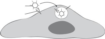
ADNOVIRUS-MEDIATED GENE TRANSFER. Adenoviruses are double-stranded DNA viruses and belong to a family composed of more than 30 serotypes. Many of the serotypes invade human cells and cause infectious disorders of the upper respiratory tract, known as cold or flu, and conjunctivae. Others infect the simian, bovine, canine, avian, and murine species. The mechanisms by which the adenovirus enters mammalian cells and causes infection remain poorly understood. It has been hypothesized that cell membrane receptors specific to adenoviral proteins may mediate the invasion of viruses (Fig. 10.8).
The genome of adenoviruses can be modified to remove replication control sequences and thus to prevent adenoviral replication and to eliminate toxic influence. The DNA genome of a typical adenovirus contains several regions, including E1 to E4. The E1 region is responsible for viral replication. The replacement of the E1 region with a foreign gene results in the generation of replication-defective viruses. Such engineered adenoviruses can be used as a gene transfer carrier. There are several advantages for the use of adenoviruses in gene transfer. First, the adenoviral gene carrier can induce a relatively high efficiency of gene transfection compared to a retroviral carrier. Second, an adenoviral carrier can infect both dividing and non-dividing cells. Third, an adenoviral vector can carry large DNA inserts [≤8 kb (kilobases)]. However, gene transfer mediated by the adenoviral vector often results in a relatively low rate of gene integration into the host cell genome and a short duration of gene expression (about several weeks). Although the toxicity of adenoviruses is lower than that of retroviruses, the viral particles can still induce infectious disorders in the host cells. Furthermore, the expressed viral proteins can induce host immune reactions, a potential factor that reduces the stability of the transfected gene and the duration of gene expression.
ADENO-ASSOCIATED VIRUS AS A GENE CARRIER. Adeno-associated virus belongs to a family of nonpathogenic human parvoviruses with a single-stranded DNA genome. This viral type is smaller than other types of viruses and cannot replicate unless associated with helper viruses, usually adenoviruses or herpesviruses. In the absence of helper viruses, the adeno-associated virus usually integrates its genome into the host chromosomal DNA and enters a latent state. Adeno-associated viruses can infect dividing as well as nondividing cells, and cause stable and long-term gene expression. Compared to the
Adenovirus |
Endosome |
Receptor
Cell
Figure 10.8. Schematic representation of adenovirus-mediated gene transfection.
DNA ENGINEERING |
441 |
adenovirus, the adeno-associated virus exhibits reduced immunogenicity and toxicity, since this virus consists of fewer genes that encode proteins harmful to the host cells. In a typical adeno-associated viral vector, the only wildtype sequences left are the inverted terminal repeats (ITRs) that flank the inserted gene cassette. These sequences are necessary for the packaging of the viral genome. Because of the lack of the replication capability, it is not necessary to modulate the genome of the adeno-associated virus for gene transfer. A desired therapeutic gene can be directly inserted into the viral genome and the viral vector can be used for gene transfer (Fig. 10.9). This type of virus has been extensively used for gene transfer in experimental models. However, adeno-associated viral vectors can accommodate relatively small gene inserts because of its limited genome size.
Rep-mediated, site- |
+ Ad or HSV |
specific integration |
|
|
Rescue and |
|
lytic infection |
(A) Wild-type virus
rep |
cap |
ITR |
|
|
Co-infection with |
Ad and AAV |
|
Ad or HSV |
||
particles |
||
|
||
Viral replication |
|
|
Vector production |
Cell lysis |
|
+Ad E1, E2, E4orf6 |
|
|
Rep and Cap |
|
(B) Recombinant expression vector
Promoter |
Transgene |
pA |
ITR |
AAV integration in chromosome 19 |
|||
Circular rAAV episomes |
|
Transduction |
|


 Randomly integrated rAAV
rAAV particles
Figure 10.9. Adeno-associated virus as a vector for gene transfection. (A) The structure of the wildtype adeno-associated virus (AAV) is shown. A single-stranded DNA genome is encompassed by palindromic inverted terminal repeats (ITRs). The rep open reading frame (ORF) encodes proteins that are involved in viral replication, and the cap ORF encodes proteins that are necessary for viral packaging. AAV integrates into the human genome at a specific locus on chromosome 19 (red) and persists in a latent form. It can exit this stage only if the cell is coor superinfected with helper virus such as adenovirus (Ad) or herpes simplex virus (HSV), which provide factors necessary for active AAV replication. (B) The generic genedelivery vector based on AAV is depicted. The viral genome is replaced by an expression cassette, which usually consists of a promoter, transgene, and polyA (pA) tail. For production of the recombinant virus (rAAV), Rep and Cap proteins as well as Ad or HSV elements (Ad E1, E2 and E4, or f6) have to be provided in trans. Examples of intracellular forms of the delivery vector that are responsible for transgene expression following transduction with rAAV (double-stranded circular episomes and randomly integrated vector genomes) are depicted in red. (Reprinted by permission from Macmillan Publishers Ltd.: Vasileva A, Jessberger R: Nature Rev Microbiol 3:837–47, copyright 2005.)

442 MOLECULAR ASPECTS OF BIOREGENERATIVE ENGINEERING
The efficiency of gene transfer mediated by adeno-associated viruses is usually lower than that mediated by adenoviruses.
HERPES SIMPLEX VIRUS-MEDIATED GENE TRANSFER. Herpes simplex virus belongs to a family of double-stranded DNA viruses that cause herpes simplex in humans. The virus can also be found in other mammalian species, such as canine, bovine, and simian. The virus can invade mammalian cells, integrate its DNA into the host genome, and mature in the host nucleus. This type of virus has been used for mediating gene transfer in experimental models. Herpes simplex virus-derived gene vectors can carry large gene fragments, infect dividing as well as non-dividing cells, and induce efficient gene transfection. However, the duration of gene expression mediated by herpes simplex viruses is often shorter (about one week) than that mediated by retroviral and adenoviral vectors. A possible reason is that herpes simplex viruses induce cytotoxic and/or immune reactions in the host cells, which shut down the expression of the transfected gene. A herpes simplex viral vector lacking the harmful genes, such as the immediately early genes, exhibits much reduced toxicity, improved gene transfection efficacy, and prolonged gene expression, as demonstrated in experimental models.
Receptor-Mediated Gene Transfer. Certain types of cell membrane receptors can be used to mediate gene transfer. A typical example is the transferrin receptor, which interacts with transferrin. The transferrin molecules can spontaneously link to a polymeric linker, such as polylysine, which can also link to DNA fragments at different sites, forming DNA–polylysine–transferrin complexes. These complexes can adhere to the cell membrane through the interaction of transferrin with the transferrin receptor (Fig. 10.10). Such an interaction activates the endocytosis mechanism of the target cells, which in turn take up the triplet DNA complexes. The transferred DNA complexes can be transported from the cytoplasm to the nucleus via endosomes.
Although the receptor-mediated gene transfer approach has been successfully used to transfer genes into mammalian cells, the transfer efficiency is low because most DNA molecules are trapped in the endosomes and cannot be released before degradation. To reduce DNA degradation and facilitate gene transport, a codelivery of replicationdefective adenoviruses can improve the efficiency of receptor-mediated gene transfer. The mechanism for this enhancement is that endocytosed adenoviruses can disrupt the endosome membrane. Thus, the cotransferred triplet DNA complexes can be released. In this preparation, the DNA fragments are not inserted into the genome of the adenovirus. The virus is merely used as a gene transfer helper.
|
Protein ligand |
DNA |
Endosome |
|
|
Polylysine |
|
|
Receptor |
|
Cell |
Figure 10.10. Schematic representation of receptor-mediated gene transfection.
444 MOLECULAR ASPECTS OF BIOREGENERATIVE ENGINEERING
phosphate is probably the safest substance among those used for mediating gene transfer and (2) calcium phosphate is easy to prepare and use. The only procedures involved in gene transfer are preparation of a calcium phosphate buffer supplemented with DNA and delivery of the DNA mix to target cells. However, the efficacy of calcium phosphatemediated gene transfer is generally lower than that mediated by viral vectors and liposomes.
Electroporation-Mediated Gene Transfer. Electroporation is electrophysical process that induces the formation of hydrophilic pores in the cell membrane under the influence of an electric field. When cells are subject to an electric field between two electrodes, an electric pulse of appropriate voltage can induce the formation of pores in the cell lipid membrane. The rate of pore formation and the size of the pores are dependent on the maximal voltage across the cell membrane. The higher is the voltage, the higher is the rate of pore formation and the larger is the pore size. The duration of the pore opening is proportional to the duration of the electric pulse delivered to cells. The duration of electrical pulse may also affect the pore size. The longer is the duration of the electrical pulse, the larger is the pore size. Pores about 10 nm in diameter can be achieved in mammalian cell membranes by selecting an appropriate voltage. Short pulses usually result in a uniform population of small pores with a short duration of pore opening.
The process of electroporation is transient and reversible. After the termination of an electric pulse, the cell membrane pores shrink gradually and sealed eventually within several minutes. During the period of pore opening, DNA fragments present in the buffer can enter the cytoplasm and migrate into the cell nucleus. There are several possible mechanisms for the entrance of DNA fragments into the cytoplasm: (1) DNA fragments may directly move into the cytoplasm through the cell membrane pores, (2) DNA may be transported into the cytoplasm by osmotic forces established with respect to the concentration gradient of the DNA molecules, (3) electroporation may enhance endocytosis of the DNA fragments attached to the cell membrane, and electroporation may impose electrophoretic influence on the DNA fragments, enhancing DNA transport into the cytoplasm.
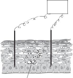
Electroporation is a highly efficient approach for gene transfer and has been extensively used in gene transfer for cultured cells. It has also been used for in vivo gene transfer into cells in accessible tissues and organs, such as the skin and skeletal muscle (Fig. 10.12). The efficacy of gene transfer mediated by electroporation is usually higher than that mediated by liposome and calcium phosphate, but lower than that mediated by viral vectors. A significant drawback for electroporation is cell injury induced by electrical pulses. In cell culture models, a standard electrical pulse (1000 V/cm in voltage), which causes effective gene transfer, may induce injury or death in about 30–50% of cells. Electroporation also causes cell injury and death for in vivo gene transfer. Thus, the voltage level for electroporation should be optimized to minimize cell injury through trial and error experiments for each specific type of cell, tissue, and organ.
Gene Gun-Mediated Gene Transfer. This is approach mediated by gene particle bombardment. Microparticles coated with the gene of interest can be accelerated by a force (e.g., a pressure) to penetrate through the cell membrane and thus to deliver the gene into the cytoplasm or the cell nucleus. The force used for gene transfer can be controlled to achieve an appropriate distance of penetration. This technique is easy to use and is efficient compared to other gene transfer-mediating approaches. However, bombardment induces cell injury and death.
DNA ENGINEERING |
445 |
Pulser
+ _
Electrode
Epidermis
Therapeutic genecontaining plasmids
Figure 10.12. Schematic representation of electroporation-mediated gene transfection.
Assessing the Expression of the Transfected Gene. One of the most important steps in molecular engineering is to assess the expression of the transferred gene. Reporter genes are often used for such a purpose. Reporter genes are recombinant genes that encode proteins exhibiting special signs for visualization and thus can be used as markers for assessing the level of gene expression. Major criteria for the construction of a reporter gene are that the genes or expressed proteins are nontoxic, do not influence the function of the host cell, can be identified, and are not expressed in the target cell. Several types of reporter genes have been established and used for gene transfer. These include the β- galactosidase (gal) gene, luciferase gene, chloramphenicol acetyltransferase (CAT) gene, and fluorescent protein genes.
The β-galactosidase gene encodes a protein enzyme known as galactosidase and is found in E. coli cells. β-Galactosidase is a hydrolase that catalyzes the hydrolysis of the terminal residues of β-galactoside, forming d-galactose. d-galactose can react with x-gal, a chemical compound, to form a blue-colored substance. This feature has been used for detecting the efficiency of gene transfer. The presence of the blue color in cells transfected with the β-gal gene indicates the expression of this gene. β-galactosidase is not expressed in mammalian cells and nontoxic to the host cells. It is commonly used as a reporter gene in experiments of gene transfer. The β-galactosidase gene can be cotransferred with a therapeutic gene into target cells. It is assumed that the expression of the β-galactosidase gene indicates the expression of the cotransferred therapeutic gene. Alternatively, the β- galactosidase gene can be inserted into a recombinant vector that carries a therapeutic gene. The expression of the β-galactosidase gene is usually associated with the expression of the therapeutic gene. The second approach is preferable for detecting the efficiency of gene transfer.