

Bioregenerative Engineering Principles and Applications - Shu Q. Liu
..pdf226 CELL SIGNALING PATHWAYS AND MECHANISMS
Signaling Mechanisms [5.15]
The NFκB signaling pathway can be activated by several factors, including physical stress, oxidative stress, chemical toxins, and bacterial and viral infection. These factors can interact with cells and activate NFκB, which in turn stimulates the expression of many inflammatory cytokines, chemokines, immune receptors, and cell surface adhesion molecules. Thus, NFκB has been considered as a central mediator for immune responses as well as stress responses induced by physical stress, oxidative stress, and chemical substances. For instance, NFκB can be activated in response to the stimulation of IL1. The binding of IL1 to the IL1 receptor activates a molecule known as NFκB-inducing kinase (NIK or serine/threonine protein kinase NIK), which in turn phosphorylates the IκB kinase. Activated IκB kinase can phosphorylate the IκB module of the IκB-NFκB complex, inducing the separation of NFκB from IκB. NFκB is a complex of p50 and p65 proetins, which serves as a transcriptional factor and regulates gene transcription. Figure 5.16 shows the mechanisms of NFκB activation in response to the stimulation of cytokines.
In addition, extracellular mitogens and growth factors can activate NFκB, which in turn regulates cell survival and proliferation. The activation of NFκB is mediated by a protein kinase known as IκB kinase (IKK), which is composed of at least three subunits: two catalytic subunits (IKKα and IKKβ) and a regulatory subunit (IKKγ). Extracellular mitogens and growth factors can activate IKK via various signaling pathways. For instance, protein kinases MEKK1, MEKK2, and MEKK3, which are activated through receptor protein tyrosine kinase signaling pathways, are capable of phosphorylating IKK. Activated IKK in turn phosphorylates two specific serine residuals on IκB. The phosphorylation of IκB leads to the ubiquitination and degradation of IκB by proteasome. The degradation of IκB liberates NFκB, which translocates to the cell nucleus, binds to target genes, and regulates gene transcription, resulting in the activation of inflammatory and mitogenic activities.
In addition to the degradation and removal of IκB, serine phosphorylation of NFκB may be required for certain NFκB family members for efficient binding to transcriptional activators and interaction with target genes. For instance, the catalytic domain of protein kinase A can bind to an inactive NFκB p65 protein in unstimulated cells. On IκB degradation, protein kinase A phosphorylates p65 on serine 276, resulting in a conformational change in the p65 protein and consequent interaction with a transcriptional activator, namely CBP (CREB-binding protein or cAMP response element-binding protein-binding protein), which increases the transcriptional activity of NFκB. Another example involves the activation of protein kinases PI3K and Atk (agammaglobulinemia tyrosine kinase), which mediate IL1and TNFα-initiated NFκB activation. In cultured cells, IL1 and TNFα induce the activation of PI3K and Atk. These protein kinases in turn phosphorylate NFκB p65, enhancing the DNA-binding capacity of NFκB.
UBIQUITIN AND PROTEASOME-MEDIATED CELL SIGNALING
Structure and Function [5.16]
Ubiquitin (see Table 5.14) and proteasome are two critical protein structures of a protease system that recognizes and degrades damaged, unfolded, and nonfunctional proteins. Ubiquitin is a 76-amino acid protein that can attach to a target protein via an isopeptide bond linking the terminal carboxyl group of the ubiquitin molecule to a ε-amino group

UBIQUITIN AND PROTEASOME-MEDIATED CELL SIGNALING |
227 |
IL-1
IL-1 receptor
|
NIK |
|
|
|
|
|
p |
|
IκB |
|
|
|
p50 p65 |
+ |
IκB |
p50 |
p65 |
|
p |
|
|
|
NFκB
NFκB
Nucleus
membrane Ubiquitinationmedaited
degradation
Cis-elements |
5' |
3' |
|
|
3'  5'
5'
Figure 5.16. Mechanisms of nuclear factor (NF)κB activation in response to the stimulation of cytokines IL6 and tumor necrosis factor (TNF)α (based on bibliography 5.15).
of a lysine of the target protein. Additional ubiquitins can be added to the linked ubiquitin, forming a polyubiquitin chain. Such a process is known as ubiquitination. The polyubiquitin chain serves as a tag for the recognition of the targeted protein by a proteasome, a multicatalytic protease that destroys the ubiquitin-tagged protein. Ubiquitination and proteasome activation are an effective means for the destruction of useless proteins, an important process for protein metabolism and recycling.
In addition to ubiquitins and proteasomes, several mediating factors are required for ubiquitination. These include an ubiquitin-activating enzyme, an ubiquitin-conjugating enzyme, and an ubiquitin/protein ligase. The ubiquitin-activating enzyme can bind to and activate ubiquitin via a thiolester bond, a process that requires ATP. The ubiquitin-
228 |
CELL SIGNALING PATHWAYS AND MECHANISMS |
|
||||
TABLE 5.14. Characteristics of Selected Ubiquitin Molecules* |
|
|||||
|
|
|
|
|
|
|
|
|
Alternative |
Amino |
Molecular |
|
|
Proteins |
|
Names |
Acids |
Weight (kDa) |
Expression |
Functions |
|
|
|
|
|
|
|
Ubiquitin B |
Polyubiquitin |
229 |
26 |
Brain, |
Regulating ATP- |
|
|
|
B, UBA52 |
|
|
dependent |
degradation of |
|
|
|
|
|
pancreas |
abnormal and |
|
|
|
|
|
|
nonfunctional |
|
|
|
|
|
|
proteins; also |
|
|
|
|
|
|
mediating gente |
|
|
|
|
|
|
expression by |
|
|
|
|
|
|
binding to histone |
|
|
|
|
|
|
H2A (note that |
|
|
|
|
|
|
ubiquitins do not |
|
|
|
|
|
|
cause histone H2A |
|
|
|
|
|
|
degradation) |
Ubiquitin C |
Polyubiquitin, |
685 |
77 |
Kidney |
Similar to functions of |
|
|
|
PolyUB |
|
|
|
ubiquitin B |
|
|
|
|
|
||
*Based on bibliography 5.16. |
|
|
|
|
||
|
|
Ubiquitin- |
|
|
|
|
|
|
conjugating |
|
|
Target |
|
|
|
enzyme |
|
|
|
|
|
|
|
Ubiquitin/ |
protein |
|
|
|
|
|
|
|
||
Ubiquitin- |
protein ligase |
|
|
|
|
activating |
|
|
enzyme |
|
Proteasome |
|
|
|
|
|
Target |
|
|
|
|
|
|
Ubiquitin |
|
protein |
|
|
|
Proteasome
Ubiquitins Degraded
protein
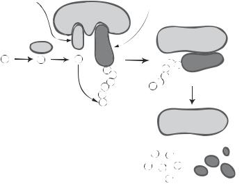
Figure 5.17. Schematic representation of the components of the ubiquitination system. (From Nakayama KI et al: Regulation of the cell cycle at the G1-S transition by proteolysis of cyclin E and p27Kip1, Biochem Biophys Res Commun 282:853–60, copyright 2001, with permission from Elsevier.)
conjugating enzyme can transfer an activated ubiquitin to a target protein with the help of the ubiquitin/protein ligase, which recognizes target proteins and promotes ubiquitin transfer and ubiquitination (Fig. 5.17). Thus, these factors play a critical role in the initiation and regulation of ubiquitination.
UBIQUITIN AND PROTEASOME-MEDIATED CELL SIGNALING |
229 |
Signaling Mechanisms [5.17]
Protein ubiquitination is regulated at the level of substrate proteins. Two processes are often involved in the regulation of ubiquitination: substrate phosphorylation and hydroxylation. Substrate phosphorylation may induce activation or inhibition of ubiquitination, depending on the nature of substrate proteins and ubiquitin ligases, whereas substrate hydroxylation activates ubiquitination. Several examples are given here, to demonstrate these regulatory mechanisms.
Stimulation of Ubiquitination by Substrate Phosphorylation. Substrate phosphorylation is required for the activation of several ubiquitin ligases, including S-phase kinaseassociated protein 1 (Skp1), Cdc53, and F-box protein, which are designated as SCF ubiquitin ligases. These ubiquitin ligases form complexes with an identical Skp1 and Cdc53 protein, but a varying F-box protein. A specified F-box protein is responsible for the recognition of a specific substrate protein and the affinity of the ligase complex to the substrate. The phosphorylation of a substrate protein can activate a SCF ligase complex, inducing ubiquitination and destruction of the substrate. Several transcriptional factors, including IκB and β-catenin, are known substrates for the SCF ligase complex.
IκB is an inhibitory molecule that binds to and sequesters NFκB in unstimulated cells. On the stimulation of NFκB-related signaling pathways, the IκB molecule becomes phosphorylated at two serine residues, which stimulate the activation of a SCF ubiquitin ligase complex, namely, SCFβ-TrCP, where β-TrCP is a specific F-box protein. Activated SCFβ-TrCP induces ubiquitination and degradation of IκB, a necessary step for the release and activation of NFκB. Activated NFκB serves as a transcriptional factor, migrates to target genes, and initiates gene transcription. In this case, ubiquitination is not only responsible for the degradation of IκB, but also contributes to the activation of NFκB.
β-Catenin is a coactivator of transcriptional factors, participating in the regulation of gene transcription. β-Catenin can be phosphorylated by glycogen synthase kinase 3β. The phosphorylation of β-catenin activates SCFβ-TrCP, which in turn induces β-catenin ubiquitination and destruction. This is an effective approach for reducing transcriptional activities mediated by β-catenin. (See Table 5.15.)
Inhibition of Ubiquitination by Substrate Phosphorylation. The phosphorylation of certain proteins may reduce the activity of ubiquitin ligases, thus suppressing ubiquitination of the substrate proteins. A typical example is p53, a tumor suppressor protein (see Table 5.18, later in this chapter for p53). Toxic stress can lead to p53 phosphorylation. Phosphorylated p53 in turn induces cell arrest during cell mitosis. Excessive p53 is usually degraded by ubiquitination, which is mediated by an ubiquitin ligase mdm2. Phosphorylation of p53 on the serine residues has been shown to prevent interaction of p53 with the ubiquitin ligase mdm2, suppressing ubiquitination. Such a process stabilizes p53 and enhances the function of p53.
Stimulation of Ubiquitination by Substrate Hydroxylation. In addition to substrate phosphorylation, substrate hydroxylation plays a role in regulating substrate ubiquitination. An example is the ubiquitination of the hypoxic response transcriptional regulator hypoxia inducible factor 1 α (HIF1α) (see Table 5.16). This factor is stabilized and activated under a hypoxic condition but degraded under a normoxic condition. Ubiquitination of HIF1α is a critical step in the degradation of the HIF1α protein. Following recovery from a

230
TABLE 5.15. Characteristics of S-Phase Kinase-Associated Protein 1A and β-Catenin*
|
|
Amino |
Molecular |
|
|
Proteins |
Alternative Names |
Acids |
Weight (kDa) |
Expression |
Functions |
|
|
|
|
|
|
S-phase kinase-associated |
SKP1, SKP1A |
163 |
19 |
Ubiquitous |
Serving as a substrate recognition component |
protein 1A |
|
|
|
|
of the SCF ubiquitin ligase complex, binding |
|
|
|
|
|
to regulatory proteins involved in ubiquitin |
|
|
|
|
|
proteolysis (e.g., cyclin F and S-phase |
|
|
|
|
|
kinase-associated protein 2); also serving as |
β-Catenin |
Catenin β 1, cadherin- |
|
|
|
an RNA polymerase II elongation factor |
781 |
86 |
Ubiquitous |
Serving as an adherens junction protein, |
||
|
associated protein β |
|
|
|
mediating cell–cell adhesion and |
|
|
|
|
|
interaction, regulating cell attachment to |
|
|
|
|
|
matrix, cell proliferation, and differentiation |
|
|
|
|
|
during embryogenesis, wound healing, and |
|
|
|
|
|
tumor cell metastasis; also serving as a |
|
|
|
|
|
coactivator of transcriptional factors |
|
|
|
|
|
|
*Based on bibliography 5.17. |
|
|
|
|
|

|
|
NUCLEAR RECEPTOR-MEDIATED CELL SIGNALING |
231 |
|||
TABLE 5.16. Characteristics of Hypoxia Inducible Factor 1a Subunit* |
|
|
||||
|
|
|
|
|
|
|
|
Alternative |
Amino |
Molecular |
|
|
|
Proteins |
Names |
Acids |
Weight (kDa) |
Expression |
Functions |
|
|
|
|
|
|
|
|
Hypoxia inducible |
HIF1α |
826 |
93 |
Ubiquitous |
Acting as a |
|
factor 1α subunit |
|
|
|
|
transcriptional |
|
factor, regulating cellular responses to reduced oxygen concentration or hypoxia, mediating inflammatory reactions, enhancing angiogenesis,
and mediating neural development
*Based on bibliography 5.17.
hypoxic condition, increased oxygen concentration stimulates the activation of a proline hydroxylase, which catalyzes the hydroxylation of a proline residue in the HIF1α protein. The hydroxylated proline mediates the interaction of HIF1α with an ubiquitin ligase complex known as the VBC complex (Von Hippel–lindau protein–elongin B–Elongin C), which is similar to the SCF complex in assembly and function. Activated VBC complex in turn induces HIF1α ubiquitination and degradation.
NUCLEAR RECEPTOR-MEDIATED CELL SIGNALING
Structure and Function [5.18]
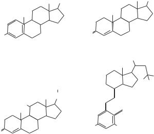
Nuclear receptors are a family of intracellular proteins that interact with steroid, thyroid, and retinoid hormones, which are lipid-soluble molecules and can diffuse through the cell membrane. These receptors are found in the cytoplasm and cell nucleus. Among the nuclear receptors, the steroid hormone receptor subfamily has been studied extensively. Common nuclear receptors include estrogen receptor (ER) α and β, glucocorticoid receptor (GR), mineralocorticoid receptor (MR), progesterone receptor (PR), androgen receptor (AR), and vitamin D receptor (VDR). These receptors interact with corresponding steroid hormones (Fig. 5.18). In addition, there are several nuclear receptors that are similar to estrogen receptors in function. These are defined as estrogen-related receptors α, β, and γ, which are also referred to as “orphan” nuclear receptors.
A typical nuclear receptor is composed of several functional domains, including a DNA-binding domain (DBD), a ligand-binding domain (LBD), an activation function 1 (AF1) domain, and an activation function 2 (AF2) domain. The DNA-binding domain is located in the central region of the receptor and composed of two zinc finger motifs that are responsible for protein–DNA interaction. The DNA-binding domain is well reserved among different nuclear receptors. The ligand-binding domain is located at the C- terminus and is composed of sequences responsible for ligand interactions, activation of nuclear transcription, binding to chaperone proteins, and dimerization with other
232 CELL SIGNALING PATHWAYS AND MECHANISMS
OH |
OH |
|
HO |
|
O |
|
|
|
|
Estrogen |
Testosterone |
|
|
OH
CH2OH
C = O
HO |
|
HO |
OH |
O |
|
Cortisol |
1,25-Dihydroxycholecalciferol |
|
(derived from Vit D3) |
Figure 5.18. Steroid hormones for nuclear receptors. (Based on bibliography 5.18).
receptors. The ligand-binding domain is moderately conserved compared with the DNAbinding domain. The AF1 and AF2 domains regulate the activation of the receptor. The difference between these two domains is that the activity of the AF1 domain is independent of ligand binding, whereas the activity of the AF2 domain is dependent on ligand binding. On the interaction with hormone ligands, the hormone–receptor complexes are activated, serve as transcriptional factors, and directly bind to corresponding cis elements in target genes, initiating gene transcription. The nuclear receptors participate in the regulation of several physiological processes, including salt balance, glucose metabolism, reproduction, and responses to environmental stress impacts. (See Table 5.17.)
Signaling Mechanisms [5.18]
In unstimulated cells, nuclear receptors are associated with “chaperones,” which suppress the activity of the receptors. On the binding of ligands to the nuclear receptors, the chaperones are dissociated from the receptors, resulting in receptor conformational changes and the exposure of the nuclear localization signals. The ligand–receptor complexes often form homodimers or heterodimers and are translocated from the cytoplasm to the nucleus, initiating gene transcription (Fig. 5.19). The activation of the nuclear receptor transcriptional factors is regulated to a large degree by the AF1 and AF2 domains of the nuclear receptor. These domains recruit and activate nuclear receptor cofactors, which are enzymes including acetylases, deacetylases, methylases, kinases, and ubiquitinases. These cofactors play a critical role in regulating the formation of the transcription-initiating complexes, the conformation of target DNA cis-acting elements, and the degradation and recycling of the nuclear receptors.

TABLE 5.17. Characteristics of Selected Molecules for Nuclear Receptor-Mediated Signaling Pathways*
|
|
Amino |
Molecular |
|
|
Proteins |
Alternative Names |
Acids |
Weight (kDa) |
Expression |
Functions |
|
|
|
|
|
|
Estrogen receptor α |
Estrogen receptor 1, ESR, |
595 |
66 |
Uterus, ovary, blood |
Acting as a transcriptional factor, |
|
ER, oestrogen receptor α |
|
|
vessel, kidney, bone |
regulating gene expression and |
|
|
|
|
|
mediating the development of female |
|
|
|
|
|
sex characters, promoting |
|
|
|
|
|
fertilization and implantation, and |
|
|
|
|
|
mediating the formation of bone |
|
|
|
|
|
matrix |
Glucocorticoid receptor |
GCR, GCCR, GR |
777 |
86 |
Ubiquitous |
Serving as a transcriptional factor, |
|
|
|
|
|
forming a homodimer or heterodimer |
|
|
|
|
|
with another protein (e.g., retinoid |
|
|
|
|
|
receptor and a heatshock protein), |
|
|
|
|
|
regulating the expression of |
|
|
|
|
|
glucocorticoid-responsive genes, |
|
|
|
|
|
promoting gluconeogenesis and |
|
|
|
|
|
elevation of blood glucose |
|
|
|
|
|
concentration, and inhibiting |
|
|
|
|
|
inflammatory and immune responses |
233

234
TABLE 5.17. Continued
|
|
Amino |
Molecular |
|
|
Proteins |
Alternative Names |
Acids |
Weight (kDa) |
Expression |
Functions |
|
|
|
|
|
|
Mineralocorticoid receptor |
MR, aldosterone receptor |
984 |
107 |
Kidney, brain, heart, |
Acting as a transcriptional factor, |
|
|
|
|
liver, intestine |
stimulating the expression of |
|
|
|
|
|
mineralocorticoid responsive genes, |
|
|
|
|
|
and mediating electrolyte and water |
|
|
|
|
|
balance via controlling ion transport |
|
|
|
|
|
in the renal tubular system, resulting |
|
|
|
|
|
in retention of sodium and water and |
|
|
|
|
|
loss of potassium |
Progesterone receptor |
PR |
933 |
99 |
Uterus, ovary, placenta, |
Acting as a transcriptional factor in |
|
|
|
|
brain, blood vessel, |
response to binding of progesterone; |
|
|
|
|
leukocytes |
also regulating the establishment and |
|
|
|
|
|
maintenance of pregnancy |
Androgen receptor |
AR, dihydrotestosterone |
920 |
99 |
Prostate, adrenal gland, |
Serving as a transcriptional factor, |
|
receptor (DHTR) |
|
|
brain |
inducing transcription of androgen |
|
|
|
|
|
responsive genes, and regulating |
|
|
|
|
|
masculinization or the development |
|
|
|
|
|
of male sex characteristics |
Vitamin D receptor |
1,25-Dihydroxyvitamin D3 |
427 |
48 |
Ubiquitous |
Serving as a transcriptional factor and |
|
receptor, VDR |
|
|
|
regulating calcium metabolism and |
|
|
|
|
|
the formation of bone matrix |
|
|
|
|
|
|
*Based on bibliography 5.18.

NUCLEAR RECEPTOR-MEDIATED CELL SIGNALING |
235 |
OH
Steroid hormone
HO
HO
OH
O
H
OH
5'
3'
HO
OH
Nuclear receptor
Nucleus membrane
HO
OH
Hormone 3' response 5' element
Figure 5.19. Mechanisms of nuclear receptor activation (based on bibliography 5.18).
When activated, the nuclear receptor transcriptional factors interact with specific DNA cis-acting elements defined as hormone response elements (HREs) with the assistance of cofactors. The HREs are located within the promoter and enhancer regions of target genes and are composed of unique sequences for different steroid ligands. For instance, the hormone response element for estrogen receptors and estrogen-related receptors contains repeating AGGTCA sequences, whereas that for glucocorticoid receptor, mineralocorticoid receptor, progesterone receptor, and androgen receptor contains AGAACA. Thus, the structure of these cis elements determines the specificity of the binding of nuclear receptor transcriptional factors to DNA. These transcriptional factors bind DNA in the form of homodimers or heterodimer.
In addition to steroid hormones, other types of molecule can activate nuclear receptors. A typical example is the activation of nuclear receptors by protein kinases, including protein kinase A, mitogen-activated protein kinases, cyclin-dependent kinases, and glycogen synthase kinases. While the exact mechanisms remain poorly understood, it appears that growth factor-induced activation of intracellular protein kinases play a role in the