

Bioregenerative Engineering Principles and Applications - Shu Q. Liu
..pdf296 FUNDAMENTAL CELLULAR FUNCTIONS
desmocollin-1,2,3. The distribution of each desmosomal cadherin varies among tissues and organs. For instance, desmoglein-2 and desmocollin-2 are ubiquitously expressed in tissues with desmosomes, whereas desmoglein-3 and desmocollin-3 are found mostly in the basal layer of stratified epithelia.
Function [6.11]. Cadherin-mediated cell adhesion plays an important role in the regulation of tissue and organ morphogenesis and development. A change in the expression pattern of major cadherins is often associated with altered morphogenetic processes. For instance, E-cadherin is highly expressed and activated in the oocyte after fertilization, while N-cadherin is not. At gastrulation, the expression and activation pattern of the E- and N-cadherins is switched; specifically, the E-cadherin is downregulated whereas the N-cadherin is upregulated. Such a switch is associated with epithelial–mesenchymal transition. Furthermore, the overexpression of N-cadherins blocks the segregation of neural crest cells from the neural tube. These observations suggest that the expression and activation of appropriate cadherins are critical to the regulation of tissue and organ morphogenesis during embryonic development.
Cadherins are involved in the regulation of cell differentiation. This function has been demonstrated in E-cadherin-null embryonic stem cells. While wildtype embryonic stem cells can differentiate into well-organized forms of specialized tissues, E-cadherin-null stem cells develop only into tissues without specialized forms. The transfer of E-cadherin gene into E-cadherin-null stem cells restores the differentiation function of the embryonic stem cells. These observations suggest that E-cadherins play a critical role in regulating the differentiation of embryonic stem cells.
Cell adhesion mediated by cadherins plays a role in the regulation of cell survival. This is supported by several lines of evidence. The lack of E-cadherins is associated with the induction of endothelial cell apoptosis. Caspases can cleave cadherins and induce cell dissociation, contributing to cell apoptosis. Thus, the presence of cadherins is essential to cell survival.
Cell Surface Heparan Sulfate Proteoglycans
Classification and Structure [6.12]. Cell surface heparan sulfate proteoglycans are composed of heparan sulfate glycosaminoglycan (GAG) chains, which are attached to core proteins in the extracellular region. These proteoglycans serve as adhesion receptors and participate in the regulation of cell–cell and cell–matrix interactions. Heparan sulfate can form various types of heparan sulfate proteoglycan, including perlecan and agrin in extracellular matrix, serglycin in the cytoplasm, and syndecans and glypicans in the cell membrane. The function of heparan sulfate proteoglycans is determined by the heparan sulfate glycosaminoglycan group.
A heparan sulfate molecule is composed of highly sulfated heparin-like domains and poorly sulfated domains with rich glucuronic acids. These domains alternate with intermediate sulfated domains. The total length and the number of each domain of a heparan sulfate vary considerably between different cell types. The differences between cell types may be due to the existence of multiple isoforms of modification enzymes. A major function of the heparan sulfate chains in a cell surface heparan sulfate proteoglycan is to bind ligands. A large number of proteins can bind to heparan sulfate. Although the amino acid sequences of these binding proteins vary widely, the binding proteins are rich in basic amino acids such as lysine and arginine.
|
|
CELL SURFACE HEPARIN SULFATE PROTEOGLYCANS |
297 |
|||
TABLE 6.8. Characteristics of Selected Heparan Sulfate Proteoglycans* |
|
|||||
|
|
|
|
|
|
|
|
Alternative |
Amino |
Molecular |
|
|
|
Proteins |
Names |
Acids |
Weight (kDa) |
Expression |
Functions |
|
|
|
|
|
|
|
|
Syndecan |
Syndecan, |
310 |
32 |
Skin, kidney, |
A transmembrane |
|
1 |
SYND1, |
|
|
placenta, |
heparan sulfate that |
|
|
SDC1, |
|
|
lymphocytes |
mediates cell |
|
|
CD138 |
|
|
|
binding, cell |
|
|
antigen, |
|
|
|
signaling, cytoskeletal |
|
|
CD138. |
|
|
|
organization, cell |
|
|
|
|
|
|
proliferation, cell |
|
|
|
|
|
|
differentiation, and |
|
|
|
|
|
|
HIV transmission to |
|
|
|
|
|
|
lymphocytes |
|
Glypican |
GPC1 |
558 |
62 |
Pancreas, |
A heparan sulfate |
|
1 |
|
|
|
intestine, |
proteoglycan that |
|
|
|
|
|
bone |
attaches to external |
|
|
|
|
|
marrow, |
surface of cell |
|
|
|
|
|
placenta |
membrane and |
|
|
|
|
|
|
regulates cell–cell |
|
|
|
|
|
|
interaction |
|
|
|
|
|
|
|
|
*Based on bibliography 6.12.
Syndecans and glypicans (see Table 6.8) are the most abundant heparan sulfate proteoglycans at the cell surface. Syndecans are transmembrane receptors with heparan sulfate chains attached to the extracellular region at the N-terminus (Fig. 6.13). Syndecans are a family of several heparan sulfate proteoglycans, including syndecan-1,2,3,4 with molecular weights 33, 22, 46, and 22 kDa, respectively. The extracellular domain of the four syndecan molecules differs considerably. Syndecan-2,-3 contain in their extracellular region primarily heparan sulfate chains, whereas syndecan-1,-4 contain chondroitin sulfates in addition to heparan sulfates. The extracellular domain of syndecans is composed of heparan sulfate attachment sites, signal peptide sites, and proteolytic cleavage sites. Syndecans can be shed from the cell membrane via protein cleavage at sites near the cell membrane. Such a process converts receptor-type to soluble syndecans. Both receptor and soluble syndecans can bind to the same type of ligands.
The transmembrane domain of syndecans is well conserved among different types of syndecan. This domain plays a role in mediating the dimerization of the syndecan molecules and localizing syndecans to appropriate membrane compartments. The cytoplasmic tail of all syndecans is highly conserved. This tail is composed of phosphorylation sites and binding sites for cytoskeletal proteins and signaling molecules.
Glypicans are globular molecules with molecular weight 60 kDa. Six types of glypican have been identified. Each glypican is composed of an N-terminal cysteine-rich domain and heparan sulfate attachment motifs. Glypicans are attached to the external surface of the cell membrane via a glycosyl phosphatidylinositol anchor, which is localized to the membrane microdomains with rich glycosphingolipids. The heparan sulfate chains are attached to the C-terminus of the core proteins near the cell membrane. Glypicans do not pass through the cell membrane.

298 FUNDAMENTAL CELLULAR FUNCTIONS
Heparan sulfate
Band 4.1
Syndecan 1 |
Glypican 4 |
Figure 6.13. Schematic representation of the structure of syndecans. Based on bibliography 6.12.
Function [6.13]. Cell surface heparan sulfate proteoglycans participate in the regulation of cell–cell adhesion and interaction. Syndecans are localized to adherens junctions and can interact with several adhesion molecules, such as L-selectin, N-CAM, and PE-CAM. The lack of syndecan-1 is associated with reduced cell aggregation, and the transfection of the syndecan-1 gene restores cell aggregation. These observations suggest that the presence of syndecans is essential to cell–cell adhesion. However, the exact mechanisms remain to be investigated.
Cell surface heparan sulfate proteoglycans play a role in the regulation of cell–matrix adhesion. Extracellular matrix contains a number of components, including collagen, elastin, fibronectin, laminin, tenascin, vitronectin, and thrombospondin, which are capable of interacting with syndecans. During development, syndecans are colocalized with extracellular matrix components. Certain types of syndecans, such as syndecan-1 and -4 are localized to the focal adhesion contacts. In heparan sulfate-deficient cells, the formation of focal adhesion contacts is impaired. These observations demonstrate the importance of cell surface heparan sulfate proteoglycans in the control of cell–matrix interaction. The role of glypicans in regulating cell–matrix adhesion remains to be determined, although glypicans have been found to bind to collagen and fibronectin.
Integrins
ClassiÞcation and Structure [6.14]. Integrins are transmembrane glycoproteins that mediate cell–matrix adhesion, providing a linkage between the cytoskeleton and extracellular matrix (Fig. 6.14). Integrins also mediate cell–cell interactions. Each integrin is a heterodimer composed of a variable α subunit and a relatively conserved β subunit. To date, at least 18 α subunits and 8 β subunits have been identified. The binding ligands of

INTEGRINS 299
von Willebrand factor A domain
 PSI
PSI
von Willebrand factor A domain
Integrin α repeats
Integrin α1 |
Integrin α2 |
Integrin β2 |
Figure 6.14. Schematic representation of the structure of integrins. PSI: domain found in plexins, semaphorins, and integrins. Based on bibliography 6.14.
integrins are determined by the combination of specific α and β subunits. Several α integrins and a common β integrin are presented in Table 6.9; some of these molecules are used for treatment of muscular dystrophy.
Both integrin α and β subunits contain an extracellular domain, a transmembrane domain, and a cytoplasmic tail. The α and β subunits contain distinct amino acid sequences. The β subunit is about 760–790 amino acids in length, whereas the α subunit is about 1000–1200 amino acids. The majority of amino acids are distributed in the extracellular region for both α and β subunits. The extracellular region of the β subunit is composed of an I-domain, which is responsible for ligand binding, and several cysteine-rich repeats. The extracellular region of the α subunit is composed of an I-domain (in at least 9 α subunits) and several repeats with a consensus sequence DxDxDGxxD (x = any amino acid). These repeats are responsible for integrin binding.
Although the cytoplasmic tail of integrins is considerably short, this region plays an important role in mediating interactions between integrins and intracellular signaling molecules, between integrins and cytoskeletal components, and between the integrin α and β subunits. The cytoplasmic tail is composed of a large number of binding sites for these interactions. The binding of integrins to the actin cytoskeleton is the basis for the formation of cell focal adhesion contacts, essential structures for the control of cell attachment to extracellular matrix and cell migration.
Each cell contains a number of different types of integrins. The β1 subunit forms a dominant subfamily of integrins with the α subunits. The β1-containing integrins are defined as β1 integrins. The ligands of β1 integrins are determined primarily by the specificities of the α subunits. For example, α1β1 and α2β1 bind to collagen and laminin, α4β1 and α5β1 bind to fibronectin, whereas α6β1 binds to laminin.

300
TABLE 6.9. Characteristics of Selected Integrins*
|
|
Amino |
Molecular |
|
|
Proteins |
Alternative Names |
Acids |
Weight (kDa) |
Expression |
Functions |
|
|
|
|
|
|
Integrin α1 Laminin and collagen receptor, |
1179 |
131 |
Ubiquitous |
Forming a heterodimer with integrin β1, serving |
|
|
very late activation protein 1, |
|
|
|
as a receptor for collagen and laminin, |
|
VLA1, CD49a |
|
|
|
regulating cell–matrix interaction, and |
|
|
|
|
|
mediating cell survival, migration, and |
|
|
|
|
|
proliferation |
Integrin α2 ITGA2, very late activation |
1181 |
129 |
Ubiquitous |
Forming a heterodimer with integrin β1 and |
|
|
protein 2 receptor α2 subunit, |
|
|
|
serving as a receptor for collagen |
|
VLA2 receptor α2 subunit, |
|
|
|
|
|
VLA2 α chain, VLAA2, |
|
|
|
|
|
CD49B, platelet glycoprotein |
|
|
|
|
|
Ia/IIa, platelet membrane |
|
|
|
|
|
glycoprotein Ia, GPIa, collagen |
|
|
|
|
|
receptor |
|
|
|
|
Integrin α3 ITGA3, CD49C, very late |
1066 |
119 |
Ubiquitous |
Forming a heterodimer with integrin β1 and and |
|
|
activation antigen 3 (VLA-3), |
|
|
|
mediating cell interaction with extracellular |
|
very common antigen 2 |
|
|
|
matrix components, such as collagen, |
|
(VCA-2), extracellular matrix |
|
|
|
fibronectin, and laminin |
|
receptor 1 (ECMR1), and |
|
|
|
|
|
galactoprotein b3 (GAPB3), |
|
|
|
|
|
VLA3 α chain |
|
|
|
|
Integrin α4 ITGA4, CD49D, very late |
1038 |
115 |
Blood vessels, blood |
Forming a heterodimer with integrin β1 regulating |
|
|
activation protein 4 receptor α4 |
|
|
cells, skin. |
cell interaction with fibronectin, and mediating |
|
subunit, VLA4 receptor α4 |
|
|
|
vascular cell–cell interaction |
subunit

Integrin α5 ITGA5, CD49e, fibronectin |
1049 |
115 |
Ubiquitous |
Forming a heterodimer with integrin β1, regulating |
receptor α subunit, very late |
|
|
|
cell interaction with fibronectin |
activation protein 5α subunit |
|
|
|
|
Integrin α6 ITGA6, CD49f, very late |
1073 |
120 |
Skin, lung, intestine, |
Forming a heterodimer with integrin β1, regulating |
activation protein 6α subunit, |
|
|
stomach, pancreas. |
cell interaction with laminin |
VLA-6 |
|
|
|
|
Integrin α7 ITGA7 |
1137 |
124 |
Heart, skeletal |
Joining with integrin β1 to form an integrin |
|
|
|
muscle, nervous |
complex, which is a major integrin complex |
|
|
|
system, lung, |
expressed in differentiated muscle cells, binding |
|
|
|
intestine, overy, |
to the extracellular matrix protein laminin-1, |
|
|
|
prostate gland |
and regulating cell attachment to extracellular |
|
|
|
|
matrix |
Integrin αv ITGAV, vitrionectin receptor α |
1048 |
116 |
Ubiquitous |
Forming heterodimers with several β integrins, |
polypeptide, CD51 |
|
|
|
such as integrin β1, 3, and 5; mediating cell |
|
|
|
|
adhesion to extracellular matrix (note that the |
|
|
|
|
integrin complex αvβ3 regulates cell adhesion |
|
|
|
|
to vitronectin), and regulating TGFβ-related |
|
|
|
|
signal transduction |
Integrin β1 ITGB1, CD29, very late activation |
825 |
92 |
Ubiquitous |
Joining with an integrin α subunit to form integrin |
protein, β polypeptide, VLAβ |
|
|
|
complexes, regulating cell adhesion to |
extracellular matrix, regulating various cellular activities, including embryogenesis, cell proliferation and migration, immune response, and metastasis of tumor cells
*Based on bibliography 6.14.
301

302 FUNDAMENTAL CELLULAR FUNCTIONS
Function [6.15]. Integrins play a critical role in the regulation of cell migration. As discussed on page 270 of this chapter, in order to induce migration, a cell must attach to a matrix substrate at the leading edge while detaching from the substrate at the trailing edge at a given time. Although the exact mechanisms are poorly understood, integrins are likely involved in the regulation of these processes. During cell migration, integrins at the leading edge must be engaged to form strong adhesion bonds between focal adhesion contacts and the matrix substrate (Fig. 6.15), whereas integrins at the trailing edge must dissociate from the matrix substrate. The location-dependent integrin activation and deactivation within a single cell remains a subject of research.
In addition to the regulation of cell membrane attachment to matrix substrate, integrins are involved in regulating the contractile activity of the actin cytoskeleton. Integrins can be activated by exposure to extracellular matrix. Activated integrins can lead to phosphorylation of the mitogen-activated protein kinases (MAPKs) via the mediation of focal adhesion kinase and adaptor proteins. MAPKs can directly phosphorylate the myosin light-chain kinase (MLCK), which in turn activates myosin light chain, inducing and enhancing actin–myosin interaction. Actin–myosin interaction generates forces necessary for cell protrusion and traction during migration.
Integrins are involved in regulating the assembly of extracellular matrix. One example is the control of basement membrane formation by β1 integrins in epithelial tissues. Embryonic stem cells derived from β1 integrin-null mice are not able to form a basement membrane in epidermal tissue. Another example is integrin-related fibronectin fibrillogenesis. The assembly of fibronectin fibrils can be initiated on the binding of fibronectin to the α5β1 integrin. The loss of this type of integrin is associated with impairment of fibronectin fibrillogenesis. Other types of integrin, such as α4β1 and αvβ3, also contribute to the regulation of fibronectin fibrillogenesis.
Integrins play a critical role in the regulation of cell differentiation and proliferation. In several experimental models, integrins have been shown to mediate the pattern of gene expression and cell differentiation. For instance, salivary gland cells can differentiate into duct and acinar epithelial cells in response to the interaction of integrins and extracellular matrix, whereas these cells cannot differentiate in the absence of extracellular matrix. Furthermore, a treatment with antibodies specific to collagen IV and integrin α6 and β1
Fibronectin
Integrin α |
Integrin β |
Figure 6.15. Schematic demonstration of interaction of integrins with fibronectin. Based on bibliography 6.15.
INTEGRINS 303
reduces the capability of cell differentiation. The attachment of cells to fibronectinand collagen-containing matrix is often associated with extensive changes in gene expression. Altered gene activities may likely contribute to integrin-initiated cell differentiation and proliferation. The loss of β1 integrins in keratinocytes is associated with a reduction in cell differentiation and proliferation. These observations suggest a critical role for integrin-matrix interaction in the regulation of cell differentiation and proliferation.
Mechanisms of Integrin-Related Activities [6.15]. The primary function of integrins is to regulate the adhesion of cells to extracellular matrix. Integrins can be activated by exposure to extracellular matrix, which induces conformational changes and clustering of integrins. The activation of integrins is associated with an increase in integrin binding affinity. Integrins are major constituents of focal adhesion contacts, structures regulating cell adhesion to extracellular matrix. Focal adhesion contacts were original identified in cultured fibroblasts by electron microscopy, which demonstrates the presence of electron dense plaques with filamentous structures. These plaques were later found to contain a number of molecules, including integrins, actin filaments, talin, filamin, α-actinin, vinculin, profilin, paxillin, tensin, focal adhesion kinase, the Src family kinases, protein tyrosine phosphatases, the Grb2 adaptor protein, and phosphoinositide 3-kinase. Integrins serve as links between the intracellular actin cytoskeleton and extracellular matrix, and transmit signals from the extracellular matrix to the actin cytoskeleton and intracellular signaling pathways.
Integrins do not possess intrinsic catalytic activity. However, the β subunit of integrins can transmit a variety of extracellular signals via their connection with intracellular signaling molecules and actin cytoskeleton-associated molecules as described above. The importance of the β subunit can be tested by selected sequence deletion and restoration. The deletion of the β subunit is associated with diminished interaction of integrins with intracellular signaling molecules. Compared with the β subunit, the α subunit binds to fewer molecules. Identified molecules that bind to the α subunit include calreticulin, guanine nucleotide exchange factor Mss4, and calcium-binding protein. The physiological function of the α subunit-binding proteins remains to be determined.
The interaction of extracellular matrix with integrins often initiates intracellular signaling events, leading to molecular activities such as phosphorylation of protein tyrosine kinases, changes in the level of cAMP and calcium, and expression of mitogenic genes. In particular, integrins can transmit signals to two nonreceptor protein tyrosine kinases: focal adhesion kinase (FAK) and Src protein tyrosine kinase. These protein tyrosine kinases are localized to focal adhesion contacts. FAK can be phosphorylated in response to interaction of integrins with fibronectin, although the mechanisms of FAK activation remain poorly understood. Phosphorylated FAK induces the recruitment of Src to FAK. Recruited Src in turn phosphorylates FAK at various sites, enhancing the activity of FAK. These activities lead to the recruitment of adaptor proteins, including Grb2 and pp130Cas, to the focal adhesion contacts. These adapter proteins link the integrin-FAK pathway to other signaling pathways, including the PI3-kinase, Ras, and MAPK pathways, which play critical roles in regulating cell proliferation and migration.
Integrin-related signaling molecules can communicate with the Rho family of small GTPases, including Rho, Cdc42, and Rac, which regulate the assembly and function of the actin cytoskeleton. The interaction of integrins with extracellular matrix can induce activation of Rho, Cdc42, and Rac. Activated Rho, Cdc42, and Rac enhance the assembly of focal adhesion contacts and the activity of related signaling molecules, including FAK.
304 FUNDAMENTAL CELLULAR FUNCTIONS
However, the exact regulatory mechanisms remain to be investigated. These investigations suggest that integrin-dependent signaling pathways can “crosstalk” to other signaling pathways. Such interaction provides a synergistic mechanism for the regulation of cell adhesion, migration, proliferation, and differentiation.
APOPTOSIS [6.16]
Apoptosis is a process of naturally occurring cell death, which eliminates malfunctioned and undesired cells during development and remodeling. During development, apoptosis and cell division together contribute to the morphogenesis of tissues and organs. While cell division contributes to the growth of cell and tissue mass, apoptosis contributes to the removal of excessive cells. These processes are unnecessary for the formation of tissues and organs. After reaching maturity, apoptosis continues to play an important role in the maintenance of the homeostasis. Cells with damaged or mutant DNA are eliminated by apoptosis. Without apoptotic elimination, these cells may develop into tumor cells. Under a physiological condition, the cell density is kept at a relatively constant level through coordinated cell proliferation and apoptosis. An increase in apoptotic activity results in tissue degeneration, whereas a decrease in apoptotic activity results in hyperplasia, both of which contribute to pathogenic disorders of tissues and organs. Thus, it is important to maintain a physiological level of apoptotic activity.
Morphological Characteristics of Apoptosis [6.16]
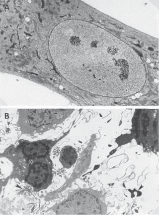
In biological research, it is important to identify apoptotic cells, which helps to understand the mechanisms of apoptosis. Apoptotic cells undergo several stages of morphological change. On the stimulation of apoptotic signals, a cell usually starts to round up and becomes spherical in shape. These morphological changes are usually associated with cell membrane budding. The next noticeable change is DNA condensation, resulting in an increase in the nucleus density and reduction in the nucleus size, which can be seen under an optical and electron microscope (Fig. 6.16). DNA condensation is followed by DNA fragmentation and nucleus disruption. The entire cell eventually disintegrates into small pieces, which are phagocytosed by macrophages or neighboring cells. These morphological features can be used to identify apoptotic cells.
Apoptosis-Inducing Factors [6.16]. Apoptosis can be induced by a variety of extracellular factors, such as depletion of growth factors and nutrients, hypoxia, UV irradiation, mechanical stress, and binding of apoptotic ligands. Intrinsic changes, such as DNA damage and disruption, and immunoreactions, such as T-lymphocyte activation, can also trigger apoptosis. In addition, cancer cells can initiate apoptosis, an important mechanism for the elimination of cancer cells. Suppression of the apoptotic function increases the possibility of tumorigenesis.
Regulation of Apoptosis [6.16]. Apoptosis can be induced and regulated by two known signaling pathways: Fas ligand (FasL)- and tumor necrosis factor (TNF)-activated pathways. FasL and TNF are apoptosis-inducing proteins. These proteins are generated in the ER, deployed to the cell membrane, and cleaved from the cell membrane to form soluble ligands. The forms of the ligands determine the effectiveness of the ligands. For the Fas ligand, the membrane-bound form is more effective than the soluble form. In
APOPTOSIS 305
Figure 6.16. Electron micrographs of apoptotic human fibroblasts. (A) A control cell and (B) cells exposed to 0.5-μM naphthazarin (5,8-dihydroxy-1,4-naphthoquinone), an apoptosis inducer, for 8 h are shown. Different stages of apoptosis can be discerned in the treated cells: reduced cell size, condensed chromatin (stars), fragmented nuclei (arrow), and apoptotic bodies (arrow heads). Reprinted from Roberg K et al., Lysosomal release of cathepsin D precedes relocation of cytochrome c and loss of mitochondrial transmembrane potential during apoptosis induced by oxidative stress, Free Radical Biol Med 27:1228–37, 1999, with permission from Elsevier.
contrast, soluble TNF is more active than the membrane-bound form of TNF. The mechanism of Fas ligand-induced apoptosis is similar to that induced by TNF. Here, the FasL signaling pathway is used as an example to demonstrate the mechanisms of apoptosis (Fig. 6.17).
A Fas ligand can interact with the Fas ligand receptor, resulting in oligomerization (often trimerization) and activation of the receptor. Activated Fas ligand receptor in turn stimulates the Fas ligand-associated death domain or FADD (note that TNF can interact with and activate TNF receptor, which stimulates the TNFR-associated death domain or TRADD). Activated FADD (or TRADD) binds to a downstream protein known as caspase 8 (cysteine aspartate protease 8), which belongs to the caspase family and possesses protease catalytic activity. In unstimulated cells, caspase 8 exists in an inactive form known as procaspase 8. In response to the stimulation of FADD (or TRADD), caspase 8 can be