

Bioregenerative Engineering Principles and Applications - Shu Q. Liu
..pdf8
EMBRYONIC ORGAN DEVELOPMENT
Early development of the pancreatic islets. By embryonic day 19 (E19), Pdx1-positive (green) and glucagon-positive (red) cell aggregates can be detected in the pancreas. Pdx1 is a marker for the pancreatic β cells, whereas glucagon is expressed in the α cells. The α cells are found primarily around the β cells. (From Jensen J: Dev Dyn 229:176–200, 2004, reprinted by permission of WileyLiss, Inc, a subsidiary of John Wiley & Sons, Inc.) See color insert.
Bioregenerative Engineering: Principles and Applications, by Shu Q. Liu
Copyright © 2007 John Wiley & Sons, Inc.
346
DEVELOPMENT OF ECTODERM-DERIVED ORGANS |
347 |
DEVELOPMENT OF ECTODERM-DERIVED ORGANS
The ectoderm is the outmost layer of the three embryonic germ layers. During the early period of embryogenesis, the ectoderm develops into a structure composed of three functional components: surface ectoderm, neural crest, and neural tube. The surface ectoderm is the origin of the epidermis, epidermal appendages (the nails, hair, and sebaceous glands), mucous membrane of the mouth and anus, tooth enamel, lens and cornea, and anterior pituitary. The neural crest gives rise to the peripheral nervous system (the peripheral ganglia, Schwann cells, and sympathetic and parasympathetic nervous systems), adrenal medulla, melanocytes, and tooth dentine. The neural tube develops into the brain, spinal cord, retina, and inner ear. In this chapter, we will focus on the development of the epidermis and the nervous system.
Epidermal Development [8.1]
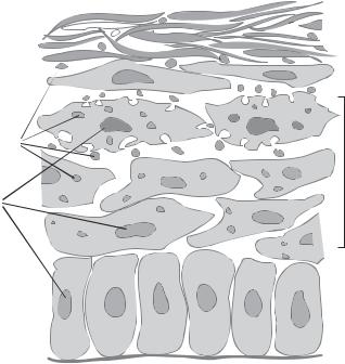
The surface ectoderm is originally a structure with a single cell layer, which further develops into a double-layered structure. The external layer forms a temporary tissue that protects the internal layer. The internal layer contains epidermal stem cells and is referred to as the basal layer. The basal layer cells can differentiate into all epidermal cell types. As a first step, basal layer cells differentiate into granular cells, which contain keratin granules in the cytoplasm. The granular cells can further differentiate into epidermal cells, also known as keratinocytes. These cells can produce a large amount of keratin. Mature keratinocytes move to the external layer of the epidermis and eventually die, forming a keratin-rich layer (Fig. 8.1). This keratin layer is about 10 cells thick and is a tough structure that protects the internal tissues and organs from physical and chemical injury. The dead, keratin-containing keratinocytes on the epidermal surface shed constantly throughout the lifespan. The keratin layer is replenished by newly generated keratinocytes from the granular cells.
The basal cells of the epidermis reside on an extracellular matrix membrane known as the basal lamina (Fig. 8.1). Underneath the basal lamina lies the dermis, a soft connective tissue. It is important to note that the dermis is not derived from the ectoderm, but from the mesoderm. A major cell type in the dermis is fibroblast. This cell type plays a role in regulating the differentiation and proliferation of the epidermal cells by releasing growth factors, such as fibroblast growth factor, transforming growth factor α, and epidermal growth factor. Keratinocytes can also produce these growth factors. The level and timing of growth factor release may determine the rate of differentiation and proliferation of the basal cells and granular cells.
The epidermal appendages, including the hair, nails, and sebaceous glands, are also derived from the basal layer stem cells at designated sites. The formation of epidermal appendages is regulated by epidermal and dermal factors. For hair formation, it is necessary to establish a hair follicle primordium, the early form of the hair follicle. The follicle primordium is derived from the basal layer. At a specific site where dermal fibroblasts are activated by autocrine regulatory factors, the basal layer cells form cell clusters, undergo shape changes, and invade the dermal layer, forming scattered cell nodes. The dermal fibroblasts release growth factors, which stimulate the node cells to divide and differentiate into hair follicle cells and keratin hair shaft. The hair shaft grows out of the dermal layer and forms hair. There are follicle stem cells that can self-renew and differentiate into hair cells and regenerate the hair shaft when hair is damaged, removed, or shed.
348 EMBRYONIC ORGAN DEVELOPMENT

 Keratin layer Keratin


Transitional cells
Keratin granules
Keratinocytes
Nuclei
Basal cells
Basal lamina
Figure 8.1. Schematic representation of the epidermis. Based on bibliography 8.1.
When the basal layer cells migrate into the dermal tissue for the formation of the hair follicle, a group of cells branches off and forms the sebaceous glands, which generate sebum, a lubricant that covers and protects the skin. When the sebaceous gland cells are injured, the follicle stem cells can be stimulated to differentiate into sebaceous cells and replenish the sebaceous glands.
Development of Neural Crest-Derived Systems [8.2]
The Neural Crest. The neural crest is an embryonic structure derived from the ectoderm. It contains stem cells that can differentiate into a number of specified cell types and tissues. These include the peripheral ganglionic neurons and glial cells, Schwann cells, sympathetic and parasympathetic neurons, medulla of the adrenal gland, epidermal pigment cells, facial bones and cartilages, corneal endothelial cells, tooth papillae, connective tissue cells and smooth muscle cells in the aortic arch, and connective tissue cells in the salivary and thyroid glands. Since these cells and structures are distributed through the entire body, the neural crest cells must migrate from the ectodermal neural crest to the periphery for a long distance. The control of the migration pattern and destination of neural crest cells as well as the specification and differentiation of these cells are major topics of developmental research.
Neurulation and Formation of Neural Crest Cells. During the stage of blastocyst formation and gastrulation, certain cells in the ectodermal region are prompted to differentiate into neuroblasts, cells that develop into nerve cells. This differentiation process is induced
|
DEVELOPMENT OF ECTODERM-DERIVED ORGANS |
349 |
Ectoderm |
Neural fold |
|
Mesoderm |
Neural groove |
|
Endoderm |
Neural plate |
|
Somite |
Notochord |
|
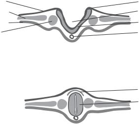
~20 days, formation of the neural groove
Neural crest
Neural tube
~23 days, completion of the neural tube
Figure 8.2. Schematic representation of the formation of the neural plate and neural tube, a process known as neurulation. Based on bibliography 8.2.
by exposing blastomeres to specific signaling molecules, such as fibroblast growth factor, noggin, and chordin. Soon following gastrulation, the narrow dorsal region of the ectoderm along the anterior–posterior (anteroposterior) axis (superior-inferior axis in the human) is transformed into a neuroblast-containing structure called neural plate. The neural plate undergoes a process called neurulation, by which the neural plate forms a neural tube (Fig. 8.2). There are two forms of neurulation: primary and secondary. Primary neurulation is the formation of the neural tube based on the ectoderm neuroblasts. This process takes place in the anterior neural plate, which develops into the brain and anterior spinal cord. In contrast, secondary neurulation is the formation of the neural tube from the mesenchymal cells of the mesoderm. The secondary process occurs near the posterior end of the neural plate, which develops into the posterior portion of the spinal cord.
Primary neurulation divides the ectoderm into three layers: the external presumptive epidermis, the middle neural crest cells, and the internal neural tube. At the beginning of the primary neurulation, cells in the neural plate elongate in the direction perpendicular to the neural plate and proliferate symmetrically with respect to the anterior–posterior axis. The neural plate thickens along its edges, bends its edges upward around the anterior–posterior axis, forming the neural folds. The groove between the two edges is defined as the neural groove, which is the centerline of the left and right central nervous system. The two neural folds bend further, meet each other at the ends, and fuse together to form a tube-shaped structure known as the neural tube. The neural tube pinches off from the ectoderm, which becomes an independent structure. At the same time, the fused ectoderm transforms to the epidermal layer at the external surface and the neural crest between the epidermal layer and the neural tube.
Secondary neurulation in the posterior portion originates from the mesenchymal cells, which develop into a tube-shaped structure underneath the ectoderm. This tube is

350 |
EMBRYONIC ORGAN DEVELOPMENT |
|
|
|
Dorsolateral route |
|
Ectodermal |
Peripheral neurons and ganglia |
|
tissues |
|
|
|
|
|
|
Schwann cells |
|
Mesodermal |
Adrenal gland cells |
|
tissues |
|
|
|
|
|
|
Pigment cells |
|
Endodermal |
|
|
tissues |
Ventral route |
Figure 8.3. Migration routes of the trunk neural crest cells. Based on bibliography 8.2.
connected to the anterior neural tube established by primary neurulation. Secondary neurulation occurs in the region of the lumbar vertebrae. This process gives rise to the posterior spinal cord.
Migration of Neural Crest Cells. During neurulation, the neural crest cells are organized into four groups: cranial neural crest, trunk neural crest, cardiac neural crest, and vagal neural crest. Cells in the cranial neural crest migrate to the craniofacial region, forming cranial neurons and glial cells, craniofacial bones, cartilages, and connective tissue. The structures of the head, except for the brain, are developed mostly from the cranial neural crest cells. The trunk neural crest cells migrate to the peripheral systems to differentiate into several cell types, including the sensory neurons, medulla of the adrenal gland, sympathetic ganglia, and melanocytes (Fig. 8.3). The cardiac neural crest cells migrate to the neck region, the ascending aorta and aortic arch, and the septum to transform into nerve cells, connective tissue cells, the medial and adventitial cells of the ascending aorta. The vagal neural crest cells migrate to peripheral systems, primarily the gastrointestinal tract, to form parasympathetic ganglia.
The migration and differentiation of neural crest cells are fundamental processes involved in the formation of almost all body systems. How neural crest cells migrate and differentiate has become a major research topic. While these processes remain poorly understood, previous investigations have provided limited information into the mechanistic aspect. For the migration of the neural crest cells, it is necessary to initiate the following actions: activation of the cell contractile apparatus, dissociation of the cell junctions, and development of extracellular matrix pathways that lead cell migration. Cell movement is initiated by activation of the actin-dependent contractile system. Soon after the neural crest is established, the cells undergo enhanced actin polymerization, resulting in the formation of contractile actin filaments. These filaments are attached to the cell membrane. Their interaction with the myosin molecules induces cell movement. In addition, actin polymerization occurs at the leading edge of migrating cells. Such a process pushes the cell leading edge forward, enhancing cell migration. At the same time, a cell–cell dissociation process is initiated by degrading adhesion molecules such as N-cadherin, which links cells together at the cell–cell junctions. Such a process frees the cells, allowing them to migrate away from the neural crest.
DEVELOPMENT OF ECTODERM-DERIVED ORGANS |
351 |
The establishment of appropriate extracellular matrix pathways is another critical factor for directing the migration of the neural crest cells. There are two types of extracellular matrix involved: stimulatory and inhibitory matrix components, which coordinately navigate the migration of neural crest cells. Stimulatory matrix components include collagen, fibronectin, laminin, tenascin, and certain proteoglycan molecules. When encountering cells, these matrix components interact with corresponding integrins on the cell membrane, activating intracellular signaling pathways and promoting cell migration. In a complex biological system composed of a variety of cellular and extracellular components, directed cell migration can only be achieved in the presence of both stimulatory and inhibitory matrix, which confines cells to the stimulatory matrix pathway. Ephrins are a family of inhibitory matrix proteins. These proteins, when interacting with their receptors on the neural crest cells, prevent cell migration. In all tissue and organ systems, stimulatory and inhibitory extracellular matrix components are coordinately organized, ensuring directed migration of neural crest cells.
Differentiation of Neural Crest Cells. Neural crest cells are pluripotent cells that can differentiate into a variety of specified cell types essentially in all organ systems. There are several factors that are known to regulate the differentiation of the neural crest cells, including (1) the local environment within the neural crest and (2) the environment of peripheral tissue to which the neural crest cells migrate. Within the neural crest, there exist heterogeneous populations of pluripotent cells. The specification of these populations may be dependent on the local presence of different soluble factors (e.g., bone morphogenetic protein and Wnts) in the neural crest. Different combinations of these factors may determine the fate of the neural crest cells. For instance, when selected trunk neural crest cells (developing into sympathetic neurons) are removed and transplanted to the location of the vagal crest cells (differentiating into parasympathetic neurons) in the chick, the trunk crest cells can generate neurons that produce parasympathetic neurotransmitters. Thus, the initial specification of the neural crest cells is predetermined not by factors within the cells, but by extracellular factors. However, it remains poorly understood how early blastomeres elect to secrete different factors at different locations. (See Table 8.1.)
When neural crest cells migrate out of the neural crest, the fate of the cells is not finally specified. Neural crest cells committed to each pathway, such as the cranial, trunk, vagal, or cardiac pathway, have the potential to develop into different cell types. The local environment of the destination tissue determines the final fate of the neural crest cells. Soluble factors secreted by local cells may play a critical role in cell specification. These factors may include bone morphogenetic factors, glial growth factor, fibroblast growth factor, epidermal growth factors, and platelet-derived growth factor. The ratio of different factors as well as the timing of factor secretion may all contribute to the regulation of cell specification and differentiation.
Development of Neural Tube-Derived Systems [8.3]
Fate of the Neural Tube. The neural tube is a straight cylindrical structure located along the anterior–posterior axis of vertebrates (superior–inferior axis in the human) and is formed during neurulation as discussed in the previous section. The neural tube gives rise to the brain and spinal cord as well as their cavities. The anterior portion of the neural tube forms three major parts of the brain: the prosencephalon (forebrain), mesencephalon

352
TABLE 8.1. Characteristics of Selected Molecules that Regulate the Development of Neural Cells*
|
|
Amino |
Molecular |
|
|
Proteins |
Alternative Names |
Acids |
Weight (kDa) |
Expression |
Functions |
|
|
|
|
|
|
Wnt 2 |
Wingless-type |
360 |
40 |
Embryo, fetus, thalamus |
Regulating embryonic development and |
|
MMTV |
|
|
|
promoting oncogenesis |
|
integration site |
|
|
|
|
|
family member |
|
|
|
|
|
2, Wnt 2 protein |
|
|
|
A member of the transforming growth factor β |
Bone morphogenetic |
BMP2 |
396 |
45 |
Embryo, fetus, bone, |
|
protein 2 |
|
|
|
cartilage |
superfamily, regulating the formation of bone |
|
|
|
|
|
and cartilage |
Bone morphogenetic |
BMP4 |
408 |
47 |
Embryo, fetus, cartilage, |
Regulating embryonic development and |
protein 4 |
|
|
|
bone, heart, lung, |
endochondral bone formation after birth |
|
|
|
|
intestine, ovary |
|
Bone morphogenetic |
BMP6, VGR1 |
513 |
57 |
Embryo, fetus, cartilage, |
Regulating embryonic development and |
protein 6 |
|
|
|
bone |
inducing bone formation after birth |
|
|
|
|
|
|
*Based on bibliography 8.2.
DEVELOPMENT OF ECTODERM-DERIVED ORGANS |
353 |
4 weeks |
|
5 weeks |
|
|
Cerebrum |
|
|
Hippocampus |
|
Telencephalon |
Olfactory |
|
Thalamus |
|
Prosencephalon |
|
|
|
Hypothalamus |
|
|
Diencephalon |
|
|
Retina |
|
|
|
|
Mesencephalon |
Mesencephalon |
Midbrain |
|
||
|
Metencephalon |
Cerebellum |
|
|
|
Rhombencephalon |
|
Pons |
|
|
|
|
Myelencephalon |
Medulla |
|
|
|
Spinal cord |
|
Spinal cord |
|
|
Figure 8.4. Schematic representation of brain development. Based on bibliography 8.3.
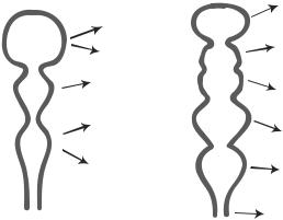
(midbrain), and rhombencephalon (hindbrain) (Fig. 8.4). The prosencephalon develops into the cerebrum, thalamus, hypothalamus, and retina. The mesencephalon gives rise to the cerebral aqueduct. The rhombencephalon forms the medulla oblongata and cerebellum. The posterior portion of the neural tube develops into the spinal cord.
Formation of Neurons and Glial Cells. There are two types of cell in the central nervous system: neurons and neuroglial cells. Neurons are the cells that can learn, memorize, sense signals from peripheral cells, and control the activity of other cell types. Each neuron is composed of a long axon, which physically connects the central and the peripheral nervous systems and is responsible for signal transduction and information transmission between the central and peripheral systems. In addition, each neuron consists of a large number of short processes known as dendrites, which contact other neurons within the central nervous system and are responsible for neuron–neuron communication.
Neuroglial cells serve to support and protect the neurons and to assist in neuronal development. There are three types of neuroglial cell: oligodendrocyte, astrocyte, and microglial cell. The oligodendrocyte can form large membrane processes called myelin sheaths, which wrap around the neuronal axons. The myelin sheaths have several functions: axon protection, molecular transport into and from the axon, maintenance of the ionic environment of the neuron, and assistance in the transmission of the action potential. The astrocyte also contributes to myelin formation and assists in neuronal function. The microglial cell behaves as a phagocyte type that degrades and removes debris from apoptotic cells.
The neuron, oligodendrocyte, and astrocyte are derived from the neural stem cells, defined as germinal neuroepithelial cells, in the neural tube. The local environment, to which a neural stem cell is migrating, is a critical factor that regulates the differentiation of the neural stem cells. It is believed that a neural tube cell can develop into either a neuron or glial cell depending on the local stimulation. However, the mechanisms remain poorly understood. Although the microglial cell appears as an interstitial cell type in the
354 EMBRYONIC ORGAN DEVELOPMENT
central nervous system, it is not derived from the ectodermal neural tube, but from the mesoderm. Mesodermal cells can migrate into the central nervous system and form microglial cells during development.
In the neural tube, neural stem cells are organized into a single-cell layer and are aligned in the direction perpendicular to the surface of the neural tube. These cells often conduct nonsymmetric division along their axial direction. The daughter cells adjacent to the luminal surface of the neural tube remain to be stem cells, whereas the daughter cells adjacent to the external surface differentiate into committed neural progenitor cells. With continuous development, the neural tube is organized into a structure with three zones: ventricular, intermediate, and marginal zones. The ventricular zone contains neural stem cells, the intermediate zone contains committed neural progenitor cells, and the marginal zone contain neurons and neuronal axons, depending on the subsystems of the brain such as spinal cord, cerebellum, or cerebrum. From the ventricular zone, the committed neural progenitor cells migrate outward into the intermediate zone. In the intermediate zone, different parts of the central nervous system are established. The patterns of cell migration, division, and organization vary considerably between the spinal cord, cerebellum, and cerebrum. The formation of these structures is briefly discussed here.
For the formation of the spinal cord, cells in the intermediate zone form axons, which extend from the intermediate zone toward the marginal zone, where the density of neurons reduces compared with the intermediate zone. With further development, established neuroglial cells migrate into the marginal zone and form myelin sheaths that enclose the axons. The neuron-containing mantle zone eventually develops into the butterflyshaped gray matter of the spinal cord, whereas the axon-containing marginal zone forms the peripheral white matter. The terms of gray matter and white matter are defined based on the color of the structures under an optical microscope. In the spinal cord, neurons localized to different regions possess distinct functions. Neurons in the dorsal half of the spinal cord are responsible for receiving and processing sensory signals from peripheral neuronal sensors, whereas the ventral neurons are for the motor control function.
For the formation of the cerebellum, the pattern of neural cell migration and organization differs from that for the spinal cord. The neuronal stem cells for the development of the cerebellum are evolved to form several types of cerebellar cells, including neurons that form cerebellar nuclei, granule neurons, and Purkinje neurons. For the formation of neuronal nuclei, the neuronal progenitor cells from the neural tube migrate out of the neural tube, establish the intermediate zone, and continue to migrate forward to enter the marginal zone, where they form neuronal nuclei, serving as relay units between different systems of the cerebellum and between the cerebellum and other systems of the brain. Another group of neuronal progenitor cells migrates through the intermediate zone, enters the marginal zone, and forms a new layer called the external granule layer adjacent to the external surface. Cells in the external granule layer make a turn and migrate inward. At the interface between the marginal and intermediate zones, these cells form the granule neurons. In addition, the neural tube stem cells differentiate into progenitor cells for the Purkinje neurons. These cells migrate to the marginal zone, forming a Purkinje cell layer. The Purkinje neurons are responsible for communications between different systems of the cerebellum.
The cerebrum is originated from the anterior portion of the neural tube and organized into a layered structure. During cerebral formation, cerebral progenitor cells are generated from the stem cells located in the ventricular zone, pass through the intermediate zone,

DEVELOPMENT OF MESODERM-DERIVED ORGANS |
355 |
Meninges
Marginal
zone
Cortical
plate
Subplate
Intermediate
zone
Subventricular
zone
Ventricular
zone
time
Figure 8.5. Migration and formation of cortical neurons. During development, neurons are generated in the ventricular zone, migrate along radially aligned glial cells through the subventricular zone, intermediate zone, and subplate, and are deployed to the cortical plate. (Reprinted by permission from Bielas S et al: Annu Rev Cell Dev Biol 20:593–618, copyright 2004 by Annual Reviews, www.annualreviews.org.)
and enter the marginal zone, where they form the cortical plate or the neocortex (Fig. 8.5). The neural progenitor cells in the cortical plate proliferate and differentiate into several layers of neurons in the vertical direction (perpendicular to the surface of the neural tube). Cells in different layers are responsible for the control of different peripheral systems. The cerebral neurons are also organized into a large umber of regions in the horizontal direction. Each region controls a corresponding peripheral system. In the cerebrum, neurons are concentrated in the cortical zone. This zone is referred to as the gray matter based on the color of the neuron-rich brain tissue. The intermediate zone is primarily composed of axons, which appear whitish under an optical microscope, and is referred to as the white matter.
DEVELOPMENT OF MESODERM-DERIVED ORGANS
The mesoderm is the middle layer of the three embryonic germ layers established during gastrulation and is the origin of a number of systems, including the cardiovascular system, lymphatic system, skeletal muscle system, bone, cartilage, connective tissue, kidney, and gonads. The mesoderm is composed of four major structures: the notochord (chordamesoderm), paraxial mesoderm, intermediate mesoderm, and lateral plate mesoderm. The organization, development, and fate of these structures are outlined here.