

Bioregenerative Engineering Principles and Applications - Shu Q. Liu
..pdf326 FUNDAMENTAL CELLULAR FUNCTIONS
Furukawa Y, Nishimura N, Furukawa Y, Satoh M, Endo H et al: Apaf-1 is a mediator of E2F-1- induced apoptosis, J Biol Chem 277:39760–8, 2002.
Honarpour N, Gilbert SL, Lahn BT, Wang X, Herz J: Apaf-1 deficiency and neural tube closure defects are found in fog mice, Proc Natl Acad Sci USA 98:9683–7, 2001.
Kim H, Jung YK, Kwon YK, Park SH: Assignment of apoptotic protease activating factor-1 gene (APAF1) to human chromosome band 12q23 by fluorescence in situ hybridization, Cytogenet Cell Genet 87:252–3, 1999.
Marsden VS, O’Connor L, O’Reilly LA, Silke J, Metcalf D et al: Apoptosis initiated by Bcl-2- regulated caspase activation independently of the cytochrome c/Apaf-1/caspase-9 apoptosome, Nature 419:634–7, 2002.
Riedl SJ, Li W, Chao Y, Schwarzenbacher R, Shi Y: Structure of the apoptotic protease-activating factor 1 bound to ADP, Nature 434:926–33, 2005.
Soengas MS, Capodieci P, Polsky D, Mora J, Esteller M et al: Inactivation of the apoptosis effector Apaf-1 in malignant melanoma, Nature 409:207–11, 2001.
Srinivasula SM, Ahmad M, Fernandes-Alnemri T, Alnemri ES: Autoactivation of procaspase-9 by Apaf-1-mediated oligomerization, Mol Cell 1:949–57, 1998.
Yoshida H, Kong YY, Yoshida R, Elia AJ, Hakem A et al: Apaf1 is required for mitochondrial pathways of apoptosis and brain development, Cell 94:739–50, 1998.
Zou H, Henzel WJ, Liu X, Lutschg A, Wang X: APAF-1, a human protein homologous to C. elegans CED-4, participates in cytochrome c-dependent activation of caspase-3, Cell 90:405–13, 1997.
Caspase 9
Hadano S, Nasir J, Nichol K, Rasper DM, Vaillancourt JP et al: Genomic organization of the human caspase-9 gene on chromosome 1p36.1-p36.3, Mam Genome 10:757–60, 1999.
Hakem R, Hakem A, Duncan GS, Henderson JT, Woo M et al: Differential requirement for caspase 9 in apoptotic pathways in vivo, Cell 94:339–52, 1998.
Kuida K, Haydar TF, Kuan CY, Gu Y, Taya C et al: Reduced apoptosis and cytochrome c-mediated caspase activation in mice lacking caspase 9, Cell 94:325–7, 1998.
Li P, Nijhawan D, Budihardjo I, Srinivasula SM, Ahmad M et al: Cytochrome c and dATPdependent formation of Apaf-1/caspase-9 complex initiates an apoptotic protease cascade, Cell 91:479–89, 1997.
Marsden VS, O’Connor L, O’Reilly LA, Silke J, Metcalf D et al: Apoptosis initiated by Bcl-2- regulated caspase activation independently of the cytochrome c/Apaf-1/caspase-9 apoptosome, Nature 419:634–7, 2002.
Srinivasula SM, Hegde R, Saleh A, Datta P, Shiozaki E et al: A conserved XIAP-interaction motif in caspase-9 and Smac/DIABLO regulates caspase activity and apoptosis, Nature 410:112–16, 2001.
Caspase 3
Fernandes-Alnemri T, Armstrong RC, Krebs J, Srinivasula SM, Wang L et al: In vitro activation of CPP32 and Mch3 by Mch4, a novel human apoptotic cysteine protease containing two FADDlike domains, Proc Natl Acad Sci USA 93:7464–9, 1996.
Fernandes-Alnemri T, Litwack G, Alnemri ES: CPP32, a novel human apoptotic protein with homology to Caenorhabditis elegans cell death protein Ced-3 and mammalian interleukin-1 beta-converting enzyme, J Biol Chem 269:30761–4, 1994.
Fernando P, Kelly JF, Balazsi K, Slack RS, Megeney LA: Caspase 3 activity is required for skeletal muscle differentiation, Proc Natl Acad Sci USA 99:11025–30, 2002.
BIBLIOGRAPHY 327
Gervais FG, Xu D, Robertson GS, Vaillancourt JP, Zhu Y et al: Involvement of caspases in proteolytic cleavage of Alzheimer’s amyloid-beta precursor protein and amyloidogenic A-beta peptide formation, Cell 97:395–406, 1999.
Jiang X, Kim HE, Shu H, Zhao Y, Zhang H et al: Distinctive roles of PHAP proteins and prothy- mosin-alpha in a death regulatory pathway, Science 299:223–6, 2003.
Kuida K, Zheng TS, Na S, Kuan C, Yang D et al: Decreased apoptosis in the brain and premature lethality in CPP32-deficient mice, Nature 384:368–72, 1996.
Mannick JB, Hausladen A, Liu L, Hess DT, Zeng M et al: Fas-induced caspase denitrosylation, Science 284:651–4, 1999.
Miura M, Chen XD, Allen MR, Bi Y, Gronthos S et al: A crucial role of caspase-3 in osteogenic differentiation of bone marrow stromal stem cells, J Clin Invest 114:1704–13, 2004.
Nasir J, Theilmann JL, Chopra V, Jones AM, Walker D et al: Localization of the cell death genes CPP32 and Mch-2 to human chromosome 4q, Mam Genome 8:56–9, 1997.
Nicholson DW, Ali A, Thornberry NA, Vaillancourt JP, Ding CK et al: Identification and inhibition of the ICE/CED-3 protease necessary for mammalian apoptosis, Nature 376:37–43, 1995.
Woo M, Hakem R, Furlonger C, Hakem A, Duncan GS et al: Caspase-3 regulates cell cycle in B cells: A consequence of substrate specificity, Nature Immun 4:1016–22, 2003.
Human protein reference data base, Johns Hopkins University and the Institute of Bioinformatics, at http://www.hprd.org/protein.
6.17. Assessment of Cell Apoptosis
Yan N, Shi Y: Mechanisms of apoptosis through structural biology, Annu Rev Cell Dev Biol 21:35–56, 2005.
Nagata S: DNA degradation in development and programmed cell death, Annu Rev Immunol 23:853–75, 2005.
Jiang X, Wang X: Cytochrome C-mediated apoptosis, Annu Rev Biochem 73:87–106, 2004.
Sancar A, Lindsey-Boltz LA, Unsal-Kacmaz K, Linn S: Molecular mechanisms of mammalian DNA repair and the DNA damage checkpoints, Annu Rev Biochem 73:39–85, 2004.
Bard JB: Growth and death in the developing mammalian kidney: Signals, receptors and conversations, Bioessays 24:72–82, 2002.
Tilly JL: Commuting the death sentence: How oocytes strive to survive, Nat Rev Mol Cell Biol 2:838–48, 2001.
Kuan CY, Roth KA, Flavell RA, Rakic P: Mechanisms of programmed cell death in the developing brain, Trends Neurosci 23:291–7, 2000.
Nijhawan D, Honarpour N, Wang X: Apoptosis in neural development and disease, Annu Rev Neurosci 23:73–87, 2000.
Hsu SY, Hsueh AJ: Tissue-specific Bcl-2 protein partners in apoptosis: An ovarian paradigm, Physiol Rev 80:593–614, 2000.
Fraser A, McCarthy N, Evan GI: Biochemistry of cell death, Curr Opin Neurobiol 6:71–8, 1996.

SECTION 3
DEVELOPMENTAL ASPECTS OF BIOREGENERATIVE ENGINEERING
7
FERTILIZATION AND EARLY
EMBRYONIC DEVELOPMENT
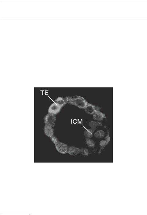
Fluorescent micrograph showing a mouse blastocyst. TE: trophoblast. ICM: inner cell mass. Cells were labeled for mitochondria (green) and nuclei (blue). (Reprinted from Houghton FD: Energy metabolism of the inner cell mass and trophectoderm of the mouse blastocyst, Differentiation 74:11–8, 2006 by permission of Blackwell Publishing.) See color insert.
Bioregenerative Engineering: Principles and Applications, by Shu Q. Liu
Copyright © 2007 John Wiley & Sons, Inc.
329
330 FERTILIZATION AND EARLY EMBRYONIC DEVELOPMENT
An animal undergoes a developmental cycle composed of a series of biological processes: the initiation, development, maturation, and reproduction of the animal. The repetition of these processes is the foundation for the continuation of the life. The initiation and development occur during the embryonic period and are collectively referred to as embryogenesis. It is in this period that a new generation of animals forms. In tradition, the embryonic development of an animal is divided into two stages: the embryonic and fetal stages based on distinct markers of functional anatomy.
The embryonic stage is defined in the human as the period from the conception to the formation of primary organs at about the eighth week. This stage is divided into two substages: germinal and embryonic development. Germinal development takes place in the human from the conception to the formation of the three germinal layers, including the ectoderm, mesoderm, and endoderm, at the end of the second week. During this period, a male gamete (sperm) fuses into a female gamete (oocyte) to form a zygote, a process known as fertilization. The fusion of the two gametes allows the integration of the genomes from both parents, an essential process for transmitting genetic information from the parents to the progeny and for initiating the development of a new individual. Fertilization triggers an early mitotic segmentation process, known as cleavage, by which a fertilized egg is divided continuously into smaller cells. When reaching a certain cell density, the cells are organized into various patterns, which undergo dynamic changes through different stages. The embryonic cells are subsequently committed to gastrulation, a process leading to the formation of a three-layered structure known as gastrula, composed of the ectoderm, mesoderm, and endoderm. The formation of a gastrula takes place during the first two weeks and is indicative of the ending of the germinal period.
Embryonic development takes place from the second to the eighth week. During this period, the basic forms of major organs are developed from the three germ layers: ectoderm, mesoderm, and endoderm. The ectoderm gives rise to the central nervous system (brain and spinal cord), peripheral nerve structures, and the epidermis of the skin, teeth, nose, and external ear. The mesoderm is the origin of the heart, vascular system, blood, muscle, bone, cartilage, and connective tissue. The endoderm develops into the gastrointestinal tract, liver, pancreas, bladder, and lung. By the end of the eighth week, tissues and organs are assembled into the primary form of a fetus. The embryonic stage is the period during which most dynamic changes take place in morphogenesis.
The fetal stage is the remaining period from the formation of the fetus to the parturition or birth of a new individual. During this stage, the fetus gains size rapidly from several centimeters to about half a meter ( 0.5 m), but the form of the tissues and organs does not change as vigorously as that during the embryonic stage. The entire embryonic period from conception to parturition is about 40 weeks.
In this chapter, the developmental processes during embryogenesis will be introduced with emphasis on the morphogenesis of embryonic structures and related regulatory mechanisms during each stage. We will see that, although bioregeneration is defined as a process occurring during the adulthood for the repair and reconstruction of lost tissues and organs, it is similar to embryonic development in many aspects.
THE SPERM [7.1]
The male and female gametes are essential components for the initiation of embryogenesis and the development of a new individual. A male gamete, known as sperm or spermato-
THE SPERM |
331 |
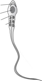
Acrosomal vesicle
Nucleus
Centriole
Mitochondria
Axoneme 
Flagellum 
Figure 7.1. Schematic representation of a sperm cell. Based on bibliography 7.1.
zoon, is composed of several systems, including a haploid nucleus, a propulsion apparatus, and protein enzymes that are required for its interaction and fusion with a female gamete. During sperm maturation, the volume of the cytoplasm is minimized to reduce the size of the sperm, but the structures that regulate the sperm–egg interaction are evolved. These include the acrosomal vesicle and the flagellum (Fig. 7.1). The acrosomal vesicle is originated from the Golgi apparatus and located in front of the nucleus. This structure contains enzymes that are necessary for degrading the external layer of the egg during sperm–egg fusion. The flagellum is developed on the basis of the centrioles and is responsible for sperm movement, which is driven by a motile apparatus known as axoneme. The motile apparatus is composed of three-dimensionally organized microtubules, consisting of dimeric tubulins, and a type of motor protein known as dynein. A dynein molecule is attached to the microtubule and serves as an enzyme that hydrolyzes ATP. The hydrolysis of ATP provides energy necessary for the motile activity of dynein molecules and the flagellar propulsion, which induces sperm movement.
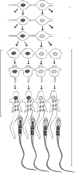
Sperms are generated from a cell type called primordial germ cell in the testes, the male reproductive organs. The process of sperm generation is referred to as spermatogenesis (Fig. 7.2). The primordial germ cells are specified during the early embryonic cell cleavage stage. A fraction of cells at the 8/16-cell stages develop into primordial germ cells. These cells usually contain mRNAs and proteins that are necessary for the development of gametes. The cytoplasm of these cells is called germ plasm. Sperms are formed via meiosis of the primordial germ cells. These cells are formed in the epiblast, an early embryonic structure that develops from the inner cell mass of a blastocyst and gives rise to the ectoderm, mesoderm, and endoderm. When the epiblast is developed into the three germ layers, the primordial germ cells are localized to the endoderm. With further development, these germ cells migrate into the genital ridges, where gonads (ovaries and testes)
332 FERTILIZATION AND EARLY EMBRYONIC DEVELOPMENT
Spermatogonia |
|
type B |
|
Primary |
|
spermatocytes |
Meiosis |
Secondary |
|
spermatocytes |
|
|
Spermatids
Differentiation
Sperm cells
Figure 7.2. Schematic demonstration of spermatogenesis. Based on bibliography 7.1.
are developed, and remain relatively quiescent in a structure called sex cord until reaching maturity.
In the sex cord, which develops into the seminiferous tubules of the testis at puberty, the primordial germ cells differentiate into type A1 spermatogonia, which can subsequently differentiate into several levels of spermatogonia, including types A2, A3, and A4 spermatogonia (type A2 differentiates into A3, and A3 into A4). These type A
THE EGG |
333 |
spermatogonia are stem cells in nature and can self-renew themselves as well as differentiate into specified cell types. Type 4 spermatogonia can further differentiate into a hierarchy of several cell types, including intermediate spermatogonia, spermatozoa, type B spermatogonia, and primary spermatocytes. Up to this point, mitosis is the basic form of cell division. The primary spermatocytes can divide to form another hierarchy of sperm progenitor cells via meiosis, including secondary spermatocytes and spermatids. The spermatids give rise to sperms (Fig. 7.2).
THE EGG [7.1]
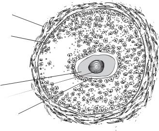
The egg is the female gamete and is also known as the ovum. A developing egg before its complete meiotic division or formation of a haploid nucleus is called an oocyte. An egg is composed of a nucleus and an enormous volume of cytoplasm. The egg stores a large amount of food and energy-producing materials in its cytoplasm for the growth and development of the embryo. In addition, the egg cytoplasm contains a variety of proteins (structural, regulatory, morphogenetic, and protective factors), rRNAs, tRNAs, and mRNAs. The egg cytoplasm is enclosed within the cell membrane. The egg cell membrane is surrounded by an extracellular matrix layer known as zona pellucida. Outside the zona pellucida, there exists a thick cellular structure called cumulus, composed of a large number of ovarian follicular cells (Fig. 7.3). The follicular cells provide soluble factors and mechanical protection to the egg cell.
The process of egg formation is referred to as oogenesis. This process is different from spermatogenesis. Whereas spermatogenesis produces a nucleus-predominant sperm with strong motility, oogenesis gives rise to eggs with materials and factors necessary for embryonic development. In the human, there exist a limited number of female primitive germ cells (about thousand) called oogonia. These cells can divide into several millions
Thecal cells
Granulosa membrane
Granulosa cells 
Antrum 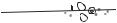
Oocyte
Nucleus
Zona pellucida
Figure 7.3. Schematic representation of an ovarian follicle. Based on bibliography 7.1.
334 FERTILIZATION AND EARLY EMBRYONIC DEVELOPMENT
of secondary germ cells via mitosis from the second to the seventh month of gestation. A large number of germ cells are committed to apoptosis afterward. The surviving germ cells give rise to primary oocytes via the first meiotic cycle. The primary oocytes are prompted to enter the prophase and metaphase of meiosis and maintain a quiescent state until puberty. When a human female individual reaches maturity, oocytes are periodically committed to meiosis and formation of mature eggs. Such an activity usually begins at the age of 13 years and disappears at the age of 50 years; these events are termed menarche and menopause, respectively. About 400 eggs can be formed through the lifespan of a female individual.
FERTILIZATION [7.1]
Fertilization is a process by which a sperm fuses into an oocyte to form a zygote, which develops into a new individual through embryogenesis. Note that the term oocyte is used here instead of egg because a sperm usually fuses with an oocyte or developing egg, which is arrested in the metaphase of meiosis and has not completed meiosis. Fertilization takes place via several steps, including: (1) attraction of sperm to an oocyte by chemotactic factors released from the oocyte, (2) interaction between the sperm and the oocyte, (3) penetration of the sperm through the extracellular layers of the oocyte, (4) the entrance of the sperm into the oocyte, and (5) fusion of the sperm with the oocyte. These steps are briefly outlined here.
Attraction of Sperm Cells to the Oocyte
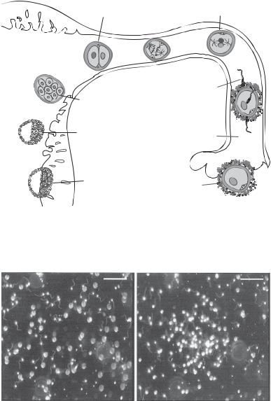
In mammals, oocytes are produced and released from the ovary and moved to the oviduct. The interaction of the oocyte with sperm occurs in the ampulla region of the oviduct, which is near the ovary (Fig. 7.4). Sperm are capable of migrating toward an oocyte. Such an activity is induced and controlled by chemotaxis, or chemical gradient-directed cell movement. An oocyte can produce and release sperm-attracting proteins, which can act on specific membrane receptors in the sperm. An example of such sperm-attracting protein is resact, discovered in the sea urchin oocytes. This molecule can induce strong chemotactic activity of the sperm in vitro (Fig. 7.5). It is important to address that the chemotactic activity of sperm is dependent on animal species. Sperm can be activated and committed to directed migration only in response to a chemoattractant released from the oocytes of the same species. Furthermore, the sperm attraction activity is dependent on the developmental stage. Sperm rarely move toward oocytes that have not yet committed to the second meiosis, since these oocytes do not release sufficient chemoattractants.
Sperm–Oocyte Interaction
When a sperm approaches an oocyte, the physical interaction of the sperm with the extracellular layer of the oocyte induces the activation of the acrosomal vesicle of the sperm. In mammals, the sperm first passes through the follicular cell layer. When reaching the zona pellucida, the acrosomal vesicle undergoes exocytosis, a process that releases proteolytic enzymes. These enzymes degrade the matrix of the zona pellucida and thus help the sperm approach the oocyte membrane. The exocytosis of the acrosomal vesicle is induced by the binding of a zona pellucida protein, zona protein 3 (ZP3), to a specific
FERTILIZATION |
335 |
2-cell stage |
Zygote |
|
Uterus
|
Sperm |
Morula |
|
Blastocyst |
Oviduct |
Implanted |
Oocyte |
blastocyst |
Figure 7.4. Schematic representation of the locations for oocyte fertilization, morula formation, and blastocyst formation and implantation in the human oviduct and uterus. Based on bibliography 7.1.
A B
Figure 7.5. Resact-mediated movement of sperm cells. (A) Dispersed control sperm cells without the release of resact. (B) Chemotactic accumulation of sperm cells in response to the release of resact. Scale bar: 100 μm. (Reproduced from Solzin J et al: J General Physiol 124:115–24, 2004 copyright by permission of The Rockefeller University Press.)
sperm surface receptor, galactosyltransferase-I. The binding activity stimulates a G- protein-mediated signaling pathway and results in the release of calcium, which in turn induces the release of proteolytic enzymes from the acrosomal vesicle. Since the structure of the ligand and receptor is specific to an animal species, sperm can interact only with the egg zona pellucida of the same species.