

Bioregenerative Engineering Principles and Applications - Shu Q. Liu
..pdfSECTION 2
REGULATORY MECHANISMS OF REGENERATION

5
CELL SIGNALING PATHWAYS AND MECHANISMS
Interaction of bone marrow-derived smooth muscle α actin+ cells with CD11b+ cells in culture. Mouse bone marrow cells were collected and cultured in DMEM with 10% FBS. Smooth muscle α actin+ cells (green) were often found on top of CD11b+ cells (red). When the CD11b+ cells were selectively removed, the number of smooth muscle α actin+ cells was reduced, suggesting that the CD11b+ bone marrow cells play a role in regulating the development of smooth muscle cells from bone marrow progenitor cells. Blue: cell nuclei. Scale: 10 μm. See color insert.
Bioregenerative Engineering: Principles and Applications, by Shu Q. Liu
Copyright © 2007 John Wiley & Sons, Inc.
147
148 CELL SIGNALING PATHWAYS AND MECHANISMS
PRINCIPLES OF CELL SIGNALING [5.1]
In a multicellular system, there exist signaling activities at the molecular and cellular levels for cell-to-cell and cell-to-matrix communications, which are essential for the function of cells, tissues, and organs. In order to conduct physiological function, a cell must communicate with other cells to achieve coordinated cellular activities. Often, synchronized molecular and cellular activities are required for tissue and organ functions. A typical example is the control of the contractile activity of skeletal muscle cells in response to an electrical stimulation from a nerve axon. In such a regulatory process, an electrical action potential is generated from a motor neuron center and transmitted through the nerve axon to the muscle cell. At the junction, or synapse, between the nerve axon and the muscle cell, the electrical signal is converted to a chemical signal, which induces the activation of an intracellular signaling cascade in the muscle cells, resulting in cell contraction. Without neuron-to-muscle cell signal transduction, the skeletal muscle cells cannot contract or relax in a controlled manner. Thus, coordinated cell signaling is a fundamental process for accomplishing complicated functional tasks. Here, the principles of cell signaling and common cell signaling pathways are discussed.
Factors Serving as Signals
There are two basic types of factors, which serve as “signals” for regulating molecular and cellular activities as well as cell-to-cell and cell-to-matrix communications: extracellular factors and intracellular factors (Fig. 5.1). Extracellular facotrs are present in extracellular space and serve as signals that initiate and control molecular and cellular activities. Intracellular factors are present in the cytoplasm, serve as elements for signaling pathways, and regulate cellular activities. In general, an extracellular signal must cooperate with intracellular signals to initiate and control a cellular activity. The extracellular signal may initiate the activation of an intracellular signaling pathway via interacting with a cell membrane or cytoplasmic signaling factor, whereas the intracellular signaling pathway relays the extracellular signal to target subcellular organelles, initiating a specified activity, such as gene expression, cell adhesion, cell proliferation, or cell migration.
Ions |
Shearing |
Fatty acids |
|
||
Proteins |
|
Steroids |
Nucleotides |
|
|
Proteins
Stretching Lipids Stretching
Steroids
Ions
Electrical signals |
Cell-cell contact |
Pressure
Figure 5.1. Extracellular and intracellular signals that regulate cell activities and functions (based on bibliography 5.1).
PRINCIPLES OF CELL SIGNALING |
149 |
Cellular activities are often initiated in response to environmental or extracellular stimulations or cues. Extracellular factors that serve as signaling factors and induce cellular activities may include biochemical molecules and substances, electrical signals, and physical factors. Biochemical molecules and substances include cell-secreted factors and extracellular matrix factors, such as ions, proteins, nucleotides, fatty acids, and steroids. Some of these factors, such as proteins (growth factors and extracellular matrix components) and nucleotides, can act on cell membrane receptors, while others, such as steroids, can diffuse through the cell membrane and interact directly with intracellular receptors, inducing activation of signaling pathways. Electrical signals are action potentials, or changes in membrane potentials. An electrical signal initiates the activation of nerve and muscular cells. Physical factors, such as mechanical forces and deformation, can serve as signaling factors. For instance, mechanical shearing and stretching forces due to bloodflow and pressure, respectively, induce various cellular activities, ranging from cell migration, proliferation to apoptosis, in the vascular system. These biochemical, electrical, and physical signals initiate and control specified cellular activities.
Intracellular signaling factors include proteins, lipids, steroids, and ions. These factors are also referred to as regulatory factors. In each cell, there exist a large number of proteins. Most of these protiens serve as signaling molecules. Common types of signaling proteins include cell membrane receptors, ion channels, protein kinases, protein phosphatases, and enzymes. Certain types of structural proteins, such as actin filaments, also play a role in signal transduction. A variety of lipid molecules in the cell membrane and endoplasmic reticulum serve as signaling factors. Ions, especially, calcium, play a critical role in signal transduction and the regulation of cellular activities. The roles of these signaling factors are discussed in the following sections.
Types of Cell Signaling
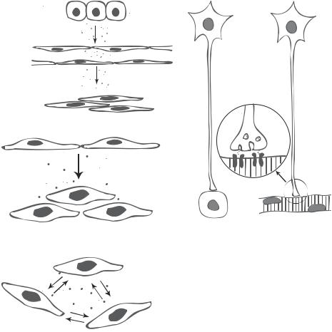
There are various cell signaling mechanisms. Based on the range of signal transduction, cell signaling events can be divided into longand short-range events (Fig. 5.2). Longrange signaling events involve cells from different systems. These events are mediated by either hormones or synapses. Hormones are biochemical factors, which are synthesized by endocrine gland cells and released into blood. These factors regulate the activities of remote cells. This type of signaling is also known as endocrine signaling. Since a hormone is delivered via the bloodflow, it can reach almost all cells in the body. The specificity of hormones is dependent on the receptors on the target cells. Synapses are subcellular structures found at the junctions between different neurons and between neurons and muscular cells. In a synapse-related signaling event, signals from a presynaptic neuron are conducted to another neuron or a peripheral muscular cell via the propagation of electrical action potentials, which stimulate the release of neurotransmitters at the terminal of the presynaptic cell. The synapse in turn delivers the neurotransmitters to the postsynaptic target cell, initiating postsynaptic cell activation. Synaptic signaling events influence target cells with great precision. In contrast to long-range signaling, short-range signaling events involve cells within a local neighborhood. These events are mediated by regional chemical factors and mechanical forces. Changes in mechanical forces often induce the activation and release of chemical factors, which in turn transmit mechanical signals to the cell. Signaling events mediated by regional chemical factors are often referred to as paracrine signaling. Chemical factors for paracrine signaling are usually
150 CELL SIGNALING PATHWAYS AND MECHANISMS
Endocrine cells
Hormone
Capillary
Hormone
Target cells
Hormone
 Mediators
Synapse
Paracrine
Mediators
Autocrine
Figure 5.2. Long-range (hormoneand synapse-mediated) and short-range (paracrine and autocrine) cell signaling (based on bibliography 5.1).
endocytosed by neighboring cells or immobilized by extracellular matrix, ensuring a local influence without spreading to remote cells.
General Mechanisms of Cell Signaling
On the stimulation of an extracellular signal, a cell responds by activating its intracellular signaling pathways, leading to specific cellular activities, such as gene expression, cell division, cell migration, cell adhesion, and cell apoptosis. Several steps are involved in the activation of the intracellular signaling pathways: interaction of an extracellular signaling factor with a corresponding receptor or factor in the target cell, transduction of the extracellular signal to the target cell, activation of intracellular biochemical and/or electrical reactions, and termination of intracellular reactions.
PROTEIN TYROSINE KINASE-MEDIATED CELL SIGNALING |
151 |
A large number of molecules, including proteins, lipids, and ions, can serve as intracellular signaling molecules and participate in signal transduction. Proteins, including receptors, enzymes, and adapters, are among the common signaling molecules. Receptors are distributed in the cell membrane or cytoplasm, and are responsible for receiving extracellular signals and transmitting the signals into intracellular signaling pathways. Enzymes involved in cell signaling primarily include protein kinases, protein phosphatases, and GTPases. Protein kinases and phosphatases are responsible for protein phosphorylation and dephosphorylation, respectively, which are critical processes for cell signaling. GTPases serve as switches for signal transduction to downstream signaling elements. Adapter proteins help to target, recruit, and co-localize proteins. Some adapter proteins serve as scaffolds or docking sites for signaling proteins.
Signaling molecules exist in two modes: inactive and active. In unstimulated cells, signaling molecules are present mostly in the inactive mode. In response to a stimulation, specified signaling molecules can be activated by various mechanisms, including chemical modification, enzymatic activation, conformational changes in signaling molecules, alterations in molecular concentration, and col/localization and clustering of molecules. In most cases, multiple mechanisms are involved for the same or various signaling molecules at a given time and location.
There are several common features for cell signaling. These include signaling specificity, the involvement of signaling cascades, and crosstalk between signaling pathways. In general, each signaling molecule reacts with specific upstream and downstream molecules, ensuring the induction of specific activities. Various signaling pathways are designed and developed for specific cellular activities. For the regulation of each cellular activity, multiple signaling molecules and pathways may be involved. In addition, each signaling molecule may exhibit multiple functions. The abundance and multifunctionality of signaling molecules may be a mechanism that ensures the accomplishment of cellular activities and functions.
Each signaling pathway is composed of a number of signaling molecules, which relay signals from extracellular space to the cytoplasm and nucleus. Such a cascade is also referred to as a signaling cascade. In each cell, there exist multiple signaling pathways. These pathways often communicate or crosstalk with each other via branching pathways, forming signaling networks. Through these crosstalk pathways, a signaling molecule can be activated by different upstream signaling molecules and can act on different downstream effectors. With such an approach, various upstream signals can be converged to a single signaling pathway, and one activated signaling molecule can initiate multiple downstream activities. In addition, signals in one pathway can influence signals in other pathways. Thus crosstalk is an effective approach for amplifying and controlling signals and facilitating signal transduction. In the following sections, common signaling pathways in mammalian cells are briefly reviewed.
PROTEIN TYROSINE KINASE-MEDIATED CELL SIGNALING [5.2]
Protein tyrosine kinases belong to the superfamily of protein kinases. Protein kinases are enzymes that catalyze the phosphorylation of, or the addition of a phosphate group to, a substrate protein. Protein phosphorylation is a major form of molecular modification, which mediates a variety of molecular activities, including enzymatic activation, cell signaling, gene transcription, activation of ion channels, and reorganization of cytoskeletal
152 CELL SIGNALING PATHWAYS AND MECHANISMS
proteins. Protein kinases represent a large family of signaling molecules. The genes encoding protein kinases constitute about 1.7% of total genes in the human. Based on the types of target amino acid, protein kinases are classified into four types: serine/threonine-, tyrosine-, histidine-, and aspartate- (or glutamate-) specific protein kinases. These protein kinases catalyze the phosphorylation of substrate proteins on serine/threonine, tyrosine, histidine, and aspartate (or glutamate), respectively. Among these protein kinases, serine/ threonine and tyrosine protein kinases can phosphorylate signaling proteins, play critical roles in signal transduction. Thus, these two types of protein kinase are the focus of this book. The protein tyrosine kinases are covered in this section. The protein serine/ threonine kinases are covered later in this chapter.
Structure and Function
Protein tyrosine kinases are enzymes that catalyze the phosphorylation of the tyrosine residues of substrate proteins. A protein tyrosine kinase is often found as an integral part of growth factor receptors in the cell membrane and is referred to as receptor protein tyrosine kinase. A receptor with a protein tyrosine kinase is known as protein tyrosine kinase receptor. Such a receptor is composed of an extracellular domain for ligand binding, a transmembrane domain for anchoring the receptor to the cell membrane, and an intracellular domain for interaction with cytoplasmic signaling molecules. The receptor protein tyrosine kinase is localized to the cytoplasmic domain of the growth factor receptor. The receptor tyrosine kinase assists the growth factor receptor in transducing signals that induce essential cell activities, such as proliferation, differentiation, and migration. There are also nonreceptor protein tyrosine kinases such as the Src family of protein tyrosine kinases. These kinases are discussed on page 180.
A signaling pathway mediated by a protein tyrosine kinase receptor is composed of several components, including the extracellular ligands, cell membrane receptors, cytoplasmic signaling elements, and transcriptional factors. Extracellular ligands are usually growth factors produced and released by cells. There are a number of growth factors that interact and activate protein tyrosine kinase receptors. These include epidermal growth factor (EGF), platelet-derived growth factors (PDGFs) A and B, nerve growth factor (NGF), basic fibroblast growth factor (bFGF), vascular endothelial growth factor (VEGF), insulin-like growth factor (ILGF), nerve growth factor (NGF), hepatocyte growth factor (HGF), and ephrins. Based on the type of ligand growth factors, growth factor receptors are classified as EGF receptor, PDGF receptor, NGF receptor, FGF receptor, VEGF receptor, insulin receptor, HGF receptor, and Eph receptor groups, respectively (see Fig. 5.3 for schematic representation of growth factor receptors). The ligand–receptor interaction is highly specific. Each growth factor only interacts with and activates a specific protein tyrosine kinase receptor.
The EGFR group is composed of several receptors, including EGFR (epidermal growth factor receptor), ERBB2 (v-erb-b2 erythroblastic leukemia viral oncogene homolog 2 or neuro/glioblastoma-derived oncogene homolog), ERBB3 (v-erb-b2 erythroblastic leukemia viral oncogene homolog 3), and ERBB4 (v-erb-a erythroblastic leukemia viral oncogene homolog 4). These receptors are expressed in various cell types, including epithelial, mesenchymal, and neural cells. The EGFR contains two extracellular cysteine-rich domains, a transmembrane domain, and a cytoplasmic tyrosine kinase domain (Fig. 5.3). This receptor interacts with EGF and similar ligands, regulating cell development, morphogenesis, regeneration, and tumorigenesis.

rich - Cysteine
Kinase
like-Ig
PROTEIN TYROSINE KINASE-MEDIATED CELL SIGNALING |
153 |
||||||
|
|
|
|
|
|
Sema |
|
rich |
like-Ig |
like-Ig |
li-Igke |
|
like-Ig |
PSI |
3Fibronectin |
- |
|
||||||
teine |
|
|
|
|
|
|
|
Cys |
|
|
|
|
|
|
|
|
|
|
|
|
|
|
|
|
|
|
|
|
|
|
|
SAM
EGFR |
PDGFR |
ILGFR |
VEGFR |
FGFR |
NGFR |
HGFR |
EphR |
Figure 5.3. Schematic representation of growth factor receptors. Sema: semaphorin domain. Ig: immunoglobulin domain. PSI: plexin, semaphorin, and integrin domain. SAM: sterile alpha motif (based on bibliography 5.2).
The PDGFR group consists of several molecules, including PDGFRα, PDGFRβ, CSF1R, KIT/SCFR, FLK2, and FLT3. Each of these receptors is composed of five IgD-like domains in the extracellular domains (Fig. 5.3). The intrinsic protein tyrosine kinase domain is split into two parts by an intervening segment. These receptors play a critical role in regulating the development of connective tissue cells and vascular smooth muscle cells.
The insulin receptor group includes three members: insulin receptor, insulin growth factor (IGF)1 receptor, and IRR, which are present in the form of dimers linked by disulfide bonds (Fig. 5.3). These receptors regulate processes related to cell survival.
The VEGFR group includes three members, including VEGFR1, VEGFR2, and VEGFR3, which are characterized by the presence of seven Ig-like domains in the extracellular domain (Fig. 5.3). These receptors are expressed primarily in vascular endothelial cells and regulate the development of endothelial cells, as well as angiogenesis and vasculogenesis.
The FGFR group consists of FGFR1, FGFR2, FGFR3, and FGFR4. The extracellular region contains three Ig-like domains (Fig. 5.3). These receptors mediate the development and morphogenesis of a variety of cell types in connective tissues and the cardiovascular system.
The nerve growth factor receptor group is composed of three members, including TrkA, TrkB, and TrkC. The extracellular domain of these receptors contains a LRD domain and two Ig-like domains, and the intracellular region contains a single protein tyrosine kinase domain (Fig. 5.3). These receptors are expressed in neurons and neural glial cells and play important roles in regulating the development and morphogenesis of nerve cells.
The HGFR group is composed of two members: Met and Ron. These receptors are expressed primarily in hepatocytes and also in other epithelial cells. The extracellular domain of the receptor is composed of a Sema (semaphorin) domain, a PSI (plexin, semaphorin, and integrin) domain, and four Ig-like domains and the intracellular domain contains a single-protein tyrosine kinase domain (Fig. 5.3). HGF receptors regulate the development and regeneration of the liver and other epithelial tissues.

154 CELL SIGNALING PATHWAYS AND MECHANISMS
The Eph receptor group includes at least 14 members, EPHA1 (8 members) and EPHB1 (6 members), and interacts with various types of ephrin. A typical Eph receptor contains two fibronectin 3 domains in the extracellular region and a tyrosine kinase domain in the cytoplasmic region. The cytoplasmic region also contains a SAM (sterile α motif) (Fig. 5.3). These receptors are expressed primarily in nerve cells and vascular endothelial cells, and play a critical role in the regulation of cell migration and morphogenesis.
Most receptors listed above interact with intracellular signaling molecules, including Src homology 2 domain-containing molecules and adapter proteins, leading to activation of a mitogenic signaling cascade composed of mitogen-activated protein kinase kinase kinases (MAPKKKs), mitogen-activated protein kinase kinases (MAPKKs), and mitogenactivated protein kinases (MAPKs). These molecules regulate cell proliferation, differentiation, and migration.
Signaling Mechanisms
The activation of protein tyrosine kinase receptor signaling pathways begins with the binding of extracellular ligands to the protein tyrosine kinase receptors (Fig. 5.4). These receptors undergo a dimerization process on the interaction with ligands, a common mechanism for activating the protein tyrosine kinase receptor signaling pathways. For various groups of protein tyrosine kinase receptors, there exist different forms of dimerization. For example, PDGF receptors are dimerized into a symmetric structure on the binding of a disulfide-bonded PDGF dimeric complex (Fig. 5.4). In contrast, EGF receptors undergo conformational changes in response to EGF binding, forming receptor– receptor complexes. The dimerization of receptors brings the intracellular domains of the two receptors together, a critical process for initiating autophosphorylation of the protein tyrosine kinase domains on tyrosine residues.
The autophosphorylation of a receptor protein tyrosine kinase activates the tyrosine kinase domain, leading to the activation of downstream signaling molecules, sucs as Src homology (SH)2 domain-containing protein tyrosine kinases and adapter proteins.
PDGF dimer
PDGF receptors
Cell membrane
PDGF receptor tyrosine kinase
Inactive |
Active |
Figure 5.4. Schematic representation of the interaction of platelet-derived growth factor (PDGF) with PDGF receptor. A PDGF molecule forms a dimer with another PDGF molecule (e.g., PDGFAA and PDGF-BB). A PDGF dimer can interact with two PDGF receptors, causing crosslink between the two PDGF receptors and autophosphorylation of the receptor protein tyrosine kinase in the cytoplasmic domain (based on bibliography 5.2).
PROTEIN TYROSINE KINASE-MEDIATED CELL SIGNALING |
155 |
A typical example of SH2 domain-containing kinases is the Src protein tyrosine kinase. The molecule possesses intrinsic enzymatic kinase activities and can induce phosphorylation on substrate proteins. Adapter proteins are proteins that serve as linkers between receptor protein kinases and downstream signaling molecules. Typical examples of adapter proteins include Grb2/sos, Crk, Nck, and Shc (these and other examples are listed in Table 5.1). These molecules do not possess intrinsic enzymatic kinase activities.
The interaction of an autophosphorylated receptor tyrosine kinase with a SH2 domaincontaining kinase or an adapter protein is a critical process for signal transduction. A phosphorylated tyrosine residue in the receptor tyrosine kinase can serve as a docking site for the SH2 domain of signaling molecules, which is organized into a pocket-like structure specific for interaction with a phosphorylated tyrosine. Each receptor tyrosine kinase is capable of interacting with multiple SH2 domain-containing molecules at various tyrosine sites. For example, the PDGF receptor tyrosine kinase contains about 12 tyrosine residues that can be phosphorylated within the intracellular domain (11 are outside and 1 is within the tyrosine kinase). Some of these phosphorylated tyrosine residues serve as docking sites for SH2 domain-containing tyrosine kinases, such as Src, GAP, SHP2, and PLCγ, and others serve as docking sites for adapter signaling proteins, such as Shc, Nck, and Grb2/Sos. The phosphorylated tyrosine at site 581 (pY581) can interact with Src and Shc, pY740 can interact with Shc and PI3-kinase, pY751 can interact with PI3-kinase and Nck, and pY771 can interact with GAP and Shc. The interaction of the tyrosine kinase domain with downstream SH2 domain-containing molecules activates downstream signaling molecules, leading to activation of mitogenic cellular activities, such as cell proliferation, differentiation, and migration. (See Table 5.2.)
Although different tyrosine kinase receptors are present in the cell membrane and responsible for transducing distinct extracellular signals into the cell, there are common mechanisms of action. Here, the PDGF receptor is used as an example to demonstrate how the protein tyrosine kinase transduces PDGF signals into intracellular signaling pathways (Fig. 5.5). On the binding of PDGF ligands, PDGF receptors undergo dimerization, inducing autophosphorylation of the PDGF receptor tyrosine kinase domain. The activated tyrosine kinase domain recruits the adapter protein complex Grb2/Sos to the pY 716 site of the protein tyrosine kinase. Grb2/Sos activates the Ras protein by stimulating the substitution of GTP for GDP in the Ras protein. Activated Ras induces the activation of at least two cascades of signaling molecules, including the extracellular signal-regulated protein kinase (ERK)1/2 cascade and the c-Jun N-terminal kinase/ stress-activated protein kinase (JNK/SAPK) cascade. Both pathways are collectively known as the mitogen-activated protein kinase (MAPK) pathways.
For the ERK1/2 pathway, Ras activates several protein kinases including Raf-1, A-Raf, and B-Raf, members of the mitogen-activated protein kinase kinase kinase (MAPKKK) family. The Raf kinases phosphorylate MAPK/ERK kinase (MEK)1/2, members of the MAPKK family. MEK1/2 in turn phosphorylates the tyrosine and threonine residues of ERK1/2, which is a protein complex belonging to the MAPK family. Activated ERK1/2 can translocate from the cytoplasm to the nucleus, where it activates transcriptional factors such as c-Fos, cAMP response element binding protein (CREB), early growth response (Egr)1, and Elk1, initiating the expression of mitogenic genes.
For the JNK/SAPK pathway, Ras can activate MEK kinase (MEKK) 1, 2, and 3, which are members of the MAPKKK family, possibly via the mediation of Rac/Cdc42 and p21activated protein kinase (PAK). Activated MEKK 1, 2, and 3 phosphorylate MEK4, a member of the MAPKK family. MEK4 in turn phosphorylates JNK/SAPKs, which belong