

Bioregenerative Engineering Principles and Applications - Shu Q. Liu
..pdf56
TABLE 3.1. Characteristics of Selected Actin Isoforms*
|
|
Amino |
Molecular |
|
|
Proteins |
Alternative Names |
Acids |
Weight (kDa) |
Expression |
Functions |
|
|
|
|
|
|
Actin α, cardiac |
Smooth muscle actin, |
377 |
42 |
Cardiomyocytes and smooth |
Forming contractile actin filaments |
|
cardiac actin α, actin α |
|
|
muscle cells |
in cardiomyocytes and smooth |
Actin α, skeletal 1 |
Actin α1 |
|
|
|
muscle cells |
377 |
42 |
Skeletal muscle |
Forming contractile actin filaments in |
||
Actin α2 |
|
|
|
|
skeletal muscle cells |
Vascular smooth muscle actin, |
377 |
42 |
Vascular smooth muscle cells |
Forming actin contractile filaments in |
|
|
vascular smooth muscle |
|
|
|
vascular smooth muscle cells |
|
actin α, vascular smooth |
|
|
|
|
|
muscle actin α2, actin 2α |
|
|
|
|
Actin β |
Cytoskeletal actin β |
375 |
42 |
Primarily nonmuscular cells |
Constituting the cytoskeleton of |
|
|
|
|
|
nonmuscular cells, regulating the |
Actin γ1 |
Cytoskeletal actin γ |
|
|
|
motility of nonmuscular cells |
375 |
42 |
Primarily nonmuscular cells |
Cytoplasmic actin found in |
||
|
|
|
|
|
nonmuscular cells, constituting |
|
|
|
|
|
cytoskeleton, and mediating cell |
Actin γ 2 enteric |
Actin α3, smooth muscle |
|
|
|
motility |
376 |
42 |
Intestinal smooth muscle cells |
Constituting the cytoskeleton of |
||
smooth muscle |
actin γ |
|
|
|
intestinal smooth muscle cells |
|
|
|
|
|
|
*Based on bibliography 3.2.
CYTOSKELETON 57
A
B
C
D
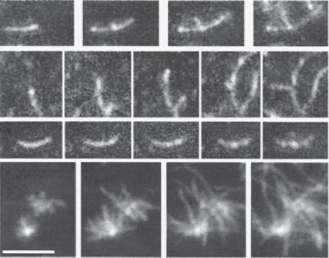
Figure 3.2. Actin filament polymerization and branching. Monomer actin molecules were prepared from rabbit skeletal muscle and labeled on Cys-374 with rhodamine. Actin polymerization was induced in the presence of 20% rhodamine-actin and observed by total internal reflection fluorescence microscopy (TIRFM). The images were subsequently captured at 100, 130, 170, and 210 s after initiating actin polymerization. Scale bar: 4 μm. (Reprinted by permission from Amann KJ et al: Proc Natl Acad Sci USA 98:15009–13, copyright 2001, National Academy of Sciences, USA.)
capping proteins, actin filament-binding proteins, actin filament-severing proteins, and actin filament crosslinking proteins.
Actin Monomer-Binding Proteins [3.3]. The family of actin monomer-binding proteins
(see Table 3.2) includes several molecules, including β-thymosins, cofilins, profilins, and formins, which bind actin monomers and regulate the activities of the actin molecules. β- Thymosins are molecules that primarily bind to and sequester ATP-actin monomers, and thus inhibit actin polymerization. Cofilins bind ADP-actin with high affinity and destabilize actin filaments. However, a controversial role of cofilins has been observed. Profilins bind to ADPand ATP-free actin monomers and play a role in sequestration of actin monomers. Profilins also inhibit nucleation and elongation at the pointed end of an actin filament, but do not influence the nucleation and elongation at the barbed end. Formin is a homodimer composed of formin homology 1 (FH1) and formin homology 2 (FH2) domains. The FH2 domain can bind to monomer actin and induce the nucleation and polymerization of actin filaments. Furthermore, The FH2 domain can bind to the barbed

58
TABLE 3.2. Characteristics of Selected Actin Monomer-Binding Proteins*
|
|
Amino |
Molecular |
|
|
Proteins |
Alternative Names |
Acids |
Weight (kDa) |
Expression |
Functions |
|
|
|
|
|
|
Cofilin 1 |
CFL1 |
166 |
19 |
Ubiquitous |
Binding and depolymerizing filamentous |
|
|
|
|
|
F-actin, inhibiting the polymerization of |
|
|
|
|
|
monomeric G-actin; however, the role of |
|
|
|
|
|
cofilins is controversial |
Cofilin 2 |
CFL2, muscle cofilin |
166 |
19 |
Skeletal muscle, heart, brain, |
Same as those for cofilins 1 |
|
|
|
|
lung, liver, pancreas, kidney |
|
Profilin |
PFN1 |
140 |
15 |
Ubiquitous |
Binding to ADPand ATP-free actin |
|
|
|
|
|
monomers, and sequestering actin |
β-Thymosin |
Thymosin β4 X chromosome |
|
|
|
monomers |
44 |
5 |
Ubiquitous |
Sequestering actin monomers, inhibiting actin |
||
|
|
|
|
|
polymerization, enhancing cardiac cell |
|
|
|
|
|
survival, migration, and regeneration |
Formin |
|
844 |
95 |
Ubiquitous |
Promoting nucleation and polymerization of |
|
|
|
|
|
actin filaments |
|
|
|
|
|
|
*Based on bibliography 3.3.
CYTOSKELETON 59
end of actin filaments and promote the elongation of the filaments. The FH1 domain can bind to the actin-binding protein profilin. This process enhances the elongation of actin filaments.
Actin Filament-Capping Proteins [3.4]. Actin filament-capping proteins (see Table 3.3) are molecules that bind either the pointed or the barbed end of actin filaments and prevent actin polymerization or depolymerization. This family of proteins includes gelsolins, heterodimeric capping proteins, the actin-related protein (Arp)2/3 complex, tropomyosin, nebulin, and tropomodulin. Gelsolins are capable of binding to the barbed end and the side of actin filaments and inhibiting actin polymerization. Heterodimeric capping proteins bind and cap the barbed end of actin filaments, and impose effects similar to those of gelsolins. Arp 2/3 is a complex of Arp 2 and Arp 3, which binds and caps the pointed end of an actin filament and promotes the attachment of the capped end to a different actin filament and the formation of actin filament branches. It has been shown that this process is regulated by the ρ family GTPases. ρ GTPases activate a protein known as the Wiskott–Aldrich syndrome protein (WASP), which in turn activates the Arp2/3 complex. Other actin filament-binding proteins, including tropomyosin, nebulin, capZ, and tropomodulin, bind to the side or ends of actin filaments and contribute to the stability of the filaments. Tropomyosin binds the side of actin filaments, induces an increase in the stiffness of the filaments, and stimulates the interaction of actin filaments with myosin. Nebulin is found in skeletal muscle cells and plays a role in the control of the length of actin filaments. Tropomodulin binds to the pointed end and enhances the stability of actin filaments.
Actin Filament-Severing Proteins. Actin filament-severing proteins include gelsolins, fragmin/severin, and cofilins. These molecules are able to sever actin filaments into short fragments and promote actin filament depolymerization. Gelsolins are also capping molecules for the barbed end of actin filaments. Cofilins can also bind to actin monomers.
Actin Filament-Crosslinking Proteins [3.5]. Actin filament crosslinking proteins (Table 3.4) include α-actinin, fimbrin, villin, and filamin. These molecules can bind simultaneously to multiple actin filaments and induce crosslink of actin filaments. α-Actinin is associated with actin stress fibers and the Z-disk of striated muscular actin fibers. In addition, α-actinin is a constituent of focal adhesion contacts, structures that mediate cell attachment and migration. This molecule possesses multiple functions. Fimbrin can bind and crosslink actin filaments in microvilli. Villin has a similar function as fimbrin. Filamin can not only crosslink actin filaments but also anchor actin filaments to integrins, major constituents of focal adhesion contacts. All these actin filament crosslinking molecules enhance the stability of actin filaments.
Regulation of Actin Assembly and Disassembly [3.6]. In mammalian cells, actin filaments undergo a dynamic turnover process, or simultaneous assembly and disassembly, under physiological conditions. The rate of turnover is dependent on cell types. Nonmuscular cells exhibit actin filament turnover at a timescale of minutes, while muscular cells demonstrate actin filament turnover at a scale of days. Actin polymerization (assembly) and depolymerization (disassembly) can be observed in living cells with fluorescent marker-tagged actin monomers. The fluorescent markers can be incorporated into actin filaments. Following photobleaching of fluorescent actin filaments, the bleached region

60
TABLE 3.3. Characteristics of Selected Actin Filament-Capping Proteins*
|
|
Amino |
Molecular |
|
|
Proteins |
Alternative Names |
Acids |
Weight (kDa) |
Expression |
Functions |
|
|
|
|
|
|
Gelsolin |
Actin depolymerizing |
782 |
86 |
Ubiquitous |
Inhibiting actin polymerization, severing |
|
factor (ADF), Brevin |
|
|
|
actin filaments, and promoting actin |
Actin capping protein α1 |
|
|
|
|
filament depolymerization |
Muscle Z-line actin filament |
286 |
33 |
Skeletal muscle, red |
Found at the Z line of muscular cells, |
|
|
capping protein α1, |
|
|
blood cells, placenta |
binding to barbed end of actin |
|
CAPZA1, F-actin capping |
|
|
|
filaments, and inhibiting actin |
|
protein α1 subunit |
|
|
|
polymerization |
Actin-related protein 2 |
ARP2, actin-like protein 2 |
394 |
45 |
Ubiquitous |
Constituting the ARP2/3 complex and |
|
|
|
|
|
participating in regulation of cell |
|
|
|
|
|
shape and motility via actin assembly |
|
|
|
|
|
and protrusion |
Actin-related protein 3 |
ARP3, actin-like protein 3 |
418 |
47 |
Ubiquitous |
Constituting the ARP2/3 complex and |
|
|
|
|
|
regulating actin assembly |
Tropomyosin 1 |
Tropomyosin skeletal |
284 |
33 |
Skeletal muscle |
Binding to actin filaments in striated |
|
muscle α, tropomyosin |
|
|
|
muscle cells, stabilizing actin |
|
1α chain, α tropomyosin |
|
|
|
filaments, and regulating calcium- |
|
|
|
|
|
dependent interaction of actin |
|
|
|
|
|
filaments with myosin molecules |
|
|
|
|
|
during muscle contraction |
Nebulin |
NEB |
6669 |
773 |
Skeletal muscle cells |
Coexisting with thick and thin filaments |
|
|
|
|
|
within sarcomeres of skeletal muscle |
|
|
|
|
|
and playing a critical role in both |
|
|
|
|
|
integrity and stability of contractile |
|
|
|
|
|
filaments |
Tropomodulin |
Tropomodulin 1, |
359 |
41 |
Skeletal muscle, heart, |
Binding to the pointed end and |
|
erythrocyte tropomodulin, |
|
|
brain, lung, liver, |
enhancing stability of actin filaments |
|
E-tropomodulin |
|
|
kidney, pancreas |
|
|
|
|
|
|
|
* Based on bibliography 3.4.

TABLE 3.4. Characteristics of Selected Actin Filament Crosslinking Proteins*
|
|
Amino |
Molecular |
|
|
Proteins |
Alternative Names |
Acids |
Weight (kDa) |
Expression |
Functions |
|
|
|
|
|
|
Actinin α1 |
ACTN1 |
892 |
103 |
Nonmuscular cells |
Interacting with actin filaments and |
|
|
|
|
|
regulating the assembly of actin |
Actinin α2 |
ACTN2, α actinin skeletal |
|
|
|
filaments and focal adhesion contacts |
894 |
104 |
Skeletal muscle, cardiomyocytes |
A muscle-specific α actinin that anchors |
||
|
muscle isoform 2, |
|
|
|
actin filaments to the Z disks |
|
F-actin-crosslinking |
|
|
|
|
|
protein |
|
|
|
|
Fimbrin |
Intestine-specific plastin, |
629 |
70 |
Intestine, lung, kidney, leukocytes |
Binding to and crosslinking actin |
|
I-plastin, plastin 1, |
|
|
|
filaments |
|
accumentin |
|
|
|
|
Villin |
Villin 1 |
827 |
93 |
Intestine, kidney |
Inducing crosslinking of actin filaments |
Filamin A |
Filamin α, filamin 1 (FLN1), |
2647 |
281 |
Primarily nonmuscular cells |
Inducing crosslinking of actin filaments |
|
actin-binding protein 280 |
|
|
|
and regulating the organization and |
|
(ABP280), nonmuscle |
|
|
|
remodeling of actin cytoskeleton by |
|
filamin, α-filamin, |
|
|
|
interacting with integrins and |
|
endothelial actin-binding |
|
|
|
transmembrane receptors |
|
protein |
|
|
|
|
Filamin B |
β filamin, filamin 1 |
2602 |
278 |
Heart, skeletal muscle, brain, |
Inducing actin filament crosslink in |
|
(actin-binding protein-280)- |
|
|
lung, liver, kidney, pancreas, |
muscular and nonmuscular cells and |
|
like, actin-binding-like |
|
|
uterus, ovary |
regulating the organization of actin |
|
protein, truncated |
|
|
|
filaments |
|
actin-binding protein |
|
|
|
|
|
Actin-binding protein |
|
|
|
|
276/278, ABP276/278 truncated actin-binding protein
* Based on bibliography 3.5.
61
62 STRUCTURE AND FUNCTION OF CELLULAR COMPONENTS
can be replaced with fluorescent actin filaments, suggesting dynamic reassembly of actin filaments. In nonmuscular cells, there exists a relatively high concentration of unpolymerized actin monomers (50–100 μM). Such a concentration allows rapid actin polymerization in response to stimulations that initiate cell adhesion and migration. Indeed, the concentration of actin monomers is a critical factor that controls actin filament assembly and disassembly.
The dynamics of actin assembly–disassembly is regulated by actin regulatory and binding proteins. Sequestration of actin monomers and the capping of actin filaments at the ends are two mechanisms that control the rate of actin filament assembly. As discussed above, profilin and thymosin can bind and sequester actin monomers and reduce the concentration of free actin monomers, suppressing the polymerization of actin filaments. Profilinor thymosin-bound actin monomers have reduced capability of initiating nucleation. An increase in the activity of actin filament-capping proteins promotes actin polymerization.
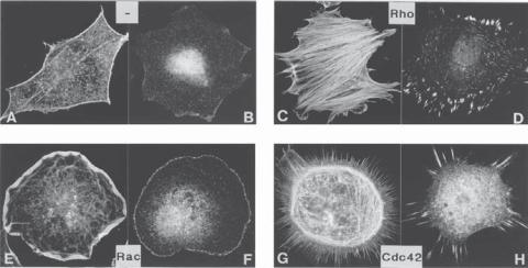
Actin filaments are found in all mammalian cells and are organized into various patterns and structures. For instance, actin filaments form a network in the cortical region of the cell, while forming fiber bundles within filopodia or microvilli. The pattern formation of actin filaments is a process that may be regulated by the Rho family of GTPases, which includes Rho, Rac, and Cdc42 (see Table 3.5). These molecules have been shown to regulate distinct processes of actin assembly. Activated Cdc42 stimulates the formation of filopodia, Rho enhances the formation of actin “stress fibers,” while Rac promotes the formation of cortical network of actin filaments (Fig. 3.3). Although the signaling path-
Figure 3.3. Influence of Rho, Rac, and Cdc42 on the organization of actin filaments and morphology of cells: (A,B) quiescent serum-starved Swiss 3T3 fibroblasts labeled for actin filaments and vinculin; (C,D) treatment of cells with lysophosphatidic acid, a growth stimulator, which activates Rho, leading to the formation of organized actin filaments or stress fibers (C) and focal adhesion contacts (D); (E,F) microinjection of Rac induces the formation of lamellipodia (E) and focal adhesion contacts (F); (G,H) microinjection of FGD1, an exchange factor for Cdc42, leads to formation of filopodia (G) and the focal adhesion contacts (H). (Reprinted by permission from Hall A: Science 279:509–14, 1998.)

TABLE 3.5. Characteristics of Selected Factors that Regulate the Formation of Actin Filaments*
|
|
Amino |
Molecular |
|
|
Proteins |
Alternative names |
Acids |
Weight (kDa) |
Expression |
Functions |
|
|
|
|
|
|
RhoA |
RHOA, aplysia Ras-related homolog 12, |
193 |
22 |
Leukocytes, platelets |
Enhancing the formation of actin stress |
|
oncogene RhoH12, RHOH12, RHO12, |
|
|
|
fibers, regulating cell migration, polarity, |
|
transforming protein RhoA, Ras |
|
|
|
and protrusion |
|
homolog gene family member A |
|
|
|
|
RhoB |
Oncogene RhoH6, RhoH6, aplysia |
196 |
22 |
Nervous system, |
Regulating the formation of actin filaments |
|
Ras-related homolog 6 |
|
|
macrophage, lung |
and assembly of focal adhesion contacts, |
|
|
|
|
|
promoting cell adhesion, vesicle |
|
|
|
|
|
trafficking, MAPK signaling, and |
|
|
|
|
|
immunity |
RhoC |
Ras homolog gene family member C, |
193 |
22 |
Leukocytes, lung, breast, |
Enhancing the formation of actin filaments, |
|
aplysia Ras-related homolog 9, |
|
|
carcinoma cells |
regulating cell motility, and mediating |
|
oncogene RhoH9, transforming |
|
|
|
tumorigenesis |
|
protein RhoC |
|
|
|
|
RAC1 |
p21-Rac1, Ras-related C3 botulinum |
211 |
23 |
Ubiquitous |
A small Ras GTP-binding protein that |
|
toxin substrate 1, Rho family small |
|
|
|
regulates cell survival, growth, |
|
GTP-binding protein RAC1, Ras-like |
|
|
|
cytoskeletal reorganization, and the |
|
protein TC25, TC-25 |
|
|
|
activation of protein kinases |
Cdc42 |
Cell division cycle 42, G25K, |
191 |
21 |
|
A small ρ GTPase that regulates cell |
|
GTP-binding protein 25 kDa |
|
|
|
morphology, migration, endocytosis, |
|
|
|
|
|
polarity, and cell cycle progression; also |
|
|
|
|
|
regulates actin polymerization via |
|
|
|
|
|
interaction with neural Wiskott–Aldrich |
|
|
|
|
|
syndrome protein (N-WASP), which |
|
|
|
|
|
subsequently activates the Arp2/3 |
|
|
|
|
|
protein complex |
|
|
|
|
|
|
* Based on bibliography 3.6.
63
64 STRUCTURE AND FUNCTION OF CELLULAR COMPONENTS
ways for these molecules remain poorly understood, these observations provide insights into the mechanisms by which actin filaments form distinct patterns.
Actin assembly and disassembly are regulated by extracellular factors. For instance, growth factors and cytokines stimulate cell attachment and migration, which are associated with increased actin assembly. These observations suggest a role for growth factors and cytokines in the regulation of actin polymerization or depolymerization. However, exact mechanisms remain poorly understood. In addition, fluid shear stress has been shown to influence actin assembly in vascular endothelial cells. In cell culture models, the introduction of fluid shear stress to endothelial cells enhances actin filament assembly, forming actin “stress fibers.” Shear stress-induced deformation of cell membrane receptors or other cell structures may play a role in the initiation of such a process. However, the signaling pathways that transduce shear stress signals remain to be identified.
Function of Actin Filaments [3.7]. Actin filaments participate in a number of functions, including cell contraction, migration, and division. In contractile cells, including skeletal, cardiac, and smooth muscle cells, actin filaments interact with myosin molecules, causing filament sliding and cell contraction, a fundamental process for force generation. In noncontractile cells, directed actin polymerization contributes to regional extension of cell membrane, a primary step in cell migration. The interaction of actin filaments and myosin molecules provide forces that induce cell traction and movement. During cell division, actin filaments form a ring-shaped structure between two premature daughter cells, known as the contractile ring, underneath the plasma membrane. Contraction of the ring is initiated following cell mitosis. Such an activity separates the mother cytoplasm into two daughter compartments. While chromosome separation is defined as mitosis, cytoplasmic separation is defined as cytokenesis.
BIBLIOGRAPHY
3.1. Cell Membrane
Mineo C, Gill GN, Anderson RG: Regulated migration of epidermal growth factor receptor from caveolae, J Biol Chem 274:30636–43, 1999.
Bretscher M: The molecules of the cell membrane, Sci Am 253:100–8, 1985.
Dowham W: Molecular basis for membrane phospholipid diversity: Why are there so many lipids? Annu Rev Biochem 66:199–212, 1997.
Englund PT: The structure and biosynthesis of glycosyl phosphatidylinositol protein anchors, Annu Rev Biochem 62:121–38, 1993.
Farazi TA, Waksman G, Gordon J: The biology and enzymology of protein N-myristoylation, Annu Rev Biochem 276:39501–64, 2001.
Petty HR: Molewlar Biology of Membrmles: Structure and Function, Plenum Press, New York, 1993.
Singer SJ: The structure and insertion of integral proteins in membranes, Annu Rev Cell Biol 6:247–96, 1990.
Singer SJ, Nicolson GL: The fluid mosaic model of the structure of cell membranes, Science 175:720–31, 1972.
Tamm LK, Arora A, Kleinschmidt JH: Structure and assembly of beta-barrel membrane proteins, J Biol Chem 276:32399–402, 2001.
Towler DA, Gordon J, Adams SP, Glaser L: The biology and enzymology of eukaryotic protein acylation, Annu Rev Biochem 57:69–99, 1988.
BIBLIOGRAPHY 65
White SH, Ladokhin AS, Jayasinghe S, Hristoya K: How membranes shape protein structure, J Biol Chem 276:32395–8, 2001.
Yeagle PL: The Membranes of Cells, 2nd ed, Academic Press, San Diego, 1993.
Zhang FL, Casey PJ: Protein prenylation: Molecular mechanisms and functional consequences, Annu Rev Biochem 65:241–69, 1996.
Simons K, Vaz WL: Model systems, lipid rafts, and cell membranes, Annu Rev Biophys Biomol Struct 33:269–95, 2004.
Vereb G, Szollosi J, Matko J, Nagy P, Farkas T et al: Dynamic, yet structured: The cell membrane three decades after the Singer-Nicolson model, Proc Natl Acad Sci USA 100:8053–8, 2003.
Edidin M: The state of lipid rafts: From model membranes to cells, Annu Rev Biophys Biomol Struct 32:257–83, 2003.
Lipowsky R: The conformation of membranes, Nature 349:475–81, 1991.
3.2. Structure and Organization of Actin Filaments
a-Actin, Cardiac
Gunning P, Ponte P, Kedes L, Eddy R, Shows T: Chromosomal location of the co-expressed human skeletal and cardiac actin genes, Proc Natl Acad Sci USA 81:1813–7, 1984.
Hamada H, Petrino MG, Kakunaga T: Molecular structure and evolutionary origin of human cardiac muscle actin gene, Proc Natl Acad Sci USA 79:5901–5, 1982.
Humphries SE, Whittall R, Minty A, Buckingham M, Williamson R: There are approximately 20 actin genes in the human genome, Nucleic Acids Res 9:4895–908, 1981.
Mogensen J, Klausen IC, Pedersen AK, Egeblad H, Bross P et al: Alpha-cardiac actin is a novel disease gene in familial hypertrophic cardiomyopathy, J Clin Invest 103:R39–43, 1999.
Olson TM, Michels VV, Thibodeau SN, Tai YS, Keating MT: Actin mutations in dilated cardiomyopathy, a heritable form of heart failure, Science 280:750–2, 1998.
Schwartz K, de la Bastie D, Bouveret P, Oliviero P, Alonso S et al: Alpha-skeletal muscle actin mRNAs accumulate in hypertrophied adult rat hearts, Circ Res 59:551–5, 1986.
Takai E, Akita H, Shiga N, Kanazawa K, Yamada S et al: Mutational analysis of the cardiac actin gene in familial and sporadic dilated cardiomyopathy, Am J Med Genet 86:325–7, 1999.
Dunwoodie SL, Joya JE, Arkell RM, Hardeman EC: Multiple regions of the human cardiac actin gene are necessary for maturation-based expression in striated muscle, J Biol Chem 269:12212– 9, 1994.
Skeletal a-Actin
Agrawal PB, Strickland CD, Midgett C, Morales A, Newburger DE et al: Heterogeneity of nemaline myopathy cases with skeletal muscle alpha-actin gene mutations, Annu Neurol 56:86–96, 2004.
Akkari PA, Eyre HJ, Wilton SD, Callen DF, Lane SA et al: Assignment of the human skeletal muscle alpha actin gene (ACTA1) to 1q42 by fluorescence in situ hybridization, Cytogenet Cell Genet 65:265–7, 1994.
Crawford K, Flick R, Close L, Shelly D, Paul R et al: Mice lacking skeletal muscle actin show reduced muscle strength and growth deficits and die during the neonatal period, Mol Cell Biol 22:5887–96, 2002.
Gunning P, Ponte P, Kedes L, Eddy R, Shows T: Chromosomal location of the co-expressed human skeletal and cardiac actin genes, Proc Natl Acad Sci USA 81:1813–7, 1984.
Gunning P, Ponte P, Okayama H, Engel J, Blau H et al: Isolation and characterization of full-length cDNA clones for human alpha-, beta-, and gamma-actin mRNAs: Skeletal but not cytoplasmic actins have an amino-terminal cysteine that is subsequently removed, Mol Cell Biol 3:787–95, 1983.