

Bioregenerative Engineering Principles and Applications - Shu Q. Liu
..pdf2
REGULATION OF GENE EXPRESSION
Mobilization of albumin-positive hepatocyte-like cells into the blood in response to the upregulation of interleukin-6 in ischemic cardiac injury. The mobilized hepatocyte-like cells can be recruited to injured cardiac tissue, promoting the proliferation of cardiomyocytes in the zone of cardiac injury. Red: albumin. Blue: cell nuclei. Sclae: 10 μm. See color insert.
Bioregenerative Engineering: Principles and Applications, by Shu Q. Liu
Copyright © 2007 John Wiley & Sons, Inc.
36
BASIC DNA ELEMENTS FOR REGULATING GENE EXPRESSION |
37 |
Gene expression is the transcription of mRNA (transfer of genetic information from DNA to mRNA) and the translation of protein from a specific mRNA (transfer of genetic information from mRNA to protein). Gene expression is regulated at several key levels, including the transcription of pre-mRNA, the conversion of pre-mRNA to mature mRNA, and the synthesis of protein. The regulation of gene expression involves basically two types of factors: the cis-acting regulatory elements and trans-acting regulatory factors. Cisacting regulatory elements are DNA-sequences that are protein-binding sequences and are found near the initiation site of gene transcription. Trans-acting regulatory factors are proteins that bind to specific cis-acting elements and control gene transcription. In addition, as we see below, the components of chromatin also participate in the regulation of gene expression.
There are several common features for the regulation of gene expression:
1.Trans-acting factors can bind only to specific cis-acting elements.
2.The binding of trans-acting factors to cis elements can induce conformational changes in the chromatin, a critical process for regulating gene transcription.
3.The binding of trans-acting factors may induce positive or negative regulation of gene transcription. Positive regulation is a process by which the binding of trans- acting factors stimulates gene transcription, whereas negative regulation exerts an opposite effect.
4.A transcription regulatory signal can be effective for only a limited time. The signal is turned off after a mission is accomplished.
The principles of gene expression regulation are briefly discussed in this section.
BASIC DNA ELEMENTS FOR REGULATING GENE EXPRESSION [2.1]
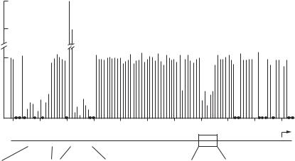
The expression of genes is controlled at the level of mRNA transcription. For each gene, there are specialized short sequences, known as cis-acting regulatory elements, which mediate the attachment and activation of RNA polymerases, necessary processes for initiating mRNA transcription. In mammalian cells, cis-acting regulatory elements include promoters, promoter-proximal elements, and enhancers. Each cis-acting element can be recognized and activated by trans-acting regulatory factors. These factors can bind to the promoter and initiate the attachment of RNA polymerase II, inducing RNA transcription. A typical promoter is composed of TATA nucleotides, known as the TATA box. The TATA box is usually found at a site about 30 bp upstream the first base pair of the transcription site. On the action of trans-acting factors, a promoter mediates the initiation of RNA transcription in coordination with promoter-proximal elements and enhancers. Typical promoter-proximal elements, which often include the CCAAT box and GC-rich segments, are located about 100–200 bp away from the TATA box. Experiments with point mutation have demonstrated that a mutation in the TATA or the promoter-proximal elements induces a decrease in the rate of RNA transcription, whereas mutation in other regions exhibits little influence (Fig. 2.1). Each gene contains several sequence motifs of enhancers, which are located at a distance, some times up to 50 kb, from the TATA box. Different motifs serve as binding sits for specific trans-acting factors. The function of promoterproximal elements and enhancers is to facilitate the activity of the promoters and enhance gene transcription.
38
Relative transcription level
REGULATION OF GENE EXPRESSION
3.5
3.0
1.0
0
|
|
|
|
|
|
|
|
GCCACACCC GGCCAATC |
ATATAA |
||||||
Figure 2.1. Identification of the promoter and promoter-proximal elements that control the expression of the β-globin gene. Point mutations were induced on most nucleotides through the promoter region of the β-globin gene, and the effect of each point mutation was tested. Mutations within the TATA promoter and the promoter-proximal elements caused a significant decrease in the level of transcription, suggesting that these cis-elements are critical to the transcription of the β-globin gene. (Reprinted by permission from Maniatis T, Goodbourn S, Fischer JA: Science 236:1237–45, 1987. Copyright 1987 AAAS.)
TRANS-ACTING REGULATORY FACTORS [2.1]
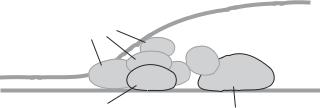
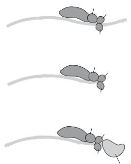
Trans-acting regulatory factors are proteins that bind to and activate target genes and regulate RNA transcription. In mammalian cells, there exist a large number of trans-acting factors. These factors, while in their inactive state, are present in the cytoplasm. Once activated in response to the stimulation of signaling molecules (see Chapter 5), a trans-acting factor can be translocated from the cytoplasm to the nucleus and bind to specific cis-acting elements, or promoters and enhancers, of target genes. A trans-acting factor, once bound to a cis-acting element, can form a complex with RNA polymerase II and other necessary regulatory factors (See Table 2.1), assisting the polymerase to initiate mRNA transcription. Each trans-acting factor contains two domains. The first domain is responsible for binding to cis-acting elements, whereas the second is for activating mRNA transcription. A typical example of trans-acting factors is the complex of transcription factors for RNA polymerase II, or TFII. Each TFII is composed of a TATA box-binding protein (TBP) and several associated proteins (Fig. 2.2). These proteins are referred to as basal trans-acting factors. Examples of other trans-acting factors include activator protein 1 and nuclear factor κ B. The functions of these factors are discussed in Chapter 5.
REGULATION OF GENE TRANSCRIPTION
At the transcription level, several mechanisms are involved in the regulation of gene expression, including the activation of trans-acting factors, modification of the chromatin structure, and DNA modulation. These mechanisms are briefly described as follows.
REGULATION OF GENE TRANSCRIPTION |
39 |
Transcriptional
factors
TATA
TATA-binding
RNA polymerase
protein
Figure 2.2. Schematic representation of controlling gene transcription by trans-acting factors (based on bibliography 2.1).
Control of the Activity of Trans-Acting factors [2.2]
In eukaryotes, there are two types of trans-acting factor: transcription-activating proteins (transcriptional activators) and transcription-inhibiting proteins (transcriptional repressors). The two types of factor regulate gene transcription via controlling the function of the RNA polymerase holoenzyme and the structure of chromatin. These factors dynamically counterbalance each other, ensuring an appropriate level of gene expression.
Trans-acting factors interact with DNA transcription apparatus, which is composed of cis-acting elements and protein coactivators. While trans-acting proteins act on specific cis elements, protein coactivators enhance the regulation of gene transcription, which is independent of specific cis elements. The binding of trans-acting factors to cis-acting DNA elements induces activation and promotes reorientation of coactivator proteins, which are necessary conditions for the initiation of gene transcription.
The activity of trans-acting proteins is controlled by three basic approaches: modification of preexisting proteins, de novo synthesis of proteins, and targeted degradation. These processes may coordinate in a timeand space-dependent manner, providing synergistic inputs for the regulation of gene transcription. Among these regulatory processes, the modification of preexisting proteins is the most important process that causes immediate activation of signaling events, resulting in prompt cellular activities. Common types of modification include phosphorylation and dimerization of proteins. Protein phosphorylation occurs primarily on the serine, threonine, and tyrosine residues, and is regulated by protein kinases and protein phosphatases, which phosphorylate and dephosphorylate target proteins, respectively. The phosphorylation of signaling molecules may lead to activation or deactivation of trans-acting factors, depending on the nature of specific signaling pathways. In most cases, phosphorylation induces activation of trans-acting factors. Such a process enhances the interaction of trans-acting factors with nuclear export machineries, which facilitate the translocation of the trans-acting factors from the cytoplasm to the nucleus. In addition, the phosphorylation of the DNA-binding domain of a trans-acting factor mediates the binding of the trans-acting factor to the target DNA. The involvement of protein phosphorylation in specific signaling pathways is described on Chapter 5.
Dimerization is another form of modification of transcriptional factors. Most trans- acting factors bind to DNA in the form of homodimer or heterodimer. The dimerization of trans-acting factors enhances their binding to target DNA and provides a mechanism for the specificity and selectivity of gene transcription. For instance, the members of the activating protein-1 family, including Jun, Fos, and activating transcription factor (ATF), form homodimers (Jun-Jun, Fos-Fos, ATF-ATF) and heterodimers (Fos-Jun, Jun-ATF,

40
TABLE 2.1. Characteristics of Selected Gene Transcription Regulators*
|
|
Amino |
Molecular |
|
|
Proteins |
Alternative Names |
Acids |
Weight (kDa) |
Expression |
Functions |
|
|
|
|
|
|
RNA polymerase II, |
RNA polymerase II 14.5-kDa |
125 |
14 |
Ubiquitous |
Forming a complex with subunits 2 and 3 and |
subunit 1 |
subunit, polymerase RNA II |
|
|
|
regulating mRNA synthesis in eukaryotes |
|
DNA-directed polypeptide I |
|
|
|
|
|
14.5 kDa, DNA-directed RNA |
|
|
|
|
|
polymerase II polypeptide I |
|
|
|
|
RNA polymerase II, |
RNA polymerase II subunit B, |
1174 |
134 |
Ubiquitous |
Forming a complex with subunits 1 and 3 and |
subunit 2 |
RNA polymerase II 140-kDa |
|
|
|
regulating mRNA synthesis |
|
subunit, DNA-directed RNA |
|
|
|
|
|
polymerase II 140-kDa |
|
|
|
|
|
polypeptide |
|
|
|
|
RNA polymerase II, |
RNA polymerase II subunit C, |
275 |
31 |
Ubiquitous |
Forming a complex with subunits 1 and 2 and |
subunit 3 |
DNA-directed RNA polymerase |
|
|
|
regulating mRNA synthesis |
|
II 33-kDa polypeptide |
|
|
|
|
TATA box-binding protein |
GTF2D, SCA17, spinocerebellar |
339 |
38 |
Ubiquitous |
Together with TATA box-binding protein- |
|
ataxia 17 |
|
|
|
associated factors, forming transcription |
|
|
|
|
|
factor IID (TFIID), which positions the |
|
|
|
|
|
RNA polymerase II to a target gene |
promoter and regulates mRNA transcription
*Based on bibliography 2.1.
REGULATION OF GENE TRANSCRIPTION |
41 |
Fos-ATF). Each type of dimer targets specific genes, thus increasing the specificity and also the capacity of transcriptional regulation. repress
In contrast to gene activation by trans-acting proteins, gene transcription can be inhibited by transcriptional protein repressors. The mechanisms of transcriptional repression are diverse. Some repressors can bind to transcription activators, reducing their activities. The TATA-box-binding protein (TBP) is a major target of transcription repressors. For instance, the binding of the repressor NC2 to the TATA-box-binding protein prevents the assembly of the RNA polymerase II holoenzyme into the initiation complex of gene transcription, thus repressing gene transcription. Another repression mechanism involves the competition of transcriptional repressors with transcriptional activators for DNA binding sites. The outcome, activation, or repression of gene transcription is determined on the basis of the relative activity of transcriptional activators and repressors as well as their affinity to target DNA binding sites.
In addition to the modification of existing trans-acting factors, de novo synthesis of trans-acting proteins contributes to the regulation of gene transcription. The synthesis of a trans-acting factor is induced by upregulating the expression of a specific gene that encodes the trans-acting factor. An increase in the level of a trans-acting factor enhances gene transcription. However, this approach is relatively slow and is not as effective as the modification of existing trans-acting factors. A trans-acting factor is usually degraded by enzymes when its task is accomplished. The degradation of trans-acting factors is an effective approach ensuring that gene transcription can be stopped promptly.
Chromatin Modification [2.3]
In addition to the regulation of gene transcription by transcriptional activators and repressors, dynamic changes in chromatin components contribute significantly to the regulation of gene transcription. Chromatin is composed of nucleosomes, the basic chromatin units. In each nucleosome, DNA is wrapped around a cylinder-like histone core. The nucleosomes are further arranged into three-dimensional structures. The structure of chromatin components can be modified via different processes, including histone phosphorylation on the serine and threonine residues, histone ubiquitination, histone acetylation on the lysine residue, and histone methylation on the lysine and arginine residues. These processes significantly influence the activities of gene transcription.
Histone phosphorylation can be induced by certain members of the mitogen-activated protein kinase family. Such a process is often a result of growth factor action and facilitates the transcription of mitogenic genes. Histone ubiquitination is a process that regulates gene transcription via degrading histone (see Chapter 5). Such a process influences the stability of chromatin and thus the activity of gene transcription. A typical example is the ubiquitination of histone H1. This type of histone is involved in the packaging of chromatin nucleosomes into a higher-ordered structure and is referred to as a linker histone. The formation of a higher-order chromatin structure usually results in the repression of gene transcription. The degradation of histone H1 thus promotes activation of gene transcription.
The acetylation of histone occurs primarily on the lysine residues at the N-terminus. This process is catalyzed by the histone acetyl transferase and can be reversed by the histone deacetylase. The acetylation of histones is thought to weaken the interaction of histone cores with DNA, increase the mobility of DNA and accessibility of trans-acting factors to cis-acting elements, and thus facilitate transcriptional activation. Furthermore,
42 REGULATION OF GENE EXPRESSION
|
NH2 |
|
DNA |
|
NH2 |
|
|
4 3N |
|
H2C |
|
|
|
5 |
|
methyltransferase |
4 |
3N |
||
|
5 |
|||||
6 |
2 |
|
|
6 |
|
2 |
|
|
|
||||
1 |
O |
1 |
||||
|
N |
|
N |
O |
||
|
H |
|
|
|
H |
|
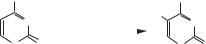
Figure 2.3. Methylation of cytosine, catalyzed by the DNA methyltransferase (DNMTs). Based on bibliography 2.4.
histone acetylation may enhance the association of trans-acting factors and cis-acting elements and facilitates gene transcription. Another means of nucleosome modification is histone methylation, which occurs usually on the lysine and arginine residues. A wellcharacterized example is the methylation of histone H3. In this type of histone, methylation is catalyzed on lysine 4 (the 4th lysine residue) and lysine 9. The outcome of methylation is dependent on the location of methylation. Methylation on the 4th lysine residue of histone H3 is associated with transcriptional activation, whereas methylation on lysine 9 suppresses gene transcription.
DNA Modification [2.4]
In addition to the modification of histones, the structure of DNA can be modified, which influences gene transcription. A well-characterized form of modification is DNA methylation at cytosines located prior to guanosines, which are referred to as the CpG dinucleotides. These dinucleotides are rare, but can be found in certain regions of a gene in the form of cluster. A cluster of CpGs is termed a CpG island, which is often found in the promoter region of a gene. DNA methylation is found in mammalian cells and is catalyzed by an enzyme known as DNA methyltransferase (DNMT), which transfers a methyl group from the donor molecule S-adenosyl-methionine to a cytosine base (Fig. 2.3). DNA methylation is often associated with inactive genes, whereas active genes are relatively demethylated, suggesting that DNA methylation suppresses gene transcription. In particular, DNA methylation in a promoter region causes gene silencing. While the mechanisms remain poorly understood, it has been hypothesized that DNA methylation may enhance the binding of methyl-CpG-binding proteins (MBPs), which are capable of recognizing and binding to methylated cytosine–guanosine groups. These methyl-CpG-binding proteins have been found to recruit histone deacetylase to chromatin, enhancing histone deacetylation. As a result, the accessibility of trans-acting factors to cis-acting elements is reduced and gene transcription is repressed. A physiological aspect of DNA methylation is that DNA methylation may help maintain certain genes in an inactive state. Such a process is highly selective and physiologically active genes may not be the target of DNA methylation.
REGULATION OF PRE-mRNA CONVERSION TO MATURE mRNA
Gene transcription is a process that produces a primary transcript known as pre-mRNA. This form of mRNA cannot be directly processed for protein translation. To translate a protein, it is necessary to convert the pre-mRNA to mature mRNA. Such a conversion is accomplished via several post-transcriptional processes, including capping at the 5′ terminus, polyadenylation at the 3′ terminus, pre-mRNA splicing, and pre-mRNA transport.
REGULATION OF PRE-mRNA CONVERSION TO MATURE mRNA |
43 |
5′-Terminal Capping and Decapping of pre-mRNA [2.5]
5′-Terminal capping is a process by which a 7-methyl guanosine is linked to the first nucleoside of the transcript with a 5′-triphosphate (Fig. 2.4). This process is necessary for efficient splicing and export of pre-mRNA as well as for the initiation of protein translation. Several enzymes are required for 5′-terminal capping, including RNA triphosphatase, RNA guanylyltransferase, and RNA methyltransferase. The RNA triphosphatase catalyzes the formation of a diphosphate from a triphosphate at the 5′-terminus of the mRNA transcript. The RNA guanylyltransferase induces the capping of the 5′-diphosphate with a GMP. The RNA methyltransferase catalyzes the methylation at the N7 position of the 5′- guanine base. These three steps are necessary for the 5′-terminal capping and take place during transcription when the transcript is about 25–30 nucleotides in length. The capping structure is required for the arrangement of mRNA in the ribosome for protein translation. The 5′ capping also stabilizes the structure of mRNA and protects mRNA from degradation by 5′-exoribonucleases.
The 5′-terminal cap of mRNA can be removed by decapping enzymes, a process leading to mRNA degradation. There are two types of decapping enzyme, including the
7-methyl guanosine |
|
||
CH2 |
O |
Cytosine |
|
N |
|
NH |
|
|
NH2 |
||
|
|
||
|
|
|
|
N N
N
5
O H2C
OH OH
NH2 |
|
|
|
|
|
|
|
N |
|
|
|
|
|
O |
O |
O |
|
|
|
|
|
|
|||||
|
|
|
|
|
|
|
|||||||
|
|
|
|
5 |
|
|
N |
O |
|
|
|
||
O P O O |
P O O P O CH2 O |
|
|
|
|||||||||
|
|
|
|
|
|
||||||||
|
|
|
|
|
4 |
|
|
1 |
|
|
|
|
|
O |
O |
O |
2 |
|
Thymine |
|
|
||||||
|
|
|
|
3 |
|
|
|
|
|||||
|
|
|
|
|
|
|
OH |
O |
|
|
|
||
|
|
|
|
|
|
|
|
|
|
|
|
|
|
|
|
|
|
|
|
|
|
H3C |
NH |
|
|
||
|
|
|
|
|
O |
|
|
|
|
|
|||
|
|
|
|
|
|
|
|
|
|
|
|
||
|
|
|
|
|
HO P |
O |
5 |
|
N |
O |
|
|
|
|
|
|
|
|
CH2 |
|
|
||||||
|
|
|
|
|
O |
|
|
|
|||||
|
|
|
|
|
|
|
|
4 |
|
1 |
|
|
|
|
|
|
|
|
O |
|
|
|
Adenine |
||||
|
|
|
|
|
|
3 |
|
2 |
|
|
|||
|
|
|
|
|
|
|
|
|
|
|
|||
|
|
|
|
|
|
|
|
|
|
OH |
|
|
NH |
|
|
|
|
|
|
|
|
O |
|
|
N |
N |
|
|
|
|
|
|
|
|
|
|
|
|
|||
|
|
|
|
|
|
|
|
|
|
5 |
|
N |
N |
|
|
|
|
|
|
|
HO |
P |
O CH2 |
O |
|||
|
|
|
|
|
|
|
|
||||||
|
|
|
|
|
|
|
|
|
|
4 |
1 |
H |
|
|
|
|
|
|
|
|
|
|
|
|
|||
|
|
|
|
|
|
|
|
O |
|
|
|||
|
|
|
|
|
|
|
|
|
|
|
|||
|
|
|
|
|
|
|
|
|
|
|
|
||
|
|
|
|
|
|
|
|
|
|
3 |
|
2 |
|
OH
O
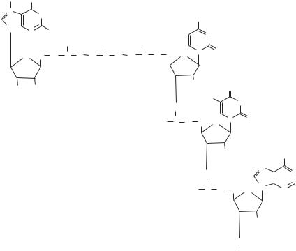
Figure 2.4. Chemical structure of the mRNA cap. The cap consists of N7-methyl guanosine linked by an inverted 5′-5′ triphosphate bridge to the first nucleoside of the mRNA chain (base N can be adenine, guanine, cytosine, or uracil). (Reprinted by permission from Gu M, Lima CD: Processing the message: Structural insights into capping and decapping mRNA, Curr Opin Struct Biol 15:99– 106, 2005.)
44 REGULATION OF GENE EXPRESSION
Dcp1/Dcp2 decapping complex and DcpS. The Dcp1/Dcp2 complex regulates mRNA cap degradation in the 5′→3′ mRNA decay pathway. Within the Dcp1/Dcp2 complex, Dcp2 can catalyze the hydrolysis of the 5′-cap, resulting in the release of GDP from the mRNA, and Dcp1 serves as a regulator that stimulates the activity of Dcp2. In contrast, DcpS regulates mRNA cap degradation in the 3′→5′ mRNA decay pathway and catalyzes residual mRNA cap hydrolysis.
Polyadenylation [2.6]
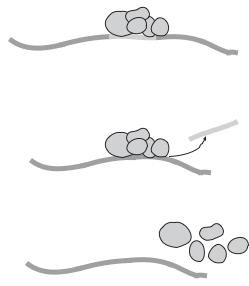
Polyadenylation is a process by which a poly-A sequence about 200 bases is added to a pre-mRNA molecule, a process necessary for the maturation of pre-mRNA. There are two basic steps for polyadenylation: cleavage of the nascent pre-mRNA transcript and addition of a poly-A sequence to the 3′ terminus. Several proteins are involved in the regulation of pre-mRNA cleavage and polyadenylation, including the cleavage and polyadenylation stimulation factor (CPSF), cleavage stimulation factor (CstF), cleavage factor I (CFI), and cleavage factor II (CFII). These factors form a complex and bind to a premRNA site that is downstream to a ubiquitous sequence AAUAAA and immediately upstream to a GU-rich sequence. The AAUAAA sequence serves as an initiation signal for the binding of the protein complex. A CPSF molecule, CPSF-160, is responsible for recognizing the AAUAAA signal. The binding of the regulatory factors induces the cleavage of the pre-mRNA sequence at the binding site. The resulting 3′-terminus by cleavage is immediately polyadenylated by a poly(A) polymerase (Fig. 2.5). The polyadenylation of pre-mRNA contributes to the stabilization of mRNA.
A
5'
B
5'
C
5'
CPSF |
|
AA |
|
U |
|
A A |
|
|
A |
CPSF |
|
AA |
|
U |
|
A A |
|
|
A |
CPSF |
|
AA |
|
U |
|
A A |
|
|
A |
CstF
CFI
3'
CFII
CstF
CFI
3'
CFII
CstF
CFI
|
|
|
3' |
|
|
A |
|
|
...A |
A |
|
AA |
A |
|
|
|
|
|
|
CFII
Poly(A) polymerase
Figure 2.5. Schematic representation of polyadenylation of pre-mRNA (based on bibliography 2.6).
REGULATION OF PRE-mRNA CONVERSION TO MATURE mRNA |
45 |
Pre-mRNA Splicing [2.7]
Pre-mRNA splicing is a process by which a transcribed pre-mRNA molecule is modified by removing the introns and rejoining the exons. This process is critical to the maturation of mRNA and the selection of various specific mRNA molecules from the same premRNA. A pre-mRNA molecule contains protein-coding exons and noncoding introns. To generate mature mRNA, the introns must be removed and the exons must be rejoined in an appropriate order. Pre-mRNA splicing takes place in a structure known as spliceosome, which consists of a number of small nuclear ribonucleoproteins (SNRNPs) and several small nuclear RNA (snRNA) molecules (note that snRNAs are involved in not only RNA splicing, but also maintenance of the telomeres or the chromosome ends). These components are thought to mediate the cleaving and rejoining of nucleotides (Fig. 2.6). In the spliceosome, multiple mature mRNA molecules with different functions can be formed by alternative splicing and selective combination of exons, which are regulated by specific regulatory small nuclear ribonucleoproteins and snRNAs. This is a mechanism for producing cellor tissue-specific proteins based on the same pre-mRNA molecule. The mechanisms of selective splicing remain poorly understood.
mRNA Transport [2.8]
mRNA transport is a process by which transcribed and processed mRNA is moved from the cell nucleus to the cytoplasm. The transcription of mRNA takes place in the cell nucleus, whereas protein translation occurs in the cytoplasm. Spliced mRNA must be transported to the cytoplasm and localized to designated sites for appropriate protein
Spliceosome
A
Exon
Exon
Intron
Spliceosome |
Intron |
B |
|
Exon |
Exon |
|
C
Exon
Figure 2.6. Schematic representation of pre-mRNA splicing (based on bibliography 2.7).