
Bioregenerative Engineering Principles and Applications - Shu Q. Liu
..pdf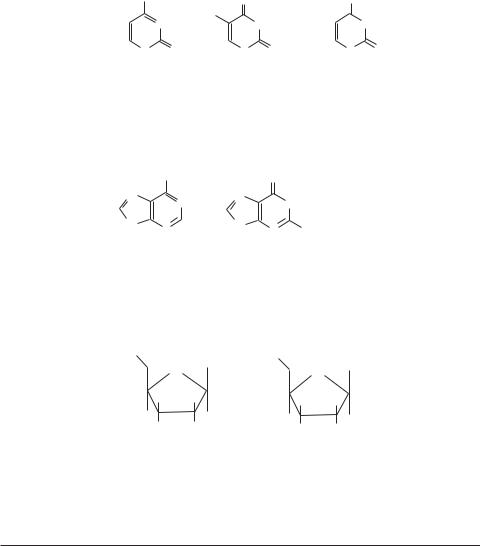
6STRUCTURE AND FUNCTION OF MACROMOLECULES
Pyrimidines
NH2 |
|
O |
O |
|
N |
|
CH3 |
|
NH |
|
|
|||
|
NH |
|
||
N |
O |
N O |
N O |
|
H |
|
H |
H |
|
Cytosine |
|
Thymidine |
Uracil |
|
Purines
|
NH2 |
|
O |
|
N |
N |
N |
|
NH |
|
|
|
||
N |
N |
N |
N |
NH2 |
|
H |
|||
|
H |
|
|
|
Adenine |
Guanine |
|
||
Figure 1.1. Chemical structure of pyrimidines (cytosine, thymine, and uracil) and purines (adenine and guanine) that constitute DNA and RNA.
HO
OH
|
O |
H |
H |
H |
H |
HO |
H |
|
Deoxyribose
HO
O |
OH |
|
|
H |
H |
H |
H |
HO |
HO |
Ribose |
|
Figure 1.2. Chemical structure of deoxyribose and ribose.
TABLE 1.1. Nomenclature of Nucleosides and Nucleotides for DNA
Types of Base |
Cytosine (C) |
Thymine (T) |
Adenine (A) |
Guanine (G) |
|
|
|
|
|
Nucleosides |
Deoxycytidine |
Deoxythymidine |
Deoxyadenosine |
Deoxyguanosine |
|
(dC) |
(dT) |
(dA) |
(dG) |
Nucleotides |
Deoxycytidine 5′ |
Deoxythymidine 5′ |
Deoxyadenosine 5′ |
Deoxyguanosine 5′ |
|
monophosphate |
monophosphate |
monophosphate |
monophosphate |
|
(dCMP) |
(dTMP) |
(dAMP) |
(dGMP) |
|
Deoxycytidine 5′ |
Deoxythymidine 5′ |
Deoxyadenosine 5′ |
Deoxyguanosine 5′ |
|
diphosphate |
diphosphate |
diphosphate |
diphosphate |
|
(dCDP) |
(dTDP) |
(dADP) |
(dGDP) |
|
Deoxycytidine 5′ |
Deoxythymidine 5′ |
Deoxyadenosine 5′ |
Deoxyguanosine 5′ |
|
triphosphate |
triphosphate |
triphosphate |
triphosphate |
|
(dCTP) |
(dTTP) |
(dATP) |
(dGTP) |
|
|
|
|
|
DEOXYRIBONUCLEIC ACIDS (DNA) |
7 |
Deoxycytidine monophosphate and triphosphate
|
|
|
|
|
NH2 |
|
|
|
|
|
|
|
NH2 |
|
|
|
|
|
|
|
|
|
|
|
|
|
|
||
O |
3 |
4 |
5N |
|
O |
O |
O |
N |
||||||
|
|
|
|
|||||||||||
|
|
|
5 |
2 |
1 |
6 |
|
|
|
|
|
|
|
N O |
|
|
|
|
N |
O |
HO |
P O |
P O P O CH2 O |
||||||
HO P O CH2 O |
|
|
||||||||||||
|
|
|
O- |
O- |
O- |
|
||||||||
|
|
- 4 |
|
1 |
|
|
|
|||||||
|
|
|
|
|
||||||||||
O |
|
|
|
|
|
|
|
|
|
|
|
|||
3 |
2 |
OH |
OH |
|
Deoxythymidine monophosphate and triphosphate
O O
|
|
|
H3C |
4 |
5 NH |
O |
O |
|
H3C |
NH |
||
O |
3 |
|
O |
|
||||||||
|
|
|
2 |
1 |
6 |
|
|
|
|
|
N |
O |
|
|
|
|
|
|
|
|
|||||
|
|
|
5 |
N |
O |
|
|
|
|
|
||
HO P O CH2 O |
HO P O P O P O CH2 O |
|
||||||||||
1 |
|
O- |
O- |
O- |
|
|||||||
|
|
- |
4 |
|
|
|||||||
|
|
|
||||||||||
O |
|
|
|
|
|
|
|
|
|
|
||
3 |
2 |
OH |
OH |
|
Deoxyadenosine monophosphate and triphosphate
NH2 NH2
O |
|
N |
5 |
6 |
O |
O |
O |
N |
N |
||||
|
8 7 |
|
1N |
|
|||||||||
|
|
5 |
9 |
4 |
2 |
|
|
|
|
|
|
|
|
|
|
3 |
|
|
|
|
|
|
|
|
|||
HO P O |
CH2 O |
N |
|
N |
|
|
|
|
O P O CH2 O |
N |
N |
||
|
HO P O P |
||||||||||||
|
|
|
|||||||||||
|
|
4 |
|
|
H |
|
|
|
|
|
|
|
H |
O- |
|
|
|
|
|
O- |
O- |
|
|
||||
|
1 |
|
|
O- |
|
|
|||||||
|
|
|
|
|
|
|
|
|
|
|
|
|
|
32
OH OH
Deoxyguanosine monophosphate and triphosphate
O O
|
|
|
N |
|
|
|
|
|
|
|
|
|
|
N |
|
O |
|
5 |
6 |
1NH |
|
|
O |
O |
NH |
||||||
|
7 |
|
|
O |
|
||||||||||
|
8 |
|
|
|
|
|
|
||||||||
|
|
|
9 |
4 |
3 |
2 |
|
|
|
|
|
|
|
|
|
|
|
5 |
N |
|
|
|
|
|
|
|
N |
|
|||
HO P O CH2 O |
|
N |
|
NH2 HO P O P |
O P O CH2 O |
N NH2 |
|||||||||
|
|
|
|
||||||||||||
O- |
4 |
1 |
|
|
|
|
O- |
O- |
O- |
|
|
||||
|
|
|
|
|
|
|
|
|
|
|
|
|
|
||
32
OH |
OH |
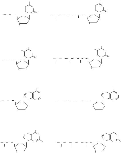
Figure 1.3. Chemical structure of the nucleotides that constitute DNA.
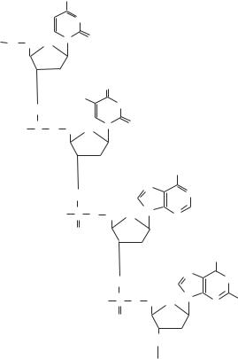
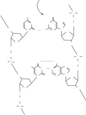
the backbone. The backbone is formed by joining the pentose molecules via covalent phosphodiester bonds. A phosphate group joins one pentose at the 5′ carbon and to the next pentose at the 3′ carbon (Fig. 1.4). The four bases are attached to the pentose– phosphate backbone and aligned on the same side of each DNA chain. The two strands of DNA are attached to each other via hydrogen bonds between the base pairs on the basis of the complementary principle, specifica, A with T and C with G. Note that a doubleringed purine base (A or G) is always paired with a single-ringed pyrimidine (T or C) (Fig. 1.5). A hydrogen bond is established between a positively charged H and a negatively
8STRUCTURE AND FUNCTION OF MACROMOLECULES
NH2
NCytosine
O |
5 |
N O |
|
CH2 O |
|||
|
41
32
O
|
H3C |
NH |
|
O |
|||
Thymine |
|||
5 |
|||
N O |
|||
HO P O CH2 |
|||
O |
|||
|
4 |
1 |
|
|
|||
O |
|
||
32
|
|
NH |
|
O |
N |
N |
Adenine |
|
|||
|
|
||
5 |
N |
N |
|
HO P O CH2 O |
|
||
|
|
||
4 |
1 |
H |
|
O |
|
|
|
|
|
|
32
|
|
|
O |
||
O |
N |
|
NH |
||
|
|||||
|
|
||||
|
|
|
|
Guanine |
|
5 |
N |
N NH2 |
|||
HO |
P O CH2 O |
||||
|
|||||
|
|
|
|||
|
4 |
1 |
|
|
|
O |
|
|
|||
|
|
|
|||
3 |
2 |
|
|
||
|
|
|
|
||
|
O |
|
|
|
|
Figure 1.4. Formation of phosphodiester bonds between pentose molecules, constituting the backbone of DNA (based on bibliography 1.1).
charged acceptor, such as an O and N. A hydrogen bond is relatively weak, about 3% of the strength of a covalent bond. However, an array of hydrogen bonds, as found in the double-stranded helical DNA molecule, can be very strong if all hydrogen bonds are aligned on the same side of a DNA strand. For a double-stranded DNA molecule, the two sugar-phosphate backbones run in opposite directions, defined as an antiparallel arrangement. One strand is defined as the 5′ → 3′ strand; the other, the 3′ → 5′ strand.
The four bases, A, G, C, and T, are organized into a series of a large number of distinct sequences, each forming an independent functional unit known as the gene. In eukaryotic cells, genes are composed of coding sequences called exons and noncoding sequences called introns. The exons contain codons for specific proteins with each codon composed of three nucleotides that specify an amino acid. In contrast, the introns are sequences for the regulation of gene transcription. The intron sequences do not contain protein-coding sequences. The different sequences or structures of DNA are defined as genotypes, which determine the chemical, physical, and functional characteristics, or phenotypes, of various living organisms.
|
DEOXYRIBONUCLEIC ACIDS (DNA) |
9 |
Hydrogen |
5' |
|
bond |
|
3'
Cytosine
O 3 2
4
1
O
5 CH2
H |
+ |
|
|
|
N H |
|
|
||
- |
|
+ |
||
|
|
|||
N |
|
|
|
HN |
|
|
|
||
NO- +HN
H
- |
O |
|
 N
N
N |
N |
|
|
|
1 |
|
2 |
O |
P |
HO |
|
O |
|
5 |
C H |
|
|
|
2 |
O |
|
|
|
4 |
|
|
3 |
|
O |
|
|
Guanine
O |
|
|
|
H |
|
HO P O |
- |
|
+ |
|
|
O |
|
HN |
|
||
H3C |
+ |
- |
N |
|
N |
Thymine |
NH |
|
|
|
|
|
|
|
|
N |
|
O 3 2 |
N O |
|
|
N |
|
|
|
|
|||
|
|
|
|
H |
1 |
4 |
|
|
|
|
|
|
|
|
|
|
|
O |
1 |
2 |
|
||
|
|
5CH2
O
HO P O
O |
P |
HO |
O |
|
5 C H |
|
2 |
Adenine |
|
|
O |
|
4 |
|
3 |
|
O |
|
3'
5'
Figure 1.5. Formation of hydrogen bonds that link two single DNA strands into a double DNA helical structure (based on bibliography 1.1).
A gene carries genetic information in a particular form that can be stored, processed, copied, transcribed to generate messenger RNA (mRNA), and transmitted from the mother cells to the progeny. The double-stranded helical structure is ideal for such purposes. Since both nucleotide strands of a DNA molecule are complementary to each other, both strands carry identical genetic information. Such an organization ensures precise information transfer during DNA replication. When a DNA molecule is ready for replication, the two strands separate. Each strand can serve as a template for the synthesis of a new strand. A newly synthesized strand is identical to the complementary counterpart of the template strand. Thus, each daughter DNA is a pair of an original template and a newly synthesized strand. The daughter DNA molecules can in turn serve as templates for further DNA replication. In such a way, the genetic information can be stored, copied, and transmitted from generation to generation endlessly.
Organization of Chromosomes [1.2]
Each DNA molecule is packaged in a nuclear structure, known as chromosome. In humans, each cell contains 46 chromosomes, including a pair of sex chromosomes (XX
10 STRUCTURE AND FUNCTION OF MACROMOLECULES
for female or XY for male), which are arranged in 23 pairs. One of each paired chromosomes is derived from each parent. Thus, each individual offspring inherits two copies of the same chromosome. The total chromosomes contain about 6.6 × 10⁹ DNA base pairs. The spacing distance is about 3.4 Å per base pair. Each human cell contains 46 DNA molecules (one DNA molecule for each chromosome) with a total length of about 2 m. Given the small size of a cell nucleus (about 5–10 μm), the DNA molecules must be folded to fit in the nucleus. The folding and packaging of DNA are accomplished with the assistance of packaging proteins (primarily histones). DNA and the packaging proteins together form a structure called chromatin, which is organized to form a chromosome. Chromatin exists in two states: heterochromatin and euchromatin. Heterochromatin is a highly condensed form of chromatin and is not involved in RNA transcription, whereas euchromatin is less condensed and is ready for RNA transcription.
There are several levels of DNA packaging, including (1) formation of nucleosomes, which are DNA coils around protein cores; (2) folding of the nucleosome DNA into a fiber structure ( 30 nm in diameter); (3) additional folding of the fiber DNA into thicker bundles (100–300 nm); and (4) formation of loop domains, each containing 15–100 kilo– base pairs (kb) (i.e., 15,000–100,000 base pairs). At the first level, a string-like DNA molecule is coiled around a series of core complexes of proteins known as histones to form nucleosomes. Each nucleosome contains about 170 base pairs (bp) of DNA. Uncoiled DNA fragment between two adjacent nucleosomes is referred to as linker DNA, which is about 30 bp in length. There are several types of histones (H), including H2A, H2B, H3, and H4, which constitute the core complex of nucleosomes. These histones contain positive charges, which neutralize the negative charges of the DNA phosphate groups, thus stabilizing the DNA–histone complex structures. Each human cell nucleus contains about 3 × 10⁷ nucleosomes.
At higher levels, a string of DNA with coiled nucleosomes is folded and condensed into chromatin fibers about 30 nm in diameter. These fibers are further folded into thicker chromatin bundles. The folded chromatins form large loops, each containing thousands to millions of base pairs of DNA. The chromatin loops are organized into a chromosomal structure by nuclear matrix proteins, also known as nonhistone chromosomal proteins, which form chromosomal scaffolds. A well-characterized complex of nuclear matrix proteins is the condensin, which can be phosphorylated by the cyclin-dependent kinase- 1/cyclin B complex and controls the final level of chromosomal condensation. With various levels of folding and confinement by nuclear matrix proteins, a DNA molecule is greatly reduced in length and well organized, allowing the fit of the molecule into a chromosome.
Functional Units of DNA [1.3]
All DNA molecules in the 23 pairs of chromosomes constitute the genome. Each DNA molecule within a chromosome is composed of several types of functional units, including the genes, a centromere, two telomeres, and numbers of replication origins (approximately 1 per 100,000 bp). A gene is a functional unit for the process and transmission of genetic information and for coding a specific protein. In total, there are more than 50,000 genes in the human genome. Each offspring individual inherits two copies of the same gene, one from the mother and the other from the father. Several terms, such as genetic locus

DEOXYRIBONUCLEIC ACIDS (DNA) |
11 |
and alleles, are often used in genetic analysis. Genetic locus is defined as the chromosomal location of the two copies of each gene. Alleles are the forms of a gene at a genetic locus. Some genes exist in two or more alternate forms. Each gene is located at a specified locus of a chromosome. When the two copies of the gene at the same locus are identical, the individual who carries the gene is defined as a homozygote. When the two gene copies are different at the same locus, the individual is said a heterozygote. In humans, about 80% genetic loci contain homozygous genes and about 20% loci contain heterozygous genes.
Each gene encodes specific information for the transcription of an mRNA, which can be translated to a specific protein. The processes of mRNA transcription and protein translation are referred to as gene expression. At a given time, only a fraction of genes is expressed in the genome. The regulation of gene expression is a complicated process, involving a variety of signaling pathways and regulatory factors. In addition to the genes, there exist a large number of DNA sequences, which contain no information for protein coding in each DNA molecule. These noncoding sequences may participate in the regulation of gene stability and function, although the exact function remains poorly understood.
The centromere is a chromosomal structure that mediates the separation of a chromosome during mitosis and meiosis. In each DNA molecule or chromosome, the centromere is located at the point where a chromosome is attached to the microtubule-based spindle (Fig. 1.6). During mitosis, the centromeric regions of the two sister chromatids separate and are pulled by microtubules toward opposite poles. Each centromere region is composed of heterochromatin, which does not contain coding genes. A centromere contains
Metaphase
Microtubule
Centromere
Chromosome
Telophase
Figure 1.6. Location of centromere and separation of chromatids during mitosis (from metaphase to telophase). Based on bibliography 1.3.
12 STRUCTURE AND FUNCTION OF MACROMOLECULES
a substructure called kinetochore, which binds microtubules and directs the movement of chromosome during mitosis. The DNA sequence of a centromere forms complexes with proteins, known as centromere proteins, including centromere protein (CENP)A, B, and C. These proteins regulate the function of the centromere. Centromere protein A [which has a molecular weight of 17 kilodaltons (17 kDa)] possibly mediates the formation of kinetochore and assists the attachment of centromere protein C to the kinetochore. Centromere protein B (65 kDa) may regulate the formation and organization of the centromeric heterochromatin. Centromere protein C (107 kDa) plays a critical role in the assembly of the kinetochore. Centromere proteins A and C are required for mitosis. The blockade of centromere protein C with neutralizing antibody, which is injected into to the cell nuclei, induces alterations in the kinetochore and cell arrest in mitosis.
Telomeres are DNA sequences found at the two ends of a DNA molecule. A telomere possesses several basic functions: (1) controlling the integrity of the DNA ends, (2) guiding the DNA replication machinery during DNA replication, and (3) providing signals that allow the DNA replication machinery to recognize the DNA ends without joining two DNA molecules mistakenly. In mammals, telomeres contain numerous repeats of the sequence 5′-TTAGGG-3′. Such an organization results in a unique structure at the two ends of each DNA molecule, rich in G in one strand while rich in C in the other strand. In mammalian cells, telomeres form complexes with nuclear proteins, such as telomere repeat factors (TRFs) 1 and 2, which play a critical role in regulating the stability and function of telomeres. Alterations in the binding of telomere repeat factor 2 to the telomeres cause an increase in the possibility of chromosome-to-chromosome fusion.
The origins of replication are sites along a DNA molecule where DNA replication is initiated. The DNA sequences of the replication origins have been characterized in lower levels of organisms, such as bacteria, yeast, and viruses. A replication origin sequence is about 300 bp in length. Such a structure allows the binding of initiator proteins and helicase, initiating the formation of a replication bubble and two replication forks. At the replication fork, the synthesis and annealing of a RNA primer activate a DNA polymerase, initiating DNA replication.
DNA Replication [1.4]
DNA replication is a process that synthesizes a copy of the entire genome based on the mother template during cell mitosis, ensuring the transmission of an identical genome from the mother to the daughter cell during mitosis. DNA replication is accomplished through several steps, including replication initiation, DNA extension, and sequence proofreading. Since Escherichia coli have been used for investigating the mechanisms of DNA replication, the process of DNA replication is described here on the basis of an E. coli model. The mechanisms of E. coli DNA replication are similar to those in eukaryotic cells.
Initiation. The replication of E. coli DNA is initiated at a specific site known as the replication origin, which is composed of several elements including a consensus 13-bp sequence and several binding sites for regulatory proteins including dnaA protein and helicase. On the binding of dnaA protein to the replication origin, a helicase binds to the replication origin and unwinds the double-stranded DNA, resulting in the formation of regionally separated single DNA strands with free bases. The two separation points flanking the replication origin are known as replication forks. With continuous separation of
DEOXYRIBONUCLEIC ACIDS (DNA) |
13 |
the DNA double strands, the replication forks are dynamically moving away from the replication origin. The separation of the replication origin and the formation of the replication forks prepare the synthesis of DNA.
The initiation of DNA synthesis requires the presence of several components in E. coli: RNA primers ( 30 bps in length) and DNA polymerases. A RNA primer is specific to the sequence of a replication origin and is synthesized by a RNA polymerase or primase. On the separation of a replication origin, a primase forms a complex with the template DNA as well as with several regulatory proteins, including dnaB, dnaT, priA, priB, and priC, leading to the synthesis of a specific RNA primer. The synthesis of a RNA primer induces the binding of a DNA polymerase. The synthesized RNA primer anneals to the replication origin, initiating DNA synthesis.
DNA Extension. DNA synthesis is a process by which the annealed RNA primer is elongated on the template DNA strand according to the base-pairing principle. Such a process requires the presence of several types of DNA polymerase and a DNA ligase. A bound, activated DNA polymerase is capable of selecting deoxynucleotides complementary to that of the template DNA and adding these deoxynucleotides to the RNA primer one at a time, resulting in the elongation of the daughter DNA molecule. The elongation of DNA occurs always in the 5′ → 3′ direction. At each replication origin, there goes bidirectional DNA synthesis. At each replication fork, DNA synthesis is conducted simultaneously along the two separated DNA strands in opposite directions, because of different polarities of the two DNA strands (Fig. 1.7). As one DNA template directs DNA elongation toward the dynamically moving replication fork, a process defined as the leading strand DNA elongation, the other DNA template directs DNA synthesis in a direction away from the fork, which is defined as lagging strand elongation. The leading strand DNA elongation is continuous, whereas the lagging strand elongation is discontinuous; thus, DNA is synthesized segment by segment, due to the constraints of the fork moving direction and the DNA synthesis direction (Fig. 1.7).
|
|
|
|
3' |
|
|
|
A T 5' 5' |
|||||||||||||||||||||
|
|
|
|
|
|
|
|||||||||||||||||||||||
Fork |
|
|
|
|
|
|
|
T A |
|
|
|
|
|
|
|
|
|
|
|
|
|
|
|
|
|||||
|
|
|
|
|
|
|
|
|
|
|
|
|
|
|
|
|
|
|
|
|
|
|
|||||||
|
|
|
|
|
|
|
|
|
|
G C |
|
|
|
|
|
|
|
|
|
|
|
|
|
|
|
|
|
||
|
|
|
|
|
|
|
|
|
|
|
|
|
|
|
|
|
|
|
|
|
|
|
|
|
|
|
|||
|
|
|
|
|
|
|
|
|
|
C G |
|
|
|
|
|
|
|
|
|
|
|
|
|
|
|
|
|
||
|
|
|
|
|
|
|
|
|
|
|
|
|
|
|
|
|
|
|
|
|
|
|
|
|
|
|
|||
|
|
|
|
|
|
|
|
|
A T |
|
|
|
|
|
|
|
|
|
|
|
|
|
|
|
|||||
|
|
|
|
|
|
|
|
T |
A |
|
|
|
|
|
|
|
|
|
|
|
|
|
|
|
|||||
|
|
|
|
|
|
G |
|
|
|
|
C |
|
|
|
|
|
|
|
|
|
|
|
|
|
|
|
|||
|
|
|
C |
|
|
|
|
|
G |
||||||||||||||||||||
|
|
|
A T |
|
|
|
5' |
|
|
|
|
|
|
|
|
A T |
|
|
|
|
|
|
|
|
|
|
|
|
|
|
|
|
|
|
|
|
|
|
|
|
|
|
|
|
|
|
|
|
|
|
|
|
|
|
|
||||
|
|
|
T A |
|
|
|
|
|
|
|
|
|
|
|
|
|
T A |
|
|
|
|
|
|
|
|
|
|
||
|
|
|
|
|
|
|
3' |
|
3' |
|
|
|
|
|
|
|
|
|
|
|
|
|
|
||||||
|
|
|
G C |
|
|
|
|
|
|
|
|
|
|
|
|
|
G C |
|
|
|
|
|
|
|
|
||||
|
|
|
|
|
|
|
|
|
|
|
|
|
|
|
|
|
|
|
|
|
|
|
|
|
|
||||
|
|
|
C |
|
|
|
|
|
|
|
|
|
|
|
|
|
C G |
|
|
|
|
|
|
||||||
|
|
|
|
|
|
|
|
|
|
|
|
|
|
|
|
|
|
|
|
|
|
|
|
||||||
|
|
|
A |
|
5' |
|
|
|
|
|
|
|
|
A T |
|
|
|
|
|
|
|||||||||
|
|
|
|
|
|
|
|
|
|
|
|
|
|
|
|
|
|||||||||||||
|
|
|
T A |
|
|
|
|
|
|
|
|
|
|
|
|
|
T A |
|
|
|
|||||||||
|
|
|
|
|
|
|
|
|
|
|
|
|
|
|
|
|
|
|
|
|
|
|
|||||||
|
|
|
G C |
|
|
|
|
3' |
|
5' |
|
|
G C |
|
|
||||||||||||||
5' |
|
|
|
|
|
|
|
|
|
|
|
|
|
|
|
|
|||||||||||||
C G |
|
|
|
|
|
C G |
|
|
|
|
|
3' |
|||||||||||||||||
|
|
|
|
|
|
|
|
|
|
|
|||||||||||||||||||
Origin |
|
|
|
|
|
|
|
|
|
|
|
|
|
|
|
|
|
|
|
|
|
|
|
|
|
|
|
||
Figure 1.7. DNA replication along the two template DNA strands (based on bibliography 1.4).
14 STRUCTURE AND FUNCTION OF MACROMOLECULES
Three types of DNA polymerase are found in E. coli: polymerases I, II, and III. DNA polymerase I possesses three functions: (1) catalyzing DNA extension in the 5′–3′ direction following a RNA primer on a DNA template, (2) serving as an exonuclease that eliminates mismatched deoxynucleotides and removes RNA primers on the lagging template following DNA extension, and (3) degrading double-stranded DNA in the 5′ → 3′ direction. DNA polymerase II is involved in the repair of damaged DNA. DNA polymerase III possesses functions similar to those of DNA polymerase I, but with different target DNA strands. DNA polymerase III can act on both DNA strands, whereas DNA polymerase I works primarily on the lagging strand, completing DNA replication based on the template segments that have not been duplicated. DNA polymerase I also removes RNA primers following the completion of DNA extension. In addition, a DNA ligase is needed to join all newly synthesized DNA segments on the lagging template by catalyzing the formation of a phosphodiester bond between the 5′ phosphate of a nucleotide and the OH group of an adjacent nucleotide.
Proofreading. During DNA synthesis, incorrect nucleotides could be mistakenly inserted into the daughter DNA. These incorrect nucleotides are removed by an enzymatic process known as DNA proofreading or DNA proofediting. The enzymes that catalyze DNA extension, including DNA polymerases I and III, can serve as nucleases, which are responsible for the removal of incorrect nucleotides. These nucleases can recognize and excise mismatched bases by hydrolysis at the 5′ end of the mismatched nucleotide. Since these enzymes act in the 3′ → 5′ direction, the excision of a nucleotide at the 5′ end will leave a free 3′ OH group in the preceding base, allowing the insertion of a correct nucleotide.
DNA Replication in Prokaryotic and Eukaryotic Cells. The processes described above are observed in prokaryotic E. coli. DNA replication is similar between prokaryotic and eukaryotic cells in many aspects, but there are differences. First, the time required for DNA replication differs between the two types of cell. A DNA replication–cell division cycle for E. coli is about 40 min, whereas that for eukaryotic cells is much longer. For instance, the cell division cycle is about 1.5 h in yeast and 24 h in mammalian cells. Second, eukaryotic cells contain usually multiple chromosomes. These cells must conduct DNA replication simultaneously in all chromosomes in a coordinated manner. An effective approach is to initiate DNA replication at multiple replication origins. Such a mechanism has been demonstrated by the existence of multiple DNA extension locations after a “pulse” exposure to radioactively labeled thymidine. In yeast, there exist about 400 replication origins in the 17 chromosomes. Third, the types of DNA polymerase are different between prokaryotic and eukaryotic cells. In eukaryotic cells, four types of DNA polymerases have been found: α, β, γ, and δ. The eukaryotic DNA polymerases α and δ are similar to prokaryotic polymerase II. DNA polymerase β may be responsible in DNA repair. The γ type may be involved in the replication of mitochondrial DNA.
RIBONUCLEIC ACID (RNA)
RNA Composition and Structure [1.5]
Ribonucleic acid (RNA) is a molecule that transmits genetic information from DNA to proteins. Similar to a DNA molecule, RNA is composed of linearly joined nucleotides,
|
|
|
RIBONUCLEIC ACID (RNA) |
15 |
|
TABLE 1.2. Nomenclature of Nucleosides and Nucleotides for RNA |
|
|
|||
|
|
|
|
|
|
Types of Base |
Cytosine (C) |
Uracil (U) |
Adenine (A) |
Guanine (G) |
|
|
|
|
|
|
|
Nucleosides |
Cytidine |
Uridine |
Adenosine |
Guanosine |
|
Nucleotides |
Cytidine |
Uridine |
Adenosine |
Guanosine |
|
|
monophosphate |
monophosphate |
monophosphate |
monophosphate |
|
|
(CMP) |
(UMP) |
(AMP) |
(GMP) |
|
|
Cytidine |
Uridine |
Adenosine |
Guanosine |
|
|
diphosphate |
diphosphate |
diphosphate |
diphosphate |
|
|
(CDP) |
(UDP) |
(ADP) |
(GDP) |
|
|
Cytidine |
Uridine |
Adenosine |
Guanosine |
|
|
triphosphate |
triphosphate |
triphosphate |
triphosphate |
|
|
(CTP) |
(UTP) |
(ATP) |
(GTP) |
|
|
|
|
|
|
|
each including a nitrogenous base, a pentose, and a phosphate group. Unlike DNA, the pentose for RNA is β-D-ribose instead of β-D-2-deoxyribose (Fig. 1.2) and the four bases are cytosine, uracil, adenine, and guanine with uracil in place of thymine (Fig. 1.1). Furthermore, RNA is a single-stranded molecule and is relatively short compared to DNA molecules. The nomenclatures for various RNA nucleosides and nucleotides are listed in Table 1.2.
RNA is synthesized via DNA transcription, a process similar to DNA replication in certain aspects. To transcribe a RNA molecule, a DNA molecule opens locally into singlestranded forms. One of the two strands serves as a template for RNA synthesis according to the base-pairing principle. That is, for a deoxynucleotide A on the template, a ribonucleotide U (but not T) is added; and for a C on the template, a G is added. The RNA molecule is elongated by adding ribonucleotides one by one. These ribonucleotides are joined together via covalent bonds. The size and type of RNA transcribed from a region of DNA is controlled by proteins called gene regulatory factors. These proteins bind to specific sites of DNA and regulate the process of RNA transcription. For any given time, some genes are activated for RNA transcription, while others are not. The selection of gene activation is controlled by gene regulatory proteins, which are activated by upstream signaling pathways. The process of RNA synthesis stops when a DNA stop codon appears.
There exist three types of RNA molecules: messenger RNA (mRNA), transfer RNA (tRNA), and ribosome RNA (rRNA). An mRNA molecule is a copy of a specific gene that carries the information or codon for a protein. Thus, mRNA directs the translation or synthesis of a specific protein. An rRNA molecule serves as a machine for protein synthesis or translation with a specific mRNA as a template. A tRNA molecule is responsible for the transfer of amino acids to an rRNA that synthesizes proteins based on an mRNA transcript. The three types of RNA work coordinately in protein translation.
RNA Transcription [1.5]
RNA transcription is a process that transfers genetic information from a gene to an mRNA, which is then translated to a protein. This sequence of processes is also known as gene expression. RNA transcription is similar to DNA replication except that (1) a different enzyme, the RNA polymerase, is used for RNA synthesis; (2) a uridine is used instead of thymidine; and (3) a single RNA strand is synthesized. Studies based on the E. coli
