

Bioregenerative Engineering Principles and Applications - Shu Q. Liu
..pdf96 STRUCTURE AND FUNCTION OF CELLULAR COMPONENTS
Hays T, Li M: Kinesin transport: Driving kinesin in the neuron, Curr Biol 11:R136–9, 2001.
Job D, Valiron O, Oakley B: Microtubule nucleation, Curr Opin Cell Biol 15:111–7, 2003.
Karsenti E, Vernos I: The mitotic spindle: A self-made machine, Science 294:543–7, 2001.
Surrey T, Nedelec F, Leibler S, Karsenti E: Physical properties determining self-organization of motors and microtubules, Science 292:1167–71, 2001.
Carazo-Salas RE, Gruss OJ, Mattaj IW, Karsenti E: Ran-GTP coordinates regulation of microtubule nucleation and dynamics during mitotic-spindle assembly, Nature Cell Biol 3:228–34, 2001.
Mitchison T, Kirschner M: Dynamic instability of microtubule growth, Nature 312:237–42, 1984.
Nogales E, Whittaker M, Milligan RA, Downing KH: High-resolution model of the microtubule, Cell 96:79–88, 1999.
Rieder CL, Salmon ED: The vertebrate cell kinetochore and its roles during mitosis, Trends Cell Biol 8:310–8, 1998.
Yildiz A, Tomishige M, Vale RD, Selvin PR: Kinesin walks hand-over-hand, Science 303:676–8, 2004.
Rogers GC, Rogers SL, Schwimmer TA, Ems-McClung SC, Walczak CE et al: Two mitotic kinesins cooperate to drive sister chromatid separation during anaphase, Nature 427:364–70, 2004.
Vale RD: The molecular motor toolbox for intracellular transport, Cell 112:467–80, 2003.
Vallee RB, Stehman SA: How dynein helps the cell find its center: A servomechanical model, Trends Cell Biol 15:288–94, 2005.
Vallee RB, Sheetz MP: Targeting of motor proteins, Science 271:1539–44, 1996.
Human protein reference data base, Johns Hopkins University and the Institute of Bioinformatics, at http://www.hprd.org/protein.
3.11. Structure and Organization of Intermediate Filaments
Keratin
Chipev CC, Korge BP, Markova N, Bale SJ, DiGiovanna JJ et al: A leucine-to-proline mutation in the H1 subdomain of keratin 1 causes epidermolytic hyperkeratosis, Cell 70:821–8, 1992.
Compton JG: Epidermal disease: Faulty keratin filaments take their toll, Nature Genet 6:6–7, 1994.
Compton JG, DiGiovanna JJ, Santucci SK, Kearns KS, Amos CI et al: Linkage of epidermolytic hyperkeratosis to the type II keratin gene cluster on chromosome 12q, Nature Genet 1:301–5, 1992.
Rothnagel JA, Dominey AM, Dempsey LD, Longley MA, Greenhalgh DA et al: Mutations in the rod domains of keratins 1 and 10 in epidermolytic hyperkeratosis, Science 257:1128–30, 1992.
Schimkat M, Baur MP, Henke J: Inheritance of some electrophoretic phenotypes of human hair, Hum Genet 85:311–4, 1990.
Sprecher E, Yosipovitch G, Bergman R, Ciubutaro D, Indelman M et al: Epidermolytic hyperkeratosis and epidermolysis bullosa simplex caused by frameshift mutations altering the V2 tail domains of keratin 1 and keratin 5, J Invest Dermatol 120:623–6, 2003.
Syder AJ, Yu QC, Paller AS, Giudice G, Pearson R et al: Genetic mutations in the K1 and K10 genes of patients with epidermolytic hyperkeratosis: Correlation between location and disease severity, J Clin Invest 93:1533–42, 1994.
Neurofilament Heavy Chain
Bucan M, Gatalica B, Nolan P, Chung A, Leroux A et al: Comparative mapping of 9 human chromosome 22q loci in the laboratory mouse, Hum Mol Genet 2:1245–52, 1993.
BIBLIOGRAPHY 97
Collard JF, Cote F, Julien JP: Defective axonal transport in a transgenic mouse model of amyotrophic lateral sclerosis, Nature 375:61–4, 1995.
Elder GA, Friedrich VL Jr, Kang C, Bosco P, Gourov A et al: Requirement of heavy neurofilament subunit in the development of axons with large calibers, J Cell Biol 143:195–205, 1998.
Hirokawa N, Takeda S: Gene targeting studies begin to reveal the function of neurofilament proteins, J Cell Biol 143:1–4, 1998.
Lees JF, Shneidman PS, Skuntz SF, Carden MJ, Lazzarini RA: The structure and organization of the human heavy neurofilament subunit (NF-H) and the gene encoding it, EMBO J 7:1947–55, 1988.
Mattei MG, Dautigny A, Pham-Dinh D, Passage E, Mattei JF et al: The gene encoding the large human neurofilament subunit (NF-H) maps to the q121-q131 region on human chromosome 22, Hum Genet 80:293–5, 1988.
Rao MV, Houseweart MK, Williamson TL, Crawford TO, Folmer J et al: Neurofilament-dependent radial growth of motor axons and axonal organization of neurofilaments does not require the neurofilament heavy subunit (NF-H) or its phosphorylation, J Cell Biol 143:171–81, 1998.
Rouleau GA, Merel P, Lutchman M, Sanson M, Zucman J et al: Alteration in a new gene encoding a putative membrane-organizing protein causes neuro-fibromatosis type 2, Nature 363:515–21, 1993.
Watson CJ, Gaunt L, Evans G, Patel K, Harris R et al: A disease-associated germline deletion maps the type 2 neurofibromatosis (NF2) gene between the Ewing sarcoma region and the leukaemia inhibitory factor locus, Hum Mol Genet 2:701–4, 1993.
Zhu Q, Lindenbaum M, Levavasseur F, Jacomy H, Julien JP: Disruption of the NF-H gene increases axonal microtubule content and velocity of neurofilament transport: Relief of axonopathy resulting from the toxin beta,beta-prime-iminodipropionitrile, J Cell Biol 143:183–93, 1998.
Vimentin
Geisler N, Plessmann U, Weber K: Amino acid sequence characterization of mammalian vimentin, the mesenchymal intermediate filament protein, FEBS Lett 163:22–4, 1983.
Colucci-Guyon E, Portier MM, Dunia I, Paulin D, Pournin S et al: Mice lacking vimentin develop and reproduce without an obvious phenotype, Cell 79:679–94, 1994.
Ferrari S, Battini R, Kaczmarek L, Rittling S, Calabretta B et al: Coding sequence and growth regulation of the human vimentin gene, Mol Cell Biol 6:3614–20, 1986.
Ferrari S, Cannizzaro LA, Battini R, Huebner K, Baserga R: The gene encoding human vimentin is located on the short arm of chromosome 10, Am J Hum Genet 41:616–26, 1987.
Gieser L, Swaroop A: Expressed sequence tags and chromosomal localization of cDNA clones from a subtracted retinal pigment epithelium library, Genomics 13:873–6, 1992.
Mathew CG, Wakeling W, Jones E, Easton D, Fisher R et al: Regional localization of polymorphic markers on chromosome 10 by physical and genetic mapping, Ann Hum Genet 54:121–9, 1990.
Mor-Vaknin N, Punturieri A, Sitwala K, Markovitz DM: Vimentin is secreted by activated macrophages, Nature Cell Biol 5:59–63, 2003.
Perreau J, Lilienbaum A, Vasseur M, Paulin D: Nucleotide sequence of the human vimentin gene and regulation of its transcription in tissues and cultured cells, Gene 62:7–16, 1988.
Zhang X, Diab IH, Zehner ZE: ZBP-89 represses vimentin gene transcription by interacting with the transcriptional activator Sp1, Nucleic Acids Res 31:2900–14, 2003.
Neurofilament Light Chain
Fabrizi GM, Cavallaro T, Angiari C, Bertolasi L et al: Giant axon and neurofilament accumulation in Charcot-Marie-Tooth disease type 2E, Neurology 62:1429–31, 2004.
98 STRUCTURE AND FUNCTION OF CELLULAR COMPONENTS
Hirokawa N, Takeda S: Gene targeting studies begin to reveal the function of neurofilament proteins, J Cell Biol 143:1–4, 1998.
Hurst J, Flavell D, Julien JP, Meijer D, Mushynski W et al: The human neurofilament gene (NEFL) is located on the short arm of chromosome 8, Cytogenet Cell Genet 45:30–2, 1987.
Nguyen MD, Lariviere RC, Julien JP: Deregulation of Cdk5 in a mouse model of ALS: toxicity alleviated by perikaryal neurofilament inclusions, Neuron 30:135–47, 2001.
Previtali SC, Zerega B, Sherman DL, Brophy PJ, Dina G et al: Myotubularin-related 2 protein phosphatase and neurofilament light chain protein, both mutated in CMT neuropathies, interact in peripheral nerve, Hum Mol Genet 12:1713–23, 2003.
Zhu Q, Couillard-Despres S, Julien JP: Delayed maturation of regenerating myelinated axons in mice lacking neurofilaments, Exp Neurol 148:299–316, 1997.
Lamin A/C
Bonne G, Di Barletta MR, Varnous S, Becane HM, Hammouda EH et al: Mutations in the gene encoding lamin A/C cause autosomal dominant Emery-Dreifuss muscular dystrophy, Nature Genet 21:285–8, 1999.
De Sandre-Giovannoli A, Bernard R, Cau P, Navarro C, Amiel J et al: Lamin A truncation in Hutchinson-Gilford progeria, Science 300:2055, 2003.
Eriksson M, Brown WT, Gordon LB, Glynn MW, Singer J et al: Recurrent de novo point mutations in lamin A cause Hutchinson-Gilford progeria syndrome, Nature 423:293–8, 2003.
Fatkin D, MacRae C, Sasaki T, Wolff MR, Porcu M, et al: Missense mutations in the rod domain of the lamin A/C gene as causes of dilated cardiomyopathy and conduction-system disease, New Engl J Med 341:1715–24, 1999.
Fisher DZ, Chaudhary N, Blobel G: cDNA sequencing of nuclear lamins A and C reveals primary and secondary structural homology to intermediate filament proteins, Proc Natl Acad Sci USA 83:6450–4, 1986.
Goldman RD, Shumaker DK, Erdos MR, Eriksson M, Goldman AE et al: Accumulation of mutant lamin A causes progressive changes in nuclear architecture in Hutchinson-Gilford progeria syndrome, Proc Natl Acad Sci USA 101:8963–8, 2004.
Lin F, Worman HJ: Structural organization of the human gene encoding nuclear lamin A and nuclear lamin C, J Biol Chem 268:16321–6, 1993.
Lloyd DJ, Trembath RC, Shackleton S: A novel interaction between lamin A and SREBP1: Implications for partial lipodystrophy and other laminopathies, Hum Mol Genet 11:769–77, 2002.
McKeon FD, Kirschner MW, Caput D: Homologies in both primary and secondary structure between nuclear envelope and intermediate filament proteins, Nature 319:463–8, 1986.
Mounkes LC, Kozlov S, Hernandez L, Sullivan T, Stewart CL: A progeroid syndrome in mice is caused by defects in A-type lamins, Nature 423:298–301, 2003.
Scaffidi P, Misteli T: Reversal of the cellular phenotype in the premature aging disease HutchinsonGilford progeria syndrome, Nature Med 11:440–5, 2005.
Shackleton S, Lloyd DJ, Jackson SNJ, Evans R, Niermeijer MF et al: LMNA, encoding lamin A/C, is mutated in partial lipodystrophy, Nature Genet 24:153–6, 2000.
Lamin B
Furukawa K, Hotta Y: cDNA cloning of a germ cell specific lamin B3 from mouse spermatocytes and analysis of its function by ectopic expression in somatic cells, EMBO J 12:97–106, 1993.
Justice MJ, Gilbert DJ, Kinzler KW, Vogelstein B, Buchberg AM et al: A molecular genetic linkage map of mouse chromosome 18 reveals extensive linkage conservation with human chromosomes 5 and 18, Genomics 13:1281–8, 1992.
BIBLIOGRAPHY 99
Lin F, Worman HJ: Structural organization of the human gene (LMNB1) encoding nuclear lamin B1, Genomics 27:230–6, 1995.
Maeno H, Sugimoto K, Nakajima N: Genomic structure of the mouse gene (Lmnb1) encoding nuclear lamin B1, Genomics 30:342–6, 1995.
Vergnes L, Peterfy M, Bergo MO, Young SG, Reue K: Lamin B1 is required for mouse development and nuclear integrity, Proc Natl Acad Sci USA 101:10428–33, 2004.
Wydner KL, McNeil JA, Lin F, Worman HJ, Lawrence JB: Chromosomal assignment of human nuclear envelope protein genes LMNA, LMNB1, and LBR by fluorescence in situ hybridization, Genomics 32:474–8, 1996.
Human protein reference data base, Johns Hopkins University and the Institute of Bioinformatics, at http://www.hprd.org/protein.
3.12. Function of Intermediate Filaments
Fuchs E, Cleveland DW: A structural scaffolding of intermediate filaments in health and disease, Science 279:514–9, 1998.
Getsios S, Huen AC, Green KJ: Working out the strength and flexibility of desmosomes, Nat Rev Mol Cell Biol 5:271–81, 2004.
Helfand BT, Chang L, Goldman RD: The dynamic and motile properties of intermediate filaments,
Annu Rev Cell Dev Biol 19:445–67, 2003.
Worman HJ, Courvalin JC: The nuclear lamina and inherited disease, Trends Cell Biol 12:591–8, 2002.
Hutchison CJ: Lamins: Building blocks or regulators of gene expression? Nat Rev Mol Cell Biol 3:848–58, 2002.
Clarke EJ, Allan V: Intermediate filaments: Vimentin moves in, Curr Biol 12:R596–8, 2002. Green KJ, Gaudry CA: Are desmosomes more than tethers for intermediate filaments? Nat Rev
Mol Cell Biol 1:208–16, 2000.
Chou YH, Helfand BT, Goldman RD: New horizons in cytoskeletal dynamics: Transport of intermediate filaments along microtubule tracks, Curr Opin Cell Biol 13:106–9, 2001.
Coulombe PA, Bousquet O, Ma L, Yamada S, Wirtz D: The “ins” and “outs” of intermediate filament organization, Trends Cell Biol 10:420–8, 2000.
Herrmann H, Aebi U: Intermediate filaments and their associates: Multi-talented structural elements specifying cytoarchitecture and cytodynamics, Curr Opin Cell Biol 12:79–90, 2000.
Stuurman N, Heins S, Aebi U: Nuclear lamins: Their structure, assembly, and interactions, J Struct Biol 122:42–66, 1998.
Herrmann H, Aebi U: Intermediate filament assembly: Fibrillogenesis is driven by decisive dimerdimer interactions, Curr Opin Struct Biol 8:177–85, 1998.
3.13. Endoplasmic Reticulum
Voelker DR: Bridging gaps in phospholipid transport, Trends Biochem Sci 30:396–404, 2005. Hebert DN, Garman SC, Molinari M: The glycan code of the endoplasmic reticulum: Asparagine-
linked carbohydrates as protein maturation and quality-control tags, Trends Cell Biol 15:364–70, 2005.
Rapoport TA, Goder V, Heinrich SU, Matlack KE: Membrane-protein integration and the role of the translocation channel, Trends Cell Biol 14:568–75, 2004.
Watanabe R, Riezman H: Differential ER exit in yeast and mammalian cells, Curr Opin Cell Biol 16:350–5, 2004.
Toyoshima C, Inesi G: Structural basis of ion pumping by Ca2+-ATPase of the sarcoplasmic reticulum, Annu Rev Biochem 73:269–92, 2004.
Pfeffer S: Membrane domains in the secretory and endocytic pathways, Cell 112:507–17, 2003.
100 STRUCTURE AND FUNCTION OF CELLULAR COMPONENTS
Venkatachalam K, van Rossum DB, Patterson RL, Ma HT, Gill DL: The cellular and molecular basis of store-operated calcium entry, Nature Cell Biol 4:E263–72, 2002.
Holthuis JC, Pomorski T, Raggers RJ, Sprong H, Van Meer G: The organizing potential of sphingolipids in intracellular membrane transport, Physiol Rev 81:1689–723, 2001.
Hirschberg CB, Robbins PW, Abeijon C: Transporters of nucleotide sugars, ATP, and nucleotide sulfate in the endoplasmic reticulum and Golgi apparatus, Annu Rev Biochem 67:49–69, 1998.
3.14. Golgi Apparatus
Machesky LM, Bornens M: Cell structure and dynamics, Curr Opin Cell Biol 15:2–5, 2003.
Puthenveedu MA, Linstedt AD: Subcompartmentalizing the Golgi apparatus, Curr Opin Cell Biol 17:369–75, 2005.
Rios RM, Bornens M: The Golgi apparatus at the cell centre, Curr Opin Cell Biol 15:60–6, 2003.
Pfeffer SR: Constructing a Golgi complex, J Cell Biol 155:873–5, 2001.
Glick BS: Organization of the Golgi apparatus, Curr Opin Cell Biol 12:450–6, 2000.
Bannykh SI, Nishimura N, Balch WE: Getting into the Golgi, Trends Cell Biol 8:21–5, 1998.
Munro S: The Golgi apparatus: defining the identity of Golgi membranes, Curr Opin Cell Biol 17:395–401, 2005.
Lee MC, Miller EA, Goldberg J, Orci L, Schekman R: Bi-directional protein transport between the ER and Golgi, Annu Rev Cell Dev Biol 20:87–123, 2004.
Altan-Bonnet N, Sougrat R, Lippincott-Schwartz J: Molecular basis for Golgi maintenance and biogenesis, Curr Opin Cell Biol 16:364–72, 2004.
de Graffenried CL, Bertozzi CR: The roles of enzyme localisation and complex formation in glycan assembly within the Golgi apparatus, Curr Opin Cell Biol 16:356–63, 2004.
Graham TR: Membrane targeting: Getting Arl to the Golgi, Curr Biol 14:R483–5, 2004.
Palmer KJ, Stephens DJ: Biogenesis of ER-to-Golgi transport carriers: Complex roles of COPII in ER export, Trends Cell Biol 14:57–61, 2004.
3.15. Endosomes and Lysosomes
Piper RC, Luzio JP: CUPpling calcium to lysosomal biogenesis, Trends Cell Biol 14:471–3, 2004.
Fevrier B, Raposo G: Exosomes: Endosomal-derived vesicles shipping extracellular messages, Curr Opin Cell Biol 16:415–21, 2004.
Miaczynska M, Pelkmans L, Zerial M: Not just a sink: Endosomes in control of signal transduction,
Curr Opin Cell Biol 16:400–6, 2004.
Gruenberg J, Stenmark H: The biogenesis of multivesicular endosomes, Nat Rev Mol Cell Biol 5:317–23, 2004.
Piddini E, Vincent JP: Modulation of developmental signals by endocytosis: Different means and many ends, Curr Opin Cell Biol 15:474–81, 2003.
Raiborg C, Rusten TE, Stenmark H: Protein sorting into multivesicular endosomes, Curr Opin Cell Biol 15:446–55, 2003.
Bonifacino JS, Traub LM: Signals for sorting of transmembrane proteins to endosomes and lysosomes, Annu Rev Biochem 72:395–447, 2003.
Sandvig K, van Deurs B: Endocytosis, intracellular transport, and cytotoxic action of Shiga toxin and ricin, Physiol Rev 76:949–66, 1996.
BIBLIOGRAPHY 101
Nixon RA, Cataldo AM: The endosomal-lysosomal system of neurons: New roles, Trends Neurosci 18:489–96, 1995.
Bomsel M, Mostov K: Sorting of plasma membrane proteins in epithelial cells, Curr Opin Cell Biol 3:647–53, 1991.
3.16. Mitochondria
Chen XJ, Butow RA: The organization and inheritance of the mitochondrial genome, Nature Rev Genet 6:815–25, 2005.
Frey TG, Mannella CA: The internal structure of mitochondria, Trends Biochem Sci 25:319–24, 2000.
Bonen L: The mitochondrial genome: So simple yet so complex, Curr Opin Genet Dev 1:515–22, 1991.
3.17. Cell Nuclei
Grunstein M: Histones as regulators of genes, Sci Am 267(4): 68–74B, 1992.
Heck MMS: Condensins, cohesins, and chromosome architecture: How to make and break a mitotic chromosome, Cell 91:5–8, 1997.
Kornberg RD: Chromatin structure: A repeating unit of histones and DNA, Science 184:868–71, 1974.
Koshland D, Strunnikov A: Mitotic chromosome condensation, Annu Rev Cell Biol 12:305–33, 1996.
Luger K, Mader AW, Richmond RK, Sargent DE, Richmond TJ: Crystal structure of the nucleosome core particle at 2.8 A resolution, Nature 389:251–60, 1997.
Paulson JR, Laemmli UK: The structure of histone-depleted metaphase chromosomes, Cell 12:817– 28, 1977.
Richmond TJ, Finch JT, Rushton B, Rhodes D, Klug A: Structure of the nucleosome core particle at 7A resolution, Nature 311:532–7, 1984.
Saitoh Y, Laemmli UK: Metaphase chromosome structure: Bands arise from a differential folding path of the highly AT-rich scaffold, Cell 76:609–22, 1994.
Van Holde KE, Zlatanovai J: Chromatin higher order structure: Chasing a mirage, J Biol Chem 270:8373–6, 1995.
4
EXTRACELLULAR MATRIX
M M
N
A
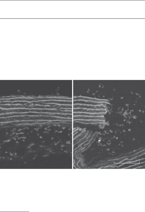
Transverse fluorescent micrographs showing the distribution of CD 11b/c-positive leukocytes in the media and adventitia of matrix-based aortic substitutes. The density of leukocytes in the elastic lamina-containing media was significantly lower than that in the collagen-containing adventitia. Note that leukocytes did not migrate into the gaps between the elastic laminae at the end of the aortic matrix substitutes (right). Red: antibody-labeled CD 11 b/c. Green: elastic laminae. Blue: Hoechst 33258-labeled cell nuclei. M, media. A, adventitia. N, neointima. Scale: 100 μm. (Reprinted from Liu SQ et al: J Biolo Chem 280:39294–301, 2005 with permission from the American Society for Biochemistry and Molecular Biology). See color insert.
Bioregenerative Engineering: Principles and Applications, by Shu Q. Liu
Copyright © 2007 John Wiley & Sons, Inc.
102
COLLAGEN MATRIX |
103 |
The extracellular matrix is the noncellular structure found in the extracellular space. This structure is composed of collagen fibers, elastic fibers or laminae, and proteoglycans. All components of extracellular matrix are produced and released by cells residing in the same tissue. Extracellular matrix plays critical roles in several aspects: (1) constituting a matrix framework that supports and organizes cells, tissues, and organs; (2) contributing to the morphogenesis and shape formation of tissues and organs; (3) providing mechanical strength to and protecting tissues and organs from injury; and (4) participating in the regulation of cell adhesion, proliferation, migration, and apoptosis. These aspects are outlined in this chapter.
The extracellular matrix can be used as biological materials for the regeneration of lost tissues and organs. Compared with synthetic polymer materials, extracellular matrix components are naturally occurring polymeric materials that are nontoxic and compatible to host cells and tissues and participate in the maintenance and regulation of cellular functions as described above. In particular, the collagen matrix has been used to construct scaffolds in experimental models for the reconstruction of a variety of tissues, such as the liver, pancreas, bones, and blood vessels. Since collagen matrix promotes cell adhesion, proliferation, and migration, collagen-based scaffolds enhance the regeneration of impaired tissues and organs. As other polymeric materials, extracellular matrix can be engineered and fabricated into various shapes and forms as desired. Thus, extracellular matrix components are preferred materials for the repair, regeneration, and engineering of malfunctioned tissues and organs.
COLLAGEN MATRIX
Composition and Formation of Collagen Matrix [4.1]
The collagen matrix is the most abundant type of extracellular matrix that is found primarily in connective tissues, such as the subcutaneous tissue, bone, and the adventitia of tubular organs, including blood vessels, airways, esophagus, stomach, and intestines. In mammalian tissues, there exist more than 20 types of collagen matrix, classified as collagen types I, II, III, and so on. Among these types of collagen, types I, II, III, IV, V, IX, XI, and XII are commonly found in connective tissue. Each type of collagen matrix is formed with one or more types of collagen molecule. A typical collagen molecule is a helical fibrillar structure composed of three peptide chains, termed α chains. A large number of collagen genes have been identified; each encodes a distinct collagen α chain. Combinations of various α chains give rise to different types of collagen fibril. Table 4.1 lists 22 types of representative collagen peptide chains.
Collagens are synthesized first as procollagen molecules in the cytoplasm of several cell types, including fibroblast, osteoblast, smooth muscle cell, and endothelial cell. Procollagen molecules are released to the extracellular space, cleaved by proteinases to remove procollagen peptides, and self-assembled into various forms of matrix structure. Collagen types I, II, III, V, and XI are organized into filamentous structures, known as collagen fibrils, with a diameter of 10–100 nm. These fibrils usually form larger collagen bundles as found in the subcutaneous tissue and the adventitia of tubular organs. Collagen types I and V are often found in the bone, skin, cornea, tendon, ligament, and internal organs, such as the lung, liver, pancreas, and kidney. Mutation of the collagen type I genes causes several disorders, including osteogenesis imperfecta, idiopathic osteoporosis, and

104
TABLE 4.1. Characteristics of Selected Collagen Molecules*
|
|
Amino |
Molecular |
|
|
Proteins |
Alternative Names |
Acids |
Weight (kDa) |
Expression |
Functions |
|
|
|
|
|
|
Collagen type I α1 |
COL1A1, collagen α1 |
1464 |
139 |
Bone, cartilage, connective tissue, |
Constituting the collagen matrix |
|
chain |
|
|
skin, cornea, tendon, ligament, |
|
Collagen type I α2 |
|
|
|
internal organs |
|
COL1A2, collagen I |
1366 |
129 |
Bone, cartilage, connective tissue, |
Constituting the collagen matrix |
|
|
α2 polypeptide |
|
|
skin, cornea, tendon, ligament, |
|
Collagen type II α1 |
|
|
|
internal organs |
|
COL2A1, chondrocalcin, |
1487 |
142 |
Cartilage, notochord, |
Constituting the collagen matrix |
|
|
collagen type XI α3 |
|
|
intervertebral disks, vitreous |
|
|
(COL11A3), cartilage |
|
|
humor of the eye |
|
Collagen type III α1 |
collagen |
|
|
|
|
COL3A1 |
1466 |
139 |
Connective tissues, skin, lung, |
Constituting the collagen |
|
Collagen type IV α1 |
|
|
|
blood vessels |
matrix |
COL4A1, collagen of |
1669 |
161 |
Brain, heart, blood vessel, liver, |
Constituting basal lamina or |
|
|
basement membrane, |
|
|
pancreas, kidney, placenta, eye |
basement membrane |
Collagen type IV α2 |
α1 chain |
|
|
|
|
COL4A2, collagen of |
1712 |
167 |
Brain, heart, blood vessel, liver, |
Constituting the basal lamina or |
|
|
basement membrane |
|
|
pancreas, kidney, placenta, eye |
basement membrane |
Collagen type V α1 |
α2 chain |
|
|
|
|
COL5A1 |
1838 |
184 |
Ubiquitous |
Present in tissues containing |
|
|
|
|
|
|
type I collagen, regulating the |
|
|
|
|
|
assembly of type I collagen |
Collagen type V α2 |
|
|
|
|
fibers |
COL5A2 |
1496 |
145 |
Bone, skin, cornea, tendon, |
Constituting the extracellular |
|
|
|
|
|
ligament, lung, liver, |
matrix and regulating the |
|
|
|
|
pancreas, kidney |
assembly of type I collagen |
|
|
|
|
|
fibers |
Collagen type VI α1 |
COL6A1 |
1028 |
109 |
Ubiquitous |
Constituting the extracellular |
|
|
|
|
|
matrix and regulating the |
Collagen type VII α1 |
|
|
|
|
integrity of tissues |
COL7A1 |
2944 |
295 |
Skin, mouth |
Found near the basement |
|
|
|
|
|
|
membrane of stratified |
|
|
|
|
|
squamous epithelia, forming |
|
|
|
|
|
fibrils that contribute to |
|
|
|
|
|
anchoring of epithelia to |
Collagen type VIII α1 |
|
|
|
|
underlying stroma |
Endothelial collagen |
744 |
73 |
Endothelial cells, skin, kidney, |
Constituting the extracellular |
|
|
|
|
|
cornea, leukocytes |
matrix, a major component |
|
|
|
|
|
of the basement membrane |
Collagen type IX α1 |
|
|
|
|
of corneal endothelium |
COL9A1, cartilage |
921 |
92 |
Cartilage, ear, eye (usually found |
Constituting the collagen matrix |
|
|
specific short |
|
|
in tissues containing type II |
|
Collagen type X α1 |
collagen |
|
|
collagen) |
|
COL10A1 |
680 |
66 |
Cartilage |
Constituting the cartilage matrix |
|
Collagen type XI α1 |
COL11A1 |
1818 |
183 |
Cartilage, cornea |
Constituting the extracellular |
Collagen type XII α1 |
|
|
|
|
matrix |
COL12A1 |
3063 |
333 |
Skin, bone |
Often found in association with |
|
|
|
|
|
|
type I collagen and regulating |
|
|
|
|
|
the interaction of collagen I |
|
|
|
|
|
fibrils with other matrix |
Collagen type XIII α1 |
|
|
|
|
components |
COL13A1 |
717 |
70 |
Eye, placenta |
Containing a transmembrane |
domain, often localized to the cell membrane, and possibly regulating cell–cell interaction and angiogenesis
105