

Bioregenerative Engineering Principles and Applications - Shu Q. Liu
..pdf76 STRUCTURE AND FUNCTION OF CELLULAR COMPONENTS
Winder SJ: Structural insights into actin-binding, branching and bundling proteins, Curr Opin Cell Biol 15:14–22, 2003.
Finer JT, Simmons RM, Spudich JA: Single myosin molecule mechanics: piconewton forces and nanometre steps, Nature 368:113–9, 1994.
Geeves MA, Holmes KC: Structural mechanism of muscle contraction, Annu Rev Biochem 68:687– 728, 1999.
Goldman YE: Wag the tail: Structural dynamics of actomyosin, Cell 93:1–4, 1998.
Huxley HE: The mechanism of muscular contraction, Science 164:1356–65, 1969.
Pantaloni D, Le Clainche C, Carlier MF: Mechanism of actin-based motility, Science 292:1502–6, 2001.
Rayment I, Smith C, Yount RG: The active site of myosin, Annu Rev Physiol 58:671–702, 1996.
Schroder RR, Manstein DJ, Jahn W, Holden H, Rayment I et al: Three-dimensional atomic model of F-actin decorated with Dictyostelium myosin S1, Nature 364:171–4, 1993.
Welch MD, Mallavarapu A, Rosenblatt J, Mitchison TJ: Actin dynamics in vivo, Curr Opin Cell Biol 9:54–61, 1997.
Rayment I, Holden HM, Whittaker M, Yohn CB, Lorenz M et al: Structure of the actin-myosin complex and its implications for muscle contraction, Science 261:58–65, 1993.
Rayment I, Rypniewski WR, Schmidt-Base K, Smith R, Tomchick DR et al: Three-dimensional structure of myosin subfragment-1: A molecular motor, Science 261:50–58, 1993.
Ruppel KM, Spudich JA: Structure-function analysis of the motor domain of myosin, Annu Rev Cell Dev Biol 12:543–73, 1996.
Small JV, Glotzer M: Cell structure and dynamics, Curr Opin Cell Biol 18:1–3, 2006.
Tan JL, Ravid S, Spudich JA: Control of nonmuscle myosins by phosphorylation, Annu Rev Biochem 61:721–59, 1992.
Cramer LP, Mitchison TJ, Theriot JA: Actin-dependent motile forces and cell motility, Curr Opin Cell Biol 6:82–6, 1994.
Theriot JA, Mitchison TJ: Actin microfilament dynamics in locomoting cells, Nature 352:126–31, 1991.
Microtubules
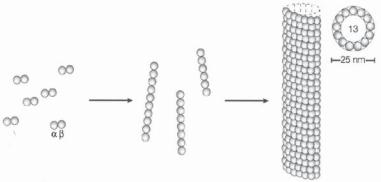
Structure and Organization of Microtubules [3.8]. Microtubules are hollow polymeric microcylinders about 20 nm in diameter and up to 20 μm in length. Microtubules are composed of dimeric tubulins. There are three types of tubulin: α, β, and γ (see Table 3.6). The α- and β-tubulins are the primary constituents of microtubules, whereas the γ- tubulin regulates the nucleation of microtubule assembly. Each tubulin molecule used for constructing the microtubules is a heterodimer of α- and β-tubulin. In mammalian cells, there are several isoforms for α- as well as for β-tubulin. These isoforms have similar structures, but are originated from different genes. All tubulin isoforms can be polymerized into microtubules. Tubulin can be found in all mammalian cells. However, the distribution of tubulin varies in different cell types. For instance, the nerve cells exhibit a higher concentration of tubulin than do other cell types. The tubulin genes are highly conserved among different species.
In microtubules, α- and β-tubulin dimers are uniformly aligned along the axis of the microtubule, forming parallel protofilaments. In each protofilament, the α- or β-tubulin subunits are always arranged in the same direction, giving a polarity to microtubules with a plus and minus end. Each microtubule is composed of 13 protofilaments (Fig. 3.4). In

TABLE 3.6. Characteristics of Selected Tubulin Isoforms*
|
Alternative |
Amino |
Molecular |
|
|
Proteins |
Names |
Acids |
Weight (kDa) |
Expression |
Functions |
|
|
|
|
|
|
Tubulin α1 |
TUBA1, Tubulin α testis-specific |
448 |
50 |
Nervous system, testis |
Constituting microtubules |
Tubulin α2 |
TUBA2 |
450 |
50 |
Thymus, leukocytes, intestine, |
Constituting microtubules |
|
|
|
|
overy, testis |
|
Tubulin α3 |
Tubulin α brain-specific, B α1 |
451 |
50 |
Brain |
Constituting microtubules in |
|
|
|
|
|
central nervous system |
Tubulin α, ubiquitous |
K-α1 |
451 |
50 |
Ubiquitous |
Constituting microtubules |
Tubulin β |
TUBB |
445 |
50 |
Brain |
Constituting microtubules |
Tubulin γ |
TUBG1, TUBG, tubulin γ1 |
451 |
51 |
Heart, lung, liver, kidney, |
Regulating the nucleation of |
|
chain, γ1 tubulin, γ tubulin |
|
|
intestine, ovary, skeletal |
microtubule assembly |
|
complex component 1, |
|
|
muscle |
|
|
tubulin γ polypeptide |
|
|
|
|
|
|
|
|
|
|
* Based on bibliography 3.8.
77
78 STRUCTURE AND FUNCTION OF CELLULAR COMPONENTS
αβ heterodimers |
Short |
Microtubule |
|
protofilaments |
|||
|
|
Figure 3.4. Formation of a microtubule from tubulin molecules. A microtubule is formed via several steps: (1) an α, β-tubulin monomer aggregates to form a tubulin heterodimer; (2) the tubulin heterodimers form short linear protofilaments; (3) 13 protofilaments are joined together laterally to organize into a microtubule. (Adapted by permission from Macmillan Publishers Ltd.: Westermann S, Weber K: Nature Rev Mol Cell Biol 4:938–48, copyright 2003.)
an interphase cell, microtubules are distributed in the radial direction with the minus end attached to the centrosome and the plus end toward the cell periphery.
It is important to note that several substances, including colchicine, colcemid, and taxol, are commonly used to modulate the assembly, structure, stability, and function of microtubules. Colchicine is an alkaloid extracted from meadow saffron. Colchicine can bind to tubulin and suppress tubulin polymerization or microtubule assembly. Since microtubules undergo continuously depolymerization, a treatment with colchicine facilitates the disassembly of microtubules. Once tubulin molecules are polymerized into microtubules, colchicine can no longer bind to tubulin. Colcemid is a substance similar to colchicine in function. Since the disassemble of microtubules interrupt cell mitosis, colchicine and colcemid are used to treat cancer. Taxol is derived from yew trees and can bind to polymerized microtubules. The binding of taxol enhances the stability of microtubules, inhibiting tubulin depolymerization. Such an effect induces cell arrest during mitosis. Taxol is also used as a drug for the treatment of cancer.
Microtubule Assembly and Disassembly [3.9]. Microtubule assembly is accomplished via tubulin polymerization, whereas its disassembly is via tubulin depolymerization. There are two critical processes, which are involved in microtubule assembly: nucleation and elongation. Nucleation is the formation of short tubulin protofilaments or oligomers, which further form a short initiating microtubule (Fig. 3.4). Elongation is the growth of microtubules based on the initial microtubule segment. Microtubule assembly can be simulated in vitro with tubulins in the presence of Mg2+ and GTP. The initial nucleation from tubulin heteodimers is a more difficult process than elongation. Thus nucleation is usually a slower process than elongation. While a microtubule is elongating via tubulin polymerization, there also exists simultaneous tubulin depolymerization. The rate of tubulin polymerization and depolymerization is dependent on the concentration of free tubulins. At a critical concentration of free tubulin, the rate of tubulin polymerization is counterbalanced by that of depolymerization, and microtubules cease growing.

MICROTUBULES 79
Microtubules are connected at their minus end to a central structure within the cell, known as the centrosome, which is located in the nucleus during the interphase. The centrosome is considered the origin where microtubules grow from. The relationship of microtubules with the centrosome can be verified by observing the growth of degraded microtubules. A treatment with colcemid induces the degradation of microtubules. In the presence of fluorescent marker-tagged tubulins, it can be found that new microtubules grow from the centrosome following the removal of colcemid. These microtubules continuously elongate toward the cell periphery until a complete microtubule network is reestablished. Each centrosome contains two cylindrical structures perpendicular to each other, known as centrioles. During the interphase, the centrosome can be split into two daughter centrosomes, which move to opposite sides of the nucleus during the early stage of cell mitosis, serving as two poles for anchoring microtubule spindles (Fig. 3.5).
A microtubule undergoes rapid assembly and disassembly. The tubulins within a microtubule could be completely replaced with new tubulins within a period as short as 20 min. Such a process can be detected by injecting fluorescent marker-tagged tubulins into a living cell and observed by fluorescence microscopy. It is interesting to note that microtubules undergo alternating growth and retraction, resulting in a dynamic change in the length of microtubules. These dynamic changes are critical for the redistribution of microtubules within a cell.
Microtubule dynamics requires the presence of GTPs, which produce energy by hydrolysis. Each α- and β-tubulin is bound with a GTP molecule, which is required for tubulin polymerization. On the polymerization of a tubulin heterodimer to a microtubule, the GTP molecule associated with the β-tubulin can be hydrolyzed to produce energy, whereas the GTP molecule associated with the α-tubulin serves as a constituent of the tubulin and cannot be hydrolyzed. The energy produced by the hydrolysis of the β-tubulin-associated GTP is used for microtubule depolymerization, but not for polymerization. This can be
Non-dividing cell
+
-
- +
-
+
+
Dividing cell
++
- |
- |
+ + |
- |
- |
|
|
|
||
- |
|
+ |
- - |
|
++
Figure 3.5. Centrosomes and microtubules in nondividing and dividing cells (based on bibliography 3.9).
80 STRUCTURE AND FUNCTION OF CELLULAR COMPONENTS
verified by using GTP analogs that cannot be hydrolyzed. Tubulins associated with GTP analogs can be polymerized. However, once incorporated into a microtubule, these tubulin molecules cannot be depolymerized, suggesting that GTP hydrolysis is critical to the depolymerization of microtubules.
A microtubule can be assembled at the plus and minus ends, but exhibits different assembly rate at these ends under a given condition. The assembly of microtubules can be observed by using in vitro experiments. Isolated microtubules from cells can grow in the presence of free tubulins. The plus end of a microtubule grows about 3 times faster than the minus end. Since microtubules are aligned in the radial direction of a cell with the plus ends pointing at the periphery, microtubules often grow from the cell center to the periphery.
Regulation of Microtubule Dynamics [3.9]. The assembly and disassembly of microtubules are processes regulated by microtubule-associated proteins (Table 3.7). Two major types of microtubule-associated proteins have been identified in nerve cells: the high-molecular-weight proteins and the τ proteins. The high-molecular-weight proteins include microtubule-associated proteins 1 and 2 with molecular weights 200 and 300 kDa, respectively. The τ proteins have molecular weights ranging from 55 to 62 kDa. Each of these microtubule-associated proteins contains two domains; the first domain is capable of binding to microtubules, and the second domain binds to other types of intracellular structures. The binding of microtubule-associated proteins to microtubules prevents microtubules from depolymerization and enhances the stability of the microtubules. The exact regulatory mechanisms, however, remain to be investigated.
Function of Microtubules [3.10]. One of the primary functions of microtubules is the control of cell polarity. Microtubules exhibit nonuniform tubulin polymerization and depolymerization through the cell. Such a nonuniform feature is critical to the controlled distribution of microtubules, potentially contributing to cell polarization. At a given time, some microtubules may undergo predominant polymerization, while others may experience depolymerization. Fast-growing microtubules may be capped or protected by capping molecules, yielding stabilized microtubules in a specified direction. Meanwhile, uncapped microtubules are not stable and cannot grow as rapidly as the capped microtubules. The rapid growth of the capped microtubules causes regional extension of the cell membrane, leading to the formation of cell polarity.
Microtubules play a critical role in the transport of intracellular organelles and vesicles, which are required for a variety of metabolic and signaling activities. The transport function is accomplished by coordinated interactions of motor proteins, including kinesin and dynein, with microtubules. Each motor molecule is composed of two heavy chains and several light chains. Each heavy chain contains a globular head and a tail. The head interacts directly with microtubules and induces the sliding of the motor protein along a microtubule, a process dependent on ATPs, whereas the tail binds to an intracellular component to be moved. The light chains also play a role in the regulation of motor protein movement.
The motor proteins kinesin and dynein (Table 3.8) are both involved in the transport of intracellular organelles and chromosome separation during mitosis. However, kinesin and dynein move in opposite directions along a microtubule. Kinesin can only move intracellular organelles from the centrosome or the minus end of the microtubule to the cell periphery or the plus end of the microtubule, whereas dynein moves toward the cen-

TABLE 3.7. Characteristics of Selected Microtubule-Associated Proteins*
|
|
Amino |
Molecular |
|
|
Proteins |
Alternative Names |
Acids |
Weight (kDa) |
Expression |
Functions |
|
|
|
|
|
|
Microtubule -associated |
Microtubule-associated protein |
2805 |
306 |
Central nerveous system |
Regulating microtubule assembly |
protein 1A |
1-like, MTAP1A |
|
|
|
|
Microtubule -associated |
MAP1B, MAP1 light chain LC1 |
2468 |
271 |
Central nerveous system |
Regulating microtubule assembly |
protein 1B |
|
|
|
|
|
Microtubule-associated |
MAP2, dendrite-specific MAP |
1858 |
203 |
Central nerveous system |
Regulating microtubule assembly |
protein-2 |
|
|
|
|
|
τ |
Microtubule-associated protein τ, |
758 |
79 |
Central nerveous system |
Regulating microtubule assembly, |
|
MAPT, MTBT1, neurofibrillary |
|
|
|
playing a critical role in both |
|
tangle protein, paired helical |
|
|
|
integrity and functionality of |
|
filament τ |
|
|
|
neuronsa |
a Note that τ mutation induces the formation of neurofibrillary tangles and causes neurodegenerative disorders such as Alzheimer’s disease, Parkinson’s disease, frontotemporal dementia, and supranuclear palsy.
*Based on bibliography 3.9.
81

82
TABLE 3.8. Characteristics of the Motor Proteins Kinesin and Dynein*
|
|
Amino |
Molecular |
|
|
Proteins |
Alternative Names |
Acids |
Weight (kDa) |
Expression |
Functions |
|
|
|
|
|
|
Kinesin heavy chain 2 |
HK2 |
679 |
77 |
Nervous system, lung, spleen, |
A constituent for the microtubule- |
|
|
|
|
stomach, testis, placenta |
associated motor protein kinesin, which |
|
|
|
|
|
mediates movement of cytoplasmic |
|
|
|
|
|
structures such as chromosomes and |
|
|
|
|
|
vesiclesa |
Kinesin light chain |
Kinesin 2 |
569 |
65 |
Nervous system |
A constituent for the microtubule- |
|
|
|
|
|
associated motor protein kinesin (see |
|
|
|
|
|
above for function) |
Dynein cytoplasmic |
Dynein cytoplasmic |
4646 |
532 |
Brain, heart, lung, pancreas, |
A constituent for the microtubule- |
heavy chain 1 |
heavy polypeptide 1 |
|
|
liver, testis |
associated motor protein dynein, which |
|
|
|
|
|
move organelles and vesicles from cell |
|
|
|
|
|
periphery or plus end of microtubule to |
|
|
|
|
|
centrosome or minus end of |
|
|
|
|
|
microtubule |
Dynein cytoplasmic |
Cytoplasmic dynein |
645 |
73 |
Brain heart, lung, pancreas, |
A constituent for the microtubule- |
intermediate chain 1 |
intermediate chain 1 |
|
|
liver, testis |
associated motor protein dynein |
|
|
|
|
|
(see above for dynein function) |
Dynein cytoplasmic |
8-kDa dynein light |
89 |
10 |
Brain, heart, lung, pancreas, |
A constituent for the microtubule- |
light chain 1 |
chain, cytoplasmic |
|
|
liver, testis |
associated motor protein dynein (see |
|
dynein light |
|
|
|
above for dynein function) |
|
polypeptide |
|
|
|
|
a Note that kinesins move organelles from the centrosome or the minus end of the microtubule to the cell periphery or the plus end of the microtubule. *Based on bibliography 3.10.
INTERMEDIATE FILAMENTS |
83 |
trosome. A kinesin molecule is a tetramer composed of two heavy and two light chains. The heavy chain is located at the N-terminus of the molecule, and the light chains are at the C-terminus. The N-terminal heavy chains form the motor domains with the microtubule-binding regions, which mediate the sliding motion of the kinesin molecule along the microtubule. The N-terminal heavy chain also possesses an ATP-binding site, which serves as an ATPase and interacts with ATP molecules to provide energy for kinesin movement. The movement caused by kinesin molecules can be readily verified by using in vitro assays with purified motor proteins and microtubules. When microtubules are mixed with kinesin-coated polystyrene beads, the beads move toward the plus end of microtubules.
Dyneins are a family of motor proteins that are divided into two groups: the axonal and cytoplasmic dyneins. The axonal dynein molecules are responsible for organelle transport within the neuronal axon. Cytoplasmic dyneins mediate intracellular motility, protein sorting, and movement of intracellular organelles such as endosomes and lysosomes. A dynein molecule is comprised of two force-generating heavy chains and several intermediate and light chains. The heavy chains contain ATPases, which interact with ATP molecules and generate energy for mechanical movement. The motility of dynein molecules can be observed by using in vitro assays with purified dynein molecules and microtubules. Dynein molecules can move intracellular organelles from the cell periphery or the plus end of the microtubule to the centrosome or the minus end of the microtubule.
Microtubules are well known for their role in regulating cell mitosis or the segregation of chromosomes. Microtubules and associated proteins constitute a key structure for cell mitosis, known as the mitotic spindle, which plays a critical role in the alignment and separation of chromosomes. During the early stage of mitosis or prophase, the centrosome is separated into two daughter centrosomes, which move toward the two opposite poles. A mitotic spindle is initiated from the two centrosomes and gradually forms a polar structure. During metaphase, chromosomes are attached to the spindle microtubules. The shortening of the microtubules induces the movement of separated daughter chromosomes from the cell center toward the two centrosome poles. The destruction of microtubules by a treatment with colchicine interrupts cell mitosis.
Intermediate Filaments
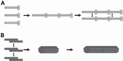
Structure and Organization of Intermediate Filaments [3.11]. Intermediate filaments are one of the three types of filamentous structures that constitute the cytoskeleton. The term “intermediate” is derived from the fact that the diameter of intermediate filaments ( 10 nm) is between the other two types of cytoskeletal filaments (Fig. 3.6), specifically, actin filaments ( 8 nm) and microtubules ( 25 nm). Intermediate filaments are composed of various molecules, including keratin, vimentin, neurofilament protein, and nuclear lamin. The constituent molecules of intermediate filaments are fibrous in shape. To form an intermediate filament, two molecules are organized into a parallel dimer with the amino termini at one end and the carboxyl termini at the other end. For most types of intermediate filaments, the two dimers in turn form an antiparallel tetramer bundle with the amino termini of one dimer arranged with the carboxyl termini of the other dimer at each end of the tetramer bundle (Fig. 3.7). The tetramers are the basic units that are assembled into helical intermediate filaments via bundle–lateral interactions. Because of the antiparallel feature of the tetramer bundles, intermediate filaments do not exhibit polarity.

84 STRUCTURE AND FUNCTION OF CELLULAR COMPONENTS
Figure 3.6. Electron micrographs of intermediate filaments at different assembly stages. (A–C) Lamin A/C. Lamin filaments can be dialyzed in pH 6.5/150 mM NaCl buffer, generating linear head-to-tail fibers (panel A). In the presence of Ca2+, lamin filaments can be dialyzed into beaded long filaments (panel B). Panel C shows assembled lamin filaments. (D–G) Assembly of recombinant human vimentin. Vimentin filament assembly was initiated by adding filament buffer and fixed with 0.1% glutaraldehyde at 10 s (panel D), 1 min (panel E), 5 min, (panel F), and 1 h (panel G). Scale bar: 100 nm. (Reprinted by permission from Herrmann H, Aebi U: Annu Rev Biochem, 73:749–89, copyright 2004 by Annual Reviews, www.annualreviews.org.)
On the basis of constituents, intermediate filaments are classified into several subtypes, including keratin filaments, vimentin filaments, neurofilaments, and lamin filaments (see list in Table 3.9), which are found in different cell types. Keratin filaments are composed of various types of keratin and are present in epithelial cells, the hair, and the nails. Individual keratin molecules are different in structure and can be grouped into to subfamilies, including types I and II keratins, based on the properties of amino acids. Type I keratins are acidic with a molecular weight 40–70 kDa, whereas type II keratins are basic or neutral with a similar molecular weight. Both type I and type II keratins are required for the constitution of keratin filaments. In a typical epithelial cell, keratin filaments are connected at the end to desmosomes, a cell junction structure that joins two neighboring cells. In addition, keratin filaments anchor to hemidesmosomes, a structure that mediates cell attachment to the basal lamina.
Vimentin filaments are present in fibroblasts, endothelial cells, and leukocytes, and contain a single type of molecule: vimentin. In addition, there exist vimentin-related filaments, which exhibit structure and properties similar to those of vimentin filaments. One type is desmin filaments, which are composed of desmin and are present primarily in
INTERMEDIATE FILAMENTS |
85 |
Figure 3.7. Schematic representation of intermediate filament assembly. (A) Lamin filament assembly. Lamin dimers are first associated into head-to-tail filaments, which are further associated laterally into complete filaments. (B) Vimentin filament assembly. Vimentin molecules first form antiparallel half-staggered double dimers (or tetramers), which form complete vimentin filaments. (Reprinted by permission from Herrmann H, Aebi U: Annu Rev Biochem, 73:749–89, copyright 2004 by Annual Reviews, www.annualreviews.org.)
muscle cells, including smooth, skeletal, and cardiac muscle cells. Desmin filaments often anchor to cell junctions. Another type is glial filaments composed of glial fibrillary acidic proteins. This type of intermediate filament is found in astrocytes of the central nervous system and Schwann cells of the peripheral nervous system. It is important to note that vimentin and vimentin-related proteins can be crosslinked together, but these proteins cannot be crosslinked with keratin-based intermediate filaments.
Neurofilaments are present in neurons, arranged primarily along the axon. There are three types of neurofilament proteins, including neurofilament-L, -M, and -H, based on low, medium, and high molecular weights, respectively. These molecular types can be found within all neurofilaments. In a typical neuronal axon, neurofilaments are uniformly spaced with a high density. These filaments are laterally crosslinked, providing mechanical strength to the axon.
Lamin filaments are found in the nuclear lamina, which is a 20-nm membrane lining the internal surface of the nuclear membrane. Lamin filaments are composed of two types of lamin: lamin A (or A/C) and lamin B. In structure, lamin is similar to other intermediate filament proteins. However, lamin contains signaling structures that direct lamin transport from the cytosol to nucleus. The lamin filaments undergo dynamic disassembly during early mitosis and reassembly during the late mitosis in coordination with chromosome reorganization and separation. In interphase cells, lamin filaments are organized into a dense lattice network. The network is interrupted at nuclear pores, which allow the transport of molecules from and to the nucleus.
Function of Intermediate Filaments [3.12]. A major function of intermediate filaments is to provide mechanical strength to cells and tissues. Such a function is supported by observations from transgenic keratin-deficient animal models. In transgenic mice with a