

Bioregenerative Engineering Principles and Applications - Shu Q. Liu
..pdf46 REGULATION OF GENE EXPRESSION
translation. The regulation of these processes involves specified RNA sequences, which are termed cis-acting RNA elements, and regulatory RNA-binding proteins, which are termed trans-acting RNA-binding factors. The trans-acting RNA-binding factors can recognize and interact with cis-acting RNA elements to form ribonucleotide protein (RNP) complexes. These complexes can move from the nucleus to the cytoplasm and interact with motor proteins, such as kinesin and dynein molecules, which further move the ribonucleotide protein complexes along the microtubules to the destination sites. The ribonucleotide protein complexes can also interact with myosin molecules, which move the complexes along the actin filaments. Once transported to destination sites, trans-acting RNA-anchoring proteins recognize and interact with the ribonucleotide protein complexes, anchoring the complexes to the specified sites. At these sites, the ribonucleotide protein complexes interact with ribosomes, initiating protein translation. It is interesting to note that unspliced mRNA cannot be transported. The exact mechanisms for selective mRNA transport remain poorly understood.
In addition to mRNA capping, splicing, and transport, the posttranscriptional stability of mRNA, which determines the concentration of mRNA, is also a critical factor for the regulation of gene expression. The mRNA concentration is controlled by the rates of RNA transcription, splicing, and transport as well as the rate of mRNA degradation. The stability of mRNA varies widely with half-life ranging from minutes to hours. The mRNAs of cytokines and growth factors, which are involved in short-term signaling events, are usually short-lived. In contrast, the mRNAs of constitutive proteins possess a relatively long-term lifespan. The stability of mRNAs is controlled by enzymes known as nucleases. These enzymes are often coupled to protein translation and can be activated when excessive proteins are produced, inducing degradation of specific mRNAs and reduction in protein translation.
REGULATION OF PROTEIN TRANSLATION [2.9]
In addition to the regulation of transcription and conversion of pre-mRNA to mature mRNA, gene expression is also controlled at the protein translation level. There are two known regulatory mechanisms at this level: modification at the 5′-terminus of mRNA and modification of initiation factors. The modification of mRNA at the 5′-terminus is usually a negative mechanism of translation regulation by RNA-binding proteins. These proteins serve as translation repressors, bind to mRNA at the 5′-terminus, and block the assembly of initiation factors and the 40 S subunit of ribosomes. Such a process results in the suppression of protein translation. The modification of translation initiation factors is another powerful means for the regulation of protein translation. A major form of modification is phosphorylation, which controls the activity of translation initiation factors and thus regulates the rate of protein translation. For example, eukaryotic initiation factor 2 (eIF2) is a protein translation initiation factor. The α subunit of this protein can be phosphorylated on the Ser 51 residue by a serine/threonine protein kinase known as RNA-dependent protein kinase (PKR), which can be activated in response to the stimulation of various factors, such as viruses and double-stranded RNA (dsRNA). The phosphorylation of the eukaryotic initiation factor 2 reduces its activity and thus results in the suppression of protein synthesis. In the case of virus infection, the suppression of protein synthesis helps to reduce viral propagation.
BIBLIOGRAPHY 47
BIBLIOGRAPHY
2.1. DNA Regulatory Elements and Transcriptional Factors
Cramer P, Bushnell DA, Fu J, Gnatt AL, Maier-Davis B et al: Architecture of RNA polymerase II and implications for the transcription mechanism, Science 288:640–8, 2000.
Acker J, Mattei MG, Wintzerith M, Roeckel N, Depetris D et al: Chromosomal localization of human RNA polymerase II subunit genes, Genomics 20:496–9, 1994.
Fanciulli M, Bruno T, Di Padova M, De Angelis R, Lovari S et al: The interacting RNA polymerase II subunits, hRPB11 and hRPB3, are coordinately expressed in adult human tissues and downregulated by doxorubicin, FEBS Lett 427:236–40, 1998.
Pati UK, Weissman SM: The amino acid sequence of the human RNA polymerase II 33-kDa subunit hRPB 33 is highly conserved among eukaryotes, J Biol Chem 265:8400–3, 1990.
Hobbs NK, Bondareva AA, Barnett S, Capecchi MR et al: Removing the vertebrate-specific TBP N terminus disrupts placental beta-2M-dependent interactions with the maternal immune system, Cell 110:43–54, 2002.
Human protein reference data base, Johns Hopkins University and the Institute of Bioinformatics, at http://www.hprd.org/protein.
Imbert G, Trottier Y, Beckmann J, Mandel JL: The gene for the TATA binding protein (TBP) that contains a highly polymorphic protein coding CAG repeat maps to 6q27, Genomics 21:667–8, 1994.
Kao CC, Lieberman PM, Schmidt MC, Zhou Q, Pei R et al: Cloning of a transcriptionally active human TATA binding factor, Science 248:1646–50, 1990.
Martianov I, Viville S, Davidson I: RNA polymerase II transcription in murine cells lacking the TATA binding protein, Science 298:1036–9, 2002.
Nikolov DB, Hu SH, Lin J, Gasch A, Hoffmann A et al: Crystal structure of TFIID TATA-box binding protein, Nature 360:40–6, 1992.
Nikolov DB, Chen H, Halay ED, Hoffman A, Roeder RG et al: Crystal structure of a human TATA box-binding protein/TATA element complex, Proc Natl Acad Sci USA, 93(10):4862–7, May 14, 1996.
Peterson MG, Tanese N, Pugh BF, Tjian R: Functional domains and upstream activation properties of cloned human TATA binding protein, Science 248:1625–30, 1990.
Rosen DR, Trofatter JA, Brown RH, Jr: Mapping of the human TATA-binding protein gene (TBP) to chromosome 6qter, Cytogenet Cell Genet 69:279–80, 1995.
Veenstra GJC, Weeks DL, Wolffe AP: Distinct roles for TBP and TBP-like factor in early embryonic gene transcription in Xenopus, Science 290:2312–4, 2000.
2.2. Control of the Activity of Trans-Acting Factors
Holmberg CI, Tran SE, Eriksson JE, Sistonen L: Multisite phosphorylation provides sophisticated regulation of transcription factors, Trends Biochem Sci 27:619–27, 2002.
Firulli AB: A HANDful of questions: The molecular biology of the heart and neural crest derivatives (HAND)-subclass of basic helix-loop-helix transcription factors, Gene 312:27–40, 2003.
Falke D, Juliano RL: Selective gene regulation with designed transcription factors: Implications for therapy, Curr Opin Mol Ther 5:161–6, 2003.
Roeder RG: Transcriptional regulation and the role of diverse coactivators in animal cells, FEBS Lett 579:909–15, 2005.
McDonnell DP, Norris JD: Connections and regulation of the human estrogen receptor, Science 296:1642–4, 2002.
48 REGULATION OF GENE EXPRESSION
Della Fazia MA, Servillo G, Sassone-Corsi P: Cyclic AMP signalling and cellular proliferation: Regulation of CREB and CREM, FEBS Lett 410:22–4, 1997.
Sassone-Corsi P: Transcription factors responsive to cAMP, Annu Rev Cell Dev Biol 11:355–77, 1995.
2.3. Chromatin Modification
van Attikum H, Gasser SM: The histone code at DNA breaks: a guide to repair? Nature Rev Mol Cell Biol 6:757–65, 2005.
Herman JG, Baylin SB: Gene silencing in cancer in association with promoter hypermethylation, N Engl J Med 349:2042–54, 2003.
Thiriet C, Hayes JJ: Chromatin in need of a fix: Phosphorylation of H2AX connects chromatin to DNA repair, Mol Cell 18:617–22, 2005.
Martin C, Zhang Y: The diverse functions of histone lysine methylation, Nature Rev Mol Cell Biol 6:838–49, 2005.
Henikoff S, Ahmad K: Assembly of variant histones into chromatin, Annu Rev Cell Dev Biol 21:133–53, 2005.
2.4. DNA Modification
Baylin SB: DNA methylation and gene silencing in cancer, Nat Clin Pract Oncol 2 (Suppl 1):S4–11, 2005.
Herman JG, Baylin SB: Gene silencing in cancer in association with promoter hypermethylation, N Engl J Med 349:2042–54, 2003.
Jones PA, Laird PW: Cancer epigenetics comes of age, Nature Genet 21:163–7, 1999.
Jones PA, Baylin SB: The fundamental role of epigenetic events in cancer, Nature Rev Genet 3:415–28, 2002.
Herman JG: Hypermethylation of tumor suppressor genes in cancer, Semin Cancer Biol 9:359–67, 1999.
Holliday R: Epigenetic inheritance based on DNA methylation, EXS 64:452–68, 1993.
2.5. 5′-Terminal Capping of Pre-mRNA
Parker R, Song H: The enzymes and control of eukaryotic mRNA turnover, Nat Struct Mol Biol 11:121–7, 2004.
Dunckley T, Parker R: The DCP2 protein is required for mRNA decapping in Saccharomyces cerevisiae and contains a functional MutT motif, EMBO J 18:5411–22, 1999.
Wang Z, Jiao X, Carr-Schmid A, Kiledjian M: The hDcp2 protein is a mammalian mRNA decapping enzyme, Proc Natl Acad Sci USA 99:12663–8, 2002.
Lykke-Andersen J: Identification of a human decapping complex associated with hUpf proteins in nonsense-mediated decay, Mol Cell Biol 22:8114–21, 2002.
van Dijk E, Cougot N, Meyer S, Babajko S, Wahle E et al: Seraphin, Human Dcp2: A catalytically active mRNA decapping enzyme located in specific cytoplasmic structures, EMBO J 21:6915– 24, 2002.
Wang Z, Kiledjian M: Functional link between the mammalian exosome and mRNA decapping, Cell 107:751–62, 2001.
Liu H, Rodgers ND, Jiao X, Kiledjian M: The scavenger mRNA decapping enzyme DcpS is a member of the HIT family of pyrophosphatases, EMBO J 21:4699–708, 2002.
Lima CD, Wang LK, Shuman S: Structure and mechanism of yeast RNA triphosphatase: An essential component of the mRNA capping apparatus, Cell 99:533–43, 1999.
BIBLIOGRAPHY 49
Changela A, Ho CK, Martins A, Shuman S, Mondragon A: Structure and mechanism of the RNA triphosphatase component of mammalian mRNA capping enzyme, EMBO J 20:2575–86, 2001.
Håkansson K, Doherty AJ, Shuman S, Wigley DB: X-ray crystallography reveals a large conformational change during guanyl transfer by mRNA capping enzymes, Cell 89:545–53, 1997.
Shuman S, Lima CD: The polynucleotide ligase/RNA capping enzyme superfamily of covalent nucleotidyltransferases, Curr Opin Struct Biol 14:757–64, 2004.
Håkansson K, Wigley DB: Structure of a complex between a cap analogue and mRNA guanylyl transferase demonstrates the structural chemistry of RNA capping, Proc Natl Acad Sci USA 95:1505–10, 1998.
Ho CK, Sriskanda V, McCracken S, Bentley D, Schwer B et al: The gaunylyltransferase domain of mammalian mRNA capping enzyme binds to the phosphorylated carboxy-terminal domain of RNA polymerase II, J Biol Chem 273:9577–85, 1998.
Bentley D: The mRNA assembly line: transcription and processing machines in the same factory,
Curr Opin Cell Biol 14:336–42, 2002.
She M, Decker CJ, Sundramurthy K, Liu Y, Chen N et al: Crystal structure of Dcp1p and its functional implications in mRNA decapping, Nat Struct Mol Biol 11:249–56, 2004.
van Dijk E, Le Hir H, Seraphin B: DcpS can act in the 5′-3′ mRNA decay pathway in addition to the 3′-5′ pathway, Proc Natl Acad Sci USA 100:12081–6, 2003.
Gu M, Lima CD: Processing the message: Structural insights into capping and decapping mRNA,
Curr Opin Struct Biol 15:99–106, 2005.
2.6. Polyadenylation
Colgan DF, Manley JL: Mechanism and regulation of mRNA polyadenylation, Genes Dev 11:2755– 66, 1997.
Zhao J, Hyman L, Moore C: Formation of mRNA 3′ ends in eukaryotes: Mechanism, regulation, and interrelationships with other steps in mRNA synthesis, Microbiol Mol Biol Rev 63:405–45, 1999.
Shatkin AJ, Manley JL: The ends of the affair: Capping and polyadenylation, Nat Struct Biol 7:838–42, 2000.
de Vries H, Ruegsegger U, Hubner W, Friedlein A, Langen H et al: Human pre-mRNA cleavage factor IIm contains homologs of yeast proteins and bridges two other cleavage factors, EMBO J 19:5895–904, 2000.
Dichtl B, Keller W: Recognition of polyadenylation sites in yeast pre-mRNAs by cleavage and polyadenylation factor, EMBO J 20:3197–209, 2001.
Zhao J, Hyman L, Moore C: Formation of mRNA 3′ ends in eukaryotes: Mechanism, regulation, and interrelationships with other steps in mRNA synthesis, Microbiol Mol Biol Rev 63:405–45, 1999.
Buratowski S: Connections between mRNA 3′ end processing and transcription termination, Curr Opin Cell Biol 17:257–61, 2005.
Proudfoot N: New perspectives on connecting messenger RNA 3′ end formation to transcription,
Curr Opin Cell Biol 16:272–8, 2004.
Proudfoot N, O’Sullivan J: Polyadenylation: A tail of two complexes, Curr Biol 12:R855–7, 2002.
2.7. Pre-mRNA Splicing
Black DL: Mechanisms of alternative pre-messenger RNA splicing, Annu Rev Biochem 72:291–336, 2003.
50 REGULATION OF GENE EXPRESSION
Chan SP, Kao DI, Tsai WY, Cheng SC: The Prp19p-associated complex in spliceosome activation, Science 302:279–82, 2003.
Fairbrother WG, Yeh RF, Sharp PA, Burge CB: Predictive identification of exonic splicing enhancers in human genes, Science 297:1007–13, 2002.
Konarska MM, Query CC: Insights into the mechanisms of splicing: More lessons from the ribosome, Genes Dev 19:2255–60, 2005.
Makarov EM, Makarova OV, Urlaub H, Gentzel M, Will CL et al: Small nuclear ribonucleoprotein remodeling during catalytic activation of the spliceosome, Science 298:2205–8, 2002.
Maniatis T, Tasic B: Alternative pre-mRNA splicing and proteome expansion in metazoans, Nature 418:236–43, 2002.
Moore MJ, Sharp PA: Evidence for two active sites in the spliceosome provided by stereochemistry of pre-mRNA splicing, Nature 365:364–8, 1993.
Nelson KK, Green MR: Mechanism for cryptic splice site activation during pre-mRNA splicing,
Proc Natl Acad Sci 87:6253–7, 1990.
Ogle JM, Ramakrishnan V: Structural insights into translational fidelity, Annu Rev Biochem 74:129–77, 2005.
Zhou Z, Licklider LJ, Gygi SP, Reed R: Comprehensive proteomic analysis of the human spliceosome, Nature 419:182–5, 2002.
Jurica MS, Moore MJ: Pre-mRNA splicing: Awash in a sea of proteins, Mol Cell 12:5–14, 2003.
Staley JP, Guthrie C: Mechanical devices of the spliceosome: Motors, clocks, springs, and things, Cell 92:315–26, 1998.
2.8. mRNA Transport
Kindler S, Wang H, Richter D, Tiedge H: RNA transport and local control of translation, Annu Rev Cell Dev Biol 21:223–45, 2005.
Ainger K, Avossa D, Diana AS, Barry C, Barbarese E et al: Transport and localization elements in myelin basic protein mRNA, J Cell Biol 138:1077–87, 1997.
Chartrand P, Singer RH, Long RM: RNP localization and transport in yeast, Annu Rev Cell Dev Biol 17:297–310, 2001.
Chennathukuzhi V, Morales CR, El-Alfy M, Hecht NB: The kinesin KIF17b and RNA-binding protein TB-RBP transport specific cAMP-responsive element modulator-regulated mRNAs in male germ cells, Proc Natl Acad Sci USA 100:15566–71, 2003.
Farina KL, Singer RH: The nuclear connection in RNA transport and localization, Trends Cell Biol 12:466–72, 2002.
Goldstein LS, Yang Z: Microtubule-based transport systems in neurons: The roles of kinesins and dyneins, Annu Rev Neurosci 23:39–71, 2000.
Hachet O, Ephrussi A: Splicing of oskar RNA in the nucleus is coupled to its cytoplasmic localization, Nature 428:959–63, 2004.
Han JR, Yiu GK, Hecht NB: Testis/brain RNA-binding protein attaches translationally repressed and transported mRNAs to microtubules, Proc Natl Acad Sci USA 92:9550–54, 1995.
Job C, Eberwine J: Localization and translation of mRNA in dendrites and axons, Nature Rev Neurosci 2:889–98, 2001.
Johnstone O, Lasko P: Translational regulation and RNA localization in Drosophila oocytes and embryos, Annu Rev Genet 35:365–406, 2001.
Kapp LD, Lorsch JR: The molecular mechanics of eukaryotic translation, Annu Rev Biochem 73:657–704, 2004.
BIBLIOGRAPHY 51
Kloc M, Zearfoss NR, Etkin LD: Mechanisms of subcellular mRNA localization, Cell 108:533–44, 2002.
Kress TL, Yoon YJ, Mowry KL: Nuclear RNP complex assembly initiates cytoplasmic RNA localization, J Cell Biol 165:203–11, 2004.
2.9. Regulation of Protein Translation
Dean KA, Aggarwal AK, Wharton RP: Translational repressors in Drosophila, Trends Genet 18:572–7, 2002.
Gray NK, Wickens M: Control of translation initiation in animals, Annu Rev Cell Dev Biol 14:399–458, 1998.
Dey M, Cao C, Dar AC, Tamura T, Ozato K et al: Mechanistic link between PKR dimerization, autophosphorylation, and eIF2alpha substrate recognition, Cell 122:901–13, 2005.
Johnstone O, Lasko P: Translational regulation and RNA localization in Drosophila oocytes and embryos, Annu Rev Genet 35:365–406, 2001.
Okabe M et al: Translational repression determines a neuronal potential in Drosophila asymmetric cell division, Nature 411:94–8, 2001.
Hershey JWB: Translational control in mammalian cells, Annu Rev Biochem 60:717–55, 1991
Holcik M, Sonenberg N: Translational control in stress and apoptosis, Nat Rev Mol Cell Biol 6:318–27, 2005.
Dar AC, Dever TE, Sicheri F: Higher-order substrate recognition of eIF2alpha by the RNAdependent protein kinase PKR, Cell 122:887–900, 2005.
Su Q, Wang S, Baltzis D, Qu LK, Wong AH et al: Tyrosine phosphorylation acts as a molecular switch to full-scale activation of the eIF2 alpha RNA-dependent protein kinase, Proc Natl Acad Sci USA 103:63–8, 2006.
3
STRUCTURE AND FUNCTION OF CELLULAR COMPONENTS
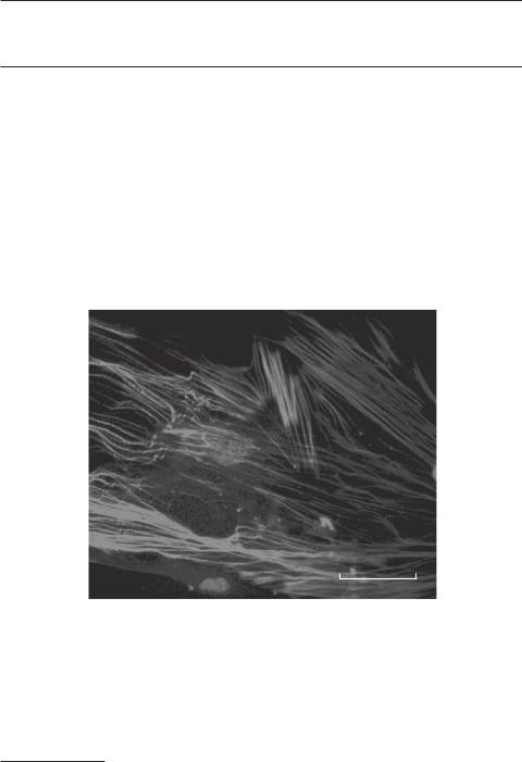
α-Actin filaments in vascular smooth muscle cells derived from the mouse aorta. Smooth muscle cells were collected from the medial layer of the mouse aorta and cultured for 10 days. The α-actin filaments were labeled with an anti-smooth-muscle α actin antibody (red in color) and observed by fluorescence microscopy. Cell nuclei were labeled with Hoechst 33258 (blue in color). Scale bar: 5 μm. See color insert.
Bioregenerative Engineering: Principles and Applications, by Shu Q. Liu
Copyright © 2007 John Wiley & Sons, Inc.
52
CELL MEMBRANE |
53 |
A mammalian cell is composed of numbers of subcellular organelles, including the cell membrane, cytoskeleton, smooth and rough endoplasmic reticulum, Golgi apparatus, lysosomes, peroxisomes, mitochondria, and nucleus. A cell membrane is a phospholipid bilayer, which encloses cell contents and separates a cell into different compartments. The cytoskeleton is constituted with three distinct elements, including actin filaments, microtubules, and intermediate filaments, which not only give a cell shape, strength, and elasticity but also regulate various cellular functions. Endoplasmic reticulum is the site where proteins and phospholipids are synthesized. The Golgi apparatus is an organelle in which proteins are processed and modulated. Lysosomes contain digestive enzymes, participating in the degradation of engulfed molecules or microorganisms. Peroxisomes contain enzymes for the mediation of oxidative reactions. Mitochondria are machineries that generate and store energy in the form of ATP. The nucleus contains chromosomes and is the center for the storage and processing of genetic information. It becomes clear that each cellular organelle possesses distinct structure and function, yet all cell organelles work together in a highly coordinated manner, ensuring appropriate regulation of cellular activities and functions. In this chapter, the structure, organization, and function of major cellular organelles and compartments are briefly reviewed.
CELL MEMBRANE [3.1]
The cell membrane is composed of lipids and proteins. As discussed in Chapter 1, lipids are amphipathic in nature (i.e., each molecule contains a polar hydrophilic and a nonpolar hydrophobic end) and can spontaneously form bilayers when mixed with an aqueous solution. The most abundant lipids are phospholipids in the cell membrane. Each phospholipid molecule contains a polar hydrophilic head and two nonpolar hydrophobic tails. In addition, cholesterol molecules can be found in a cell membrane. The membrane of a mammalian cell contains about 1 × 10⁹ lipid molecules. Lipid molecules constitute about half of the membrane mass, while the remaining half is primarily proteins. The lipid composition is asymmetric between the two lipid layers of the cell membrane. For instance, glycosphingolipids are found primarily in the external layer, whereas phosphatidylserine is in the internal layer. The primary functions of cell membranes are to separate cellular contents from the extracellular space, create a suitable internal environment for intracellular activities, and establish subcellular compartments for various metabolic and signaling processes.
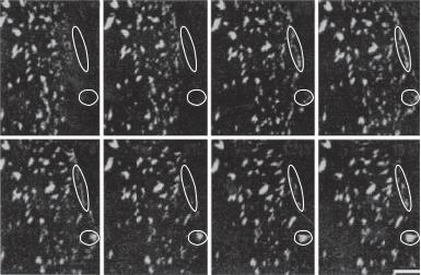
A lipid bilayer is a fluid-like structure. Lipid molecules can move laterally or diffuse within a lipid monolayer, but cannot change the molecular polarity or flip from one lipid layer to the other. The fluid-like feature of lipid bilayers is dependent on the composition of the cell membrane. For instance, cholesterol molecules reduce the fluidity of cell membranes, and thus enhance the membrane rigidity. The fluidity of a cell membrane ensures dynamic movement of membrane components, including not only lipids but also proteins. The movement of membrane molecules is critical to the function of these molecules as well as the cell. For instance, integrins move toward the leading edge of cell migration and participate in the construction of focal adhesion contacts, regulating cell attachment to the substrate (Fig. 3.1). Growth factor receptors move dynamically, resulting in the redistribution of the receptors to regions that require increased signal inputs from growth factors.
54 STRUCTURE AND FUNCTION OF CELLULAR COMPONENTS
0'1' |
1'2' |
4'5' |
5'6' |
7'8' |
9'10' |
12'13' |
13'14' |
Figure 3.1. Dynamic formation of β3 integrin complexes in porcine arterial endothelial cells. Endothelial cells were transfected with a GFP-β3 integrin gene and cultured to confluence. Cell wound was created by mechanical scraping, which induces cell migration. The images were taken from migrating endothelial cells. Note that new integrin aggregates form at the leading edge of the migrating cells (within the ovals). The times of the sequential images are indicated at the upper right corners. Scale bar: 5 μm. (Reprinted from Zaidel-Bar R et al: J Cell Sci 116:4605–13, 2003 by permission of The Company of Biologists Ltd.)
The cell membrane contains various types and amounts of proteins, depending on the type and function of the cell. For instance, a myelin membrane, which encloses and protects the nerve axon, contains proteins about 25% of the membrane mass, whereas a cell membrane that is involved in extensive molecular transport and ligand–receptor interaction may contain up to 75% proteins. Cell membrane proteins may serve as ligand receptors, ion pumps, water and ion channels, or molecule carriers. Membrane proteins can be divided into several classes based on the structure and relationship with the lipid bilayer. One type is transmembrane proteins, which pass through the cell membrane and consist of three domains: the extracellular, transmembrane, and intracellular domains. The extracellular and intracellular domains are usually hydrophilic, whereas the transmembrane domain is hydrophobic. The hydrophilic domains can interact with water-soluble proteins, while the hydrophobic domain interacts with the fatty acid tails of membrane lipids via covalent bonds, serving as an anchoring structure for the protein. The second type of membrane protein is found at the external surface of a cell membrane. These proteins attach to the lipid bilayer via the linkage of oligosaccharides. The third type of protein attaches to the intracellular side of the cell membrane via covalent bonds with fatty acids. In addition, some proteins attach to membrane proteins via noncovalent bonds. The structural relationship between a protein and the cell membrane usually determines the protein function. For instance, transmembrane proteins are responsible for molecular transport across the cell membrane and signal transduction from extracellular ligands to intracellular signaling pathways. Proteins attached to the cytosolic side of the cell membrane usually serve as signaling molecules, which relay signals from transmembrane protein receptors.
CYTOSKELETON 55
CYTOSKELETON
The cell contains a filamentous framework, known as the cytoskeleton. There are three cytoskeletal elements: the actin filaments, intermediate filaments, and microtubules. These filaments not only determine the shape and mechanical strength but also participate in the regulation of cellular activities, such as cell adhesion, division, migration, and apoptosis. The structure and function of these filaments are briefly discussed here.
Actin Filaments
Structure and Organization of Actin Filaments [3.2]. An actin filament is a helical structure of 8 nm in diameter and is established via polymerization of actin monomers. Each actin monomer contains about 375 amino acid residues with a molecular size about 43 kDa. In mammalian cells, there exist several isoforms of actin (see examples listed in Table 3.1), including the α and β isoforms in muscular cells and β and γ isoforms in nonmuscular cells. The α type of actin constitutes the contractile actin filaments in skeletal, cardiac, and smooth muscle cells. The β and γ types of actin participate in the constitution of the cytoskeleton. Actin filaments with various actin isoforms are localized to different compartments in both muscular and nonmuscular cells. For instance, in nonmuscular cells, β-actin is primarily found near the edge of the cell membrane, whereas γ-actin constitutes stress fibers, which are distributed more uniformly. An actin filament is a polarized structure. When an actin filament is bound with myosin molecules, an array of asymmetric arrowhead-like structures appears under an electron microscope. The end of an actin filament consistent with the arrowhead is defined as the pointed end, whereas the other end is defined the barbed end.
Actin monomers can be self-assembled or polymerized into actin filaments through biochemical reactions (Fig. 3.2). Actin polymerization is accomplished in several steps, including actin nucleation, filament growth, and ATP hydrolysis. Actin nucleation is a process that induces the formation of actin trimers. These trimeric actin structures, known as actin nuclei, serve as initiators for actin polymerization or filament growth. In addition, actin polymerization can be initiated from the barbed end of grown actin filaments or random sites along the side of actin filaments (Fig. 3.2). The addition of an ATP-actin to an actin nucleus or an actin filament triggers hydrolysis of ATP into ADP and phosphate. The phosphate group dissociates from the actin, leaving a newly added actin molecule with a tightly bound ADP.
An actin filament can be simultaneously polymerized and depolymerized at both ends. Under a steady physiological condition, the addition of actin subunits to the barbed end of an actin filament is counterbalanced by the dissociation of actin subunits from the pointed end, resulting in a relatively constant density for actin monomers and filaments. However, the rate of polymerization and depolymerization may change in response to environmental alterations. For instance, an increase in the concentration of ATP and the presence of cations lower the critical level of actin monomers, enhancing actin polymerization. Actin monomers above a critical concentration can be all assembled into actin filaments.
Actin-Binding Proteins. Actin polymerization and depolymerization are regulated by numbers of actin-binding proteins. These proteins are classified into various groups on the basis of their functions, including actin monomer-binding proteins, actin filament-