

Bioregenerative Engineering Principles and Applications - Shu Q. Liu
..pdf16 STRUCTURE AND FUNCTION OF MACROMOLECULES
system have established that RNA transcription is accomplished through several steps, including initiation, elongation, and termination. Similar processes are found in eukaryotic RNA transcription with several exceptions (see below). Here, the E. coli system is used to describe the mechanisms of RNA transcription.
Initiation. A RNA molecule is transcribed from a specific gene and encodes information for the sequence of amino acids of a protein. RNA transcription is initiated at a DNA site next to a special DNA sequence known as a promoter, which is found in each gene and activated in response to the binding of transcription factors to specific DNA sequences known as enhancers. There are two types of promoter in the E. coli genome with consensus sequences TTGACAT and TATAAT, which are located at the −35 and −10 sites relative to the first base pair (+1) to be transcribed. On the binding of transcription factors, a RNA polymerase binds to a gene at the promoter region and unwinds the double-stranded DNA at the −10 site, forming an open promoter complex or “transcription bubble” (Fig. 1.8). Such a process initiates RNA transcription.
Elongation. The elongation of RNA is catalyzed by RNA polymerase, a complex enzyme that contains four subunits: α, β, β′, and σ subunits. The subunits can be separated from each another and bound to the other units. A complete complex of RNA polymerase subunits is required for the initiation of RNA transcription. RNA polymerase can recruit nucleotides and synthesize RNA based on a template gene according to the base-pairing principle. The enzyme can catalyze the addition of a nucleoside monophosphate to an existing RNA fragment while a diphosphate group is released from a recruited nucleoside triphosphate. The release of the diphosphate provides energy for the synthetic process. The “transcription bubble” continues to extend when RNA is synthesized (Fig. 1.8). RNA synthesis occurs in the 5′ → 3′ direction.
Termination. RNA synthesis is terminated when the RNA polymerase recognizes a termination sequence on the DNA template strand. This sequence contains a GC-rich segment followed by a stretch of six or more adenosine nucleotides. The GC-rich segment of the synthesized RNA can fold regionally, forming a short, hairpin-shaped, double-stranded RNA segment. This segment is followed by U-rich segment. These structures serve as a termination signal. The RNA polymerase is then released and RNA synthesis terminated when the enzyme recognizes these structures.
RNA Transcription and Processing in Eukaryotes. While RNA transcription in eukaryotes is similar to that in prokaryotes in principle, there are several differences. Unlike prokaryotes, eukaryotes express at least three RNA polymerases, including polymerase I,
ATCGATCGATCGAT CG |
A T |
C G AT C G ATCGATC |
GA |
||
|
C G |
|
A |
T CGATCGATCGATCG |
|
TAGCTAGCTAGCTAG CTA |
|
|
|||
|
AUC GAUC G UCGTA GCTAGCTAGCTAGC |
||||
|
|
G C TA |
CTAG |
C |
|
|
|
A |
|||
|
|
|
G C TAG |
|
U |
|
|
|
|
|
C |
|
|
|
|
|
G |
RNA polymerase |
|
|
|
|
mRNA |
|
|
|
|
|
|
Figure 1.8. Schematic representation of RNA transcription (based on bibliography 1.5).
PROTEINS 17
II, and III. These polymerases possess distinct functions. Polymerase I is for the synthesis of rRNA, polymerase II is for mRNA, whereas polymerase III is for tRNA.
In addition, transcribed RNAs in the nucleus are processed in eukaryotes before being delivered to the cytoplasm for protein translation. Immediately after the transcription of an mRNA molecule, an enzyme called guanyltransferase is activated, adding a 7- methylguanosine residue to the 5′ end of the mRNA transcript. A sequence at the 3′ end of the transcript, usually AAUAAA, activates an endonuclease, which cleaves the mRNA molecule ( 20 bp) at the 3′ end. This step is followed by the activation of a poly(A) polymerase, inducing the addition of a poly(A) tail of 150–200 adenosine residues to the 3′ end of the mRNA. These processes generate a complete primary structure of an mRNA molecule.
Unlike prokaryotes, eukaryotes contain primary mRNAs that are composed of two types of sequence known as exons and introns. The exons contain protein coding regions and are used for protein translation, whereas the introns are sequences that interrupt the exons and do not contain protein coding information. An mRNA transcript must be spliced to remove the introns before protein translation occurs. A mature mRNA molecule is produced after the removal of the introns and is ready for protein translation. In eukaryotes, mRNA splicing occurs in a structure called splicesome, which is composed of splicing enzymes and associated factors. In addition to mRNAs, tRNAs, and rRNAs are also processed by splicing to remove introns.
PROTEINS
Protein Composition and Structure [1.6]
Proteins are molecules constituted with amino acids and are major components of living cells, constituting about 50% of the cell dry weight. There are two types of proteins in the cell: structural and regulatory proteins. Structural proteins participate in cell construction, giving a cell the shape, strength, and elasticity, whereas regulatory proteins control biological processes, such as cell-to-cell and cell-to-matrix communication, intracellular enzyme activation, signal transduction, control of gene expression, transport of necessary compounds for cell metabolism, cell division, cell differentiation, cell migration, and cell apoptosis. It is important to note that structural proteins are also involved in the regulation of cellular activities, which become clear in the following examples. Structural proteins include, for example, actin filaments, microtubules, and intermediate filaments, which form an integrated structure called cytoskeleton. While these proteins provide the cell with shape and strength, they play a critical role in the regulation of cell mitosis, migration, and adhesion. Regulatory proteins are found in the cell membrane, cytoplasm, and nucleus. Cell membrane proteins, such as growth factor receptors, adhesion molecules, and integrins, are responsible for cell-to-cell and cell-to-matrix interactions. Cytoplasmic proteins are mostly enzymes. Nuclear proteins are involved in the regulation of chromosomal organization and gene expression.
A protein consists of one or more peptides, which are constituted with 20 types of amino acid with distinct structure and chemical properties (Table 1.3). The combination of these amino acids gives rise to a variety of different peptides. The sequence of amino acids is specified by a corresponding gene. The length of a peptide varies widely, ranging from several amino acid residues, such as oxytocin, to about 25,000 residues, including titin. Most peptides are composed of 100–1000 amino acids.

18 STRUCTURE AND FUNCTION OF MACROMOLECULES
TABLE 1.3. Amino Acids Found in Humans and Animals*
|
|
|
|
|
Three-Letter |
One-Letter |
Amino Acids |
|
Chemical Forms |
Abbreviation |
Abbreviation |
||
|
|
|
|
|
||
Alanine |
CH3CHCOOH |
|
Ala |
A |
||
|
|
| |
|
|
|
|
|
NH2 |
|
|
|
|
|
Arginine |
NH |
|
|
Arg |
R |
|
|
|
|| |
|
|
|
|
|
H2NCNHCH2CH2CH2CHCOOH |
|
|
|||
|
|
|
|
| |
|
|
|
|
|
|
NH2 |
|
|
Asparagine |
O |
|
|
Asn |
N |
|
|
|
|| |
|
|
|
|
|
N2NCCH2CHCOOH |
|
|
|||
|
|
| |
|
|
|
|
|
|
NH2 |
|
|
|
|
Aspartic acid |
HOOCCH2CHCOOH |
Asp |
D |
|||
|
|
| |
|
|
|
|
|
|
NH2 |
|
|
|
|
Cysteine |
HSCH2CHCOOH |
Cys |
C |
|||
|
|
| |
|
|
|
|
|
|
NH2 |
|
|
|
|
Glutamic acid |
HOOCCH2CH2CHCOOH |
Glu |
E |
|||
|
|
|
| |
|
|
|
|
|
|
NH2 |
|
|
|
Glutamine |
O |
|
|
Gln |
Q |
|
|
|
|| |
|
|
|
|
|
N2NCCH2CH2CHCOOH |
|
|
|||
|
|
|
|
| |
|
|
|
|
|
NH2 |
|
|
|
Glycine |
HCHCOOH |
|
|
Gly |
G |
|
|
| |
|
|
|
|
|
|
NH3 |
+ |
|
|
|
|
Histidine |
|
CH2 |
CH COOH |
His |
H |
|
|
HN |
N: |
|
NH2 |
|
|
|
|
|
|
|
||
Isoleucine |
|
CH3 |
|
|
Ile |
I |
|
|
| |
|
|
|
|
|
CH3CH2CHCHCOOH |
|
|
|||
|
|
|
| |
|
|
|
|
|
NH2 |
|
|
|
|
Leucine |
CH2CHCH2CHCOOH |
Leu |
L |
|||
|
|
| |
|
| |
|
|
|
CH3 |
NH2 |
|
|
||
|
|
|
|
|
PROTEINS 19 |
TABLE 1.3. Continued |
|
|
|
|
|
|
|
|
|
|
|
|
|
|
|
Three-Letter |
One-Letter |
Amino Acids |
|
Chemical Forms |
Abbreviation |
Abbreviation |
|
|
|
|
|
||
Lysine |
H2NCH2CH2CH2CH2CHCOOH| |
Lys |
K |
||
|
|
|
NH2 |
|
|
Methionine |
CH3SCH2CH2CHCOOH |
Met |
M |
||
|
|
|
| |
|
|
|
|
|
NH2 |
|
|
Phenylalanine |
|
CH2 CH COOH |
Phe |
F |
|
|
|
|
NH2 |
|
|
Proline |
+ |
COOH |
Pro |
P |
|
N |
|||||
|
H |
H |
|
|
|
Serine |
HOCH2CHCOOH |
Ser |
S |
||
|
|
| |
|
|
|
|
|
NH2 |
|
|
|
Threonine |
OH |
|
Thr |
T |
|
|
|
| |
|
|
|
|
CH3CHCHCOOH |
|
|
||
|
|
| |
|
|
|
|
|
NH2 |
|
|
|
Tryptophan |
|
|
CH2 CH COOH |
Trp |
W |
|
|
NH2 |
|||
|
|
N |
|
|
|
|
|
H |
|
|
|
Tyrosine |
HO |
|
CH2 CH COOH |
Tyr |
Y |
|
|
|
NH2 |
|
|
Valine |
(CH3)2CHCHCOOH |
Val |
V |
||
|
|
| |
|
|
|
NH2
*Based on bibliography 1.6.
With the exception of one amino acid, proline, all amino acids are formed on the basis of a common structure, containing a central α-carbon atom bound with an amino group, a carboxyl group, a hydrogen atom, and a sidechain, the R group. Each amino acid has a distinct sidechain (Table 1.3). It is the sidechain that determines the unique chemical and physical properties of an amino acid. Proline has a cyclic sidechain linking the α carbon and the nitrogen, forming an imino group. Based on the structure and properties of the sidechains, amino acids can be classified into two major groups: charged and uncharged amino acids.
Charged amino acids include those with positive and negative charges. Amino acids with positive charges include arginine, histidine, and lysine, which are also known as basic amino acids. Those with negative charges include aspartic acid and glutamic acid,
20 STRUCTURE AND FUNCTION OF MACROMOLECULES
also known as acidic amino acids. The charged amino acids are all hydrophilic in nature, capable of interacting with water, which determines the hydrophilic features of proteins.
The remaining 15 amino acids are all uncharged. Among these amino acids, five are polar amino acids, including serine, threonine, tyrosine, asparagines, and glutamine. The sidechains of these amino acids contain either polar hydrogen bond donors or acceptors, capable of interacting with water. These are considered hydrophilic amino acids. The remaining 10 uncharged amino acids possess nonpolar sidechains and interact poorly with water. These amino acids include glycine, alanine, valine, leucine, cysteine, methionine, praline, isoleucine, phenylalanine, and tryptophan. These are considered hydrophobic amino acids. Because of the versatility of the amino acids, a large number of proteins can be constructed.
Several amino acids can be modified by the addition of various chemical groups under the action of specific enzymes. For instance, a phosphate group can be added to serine, threonine, tyrosine, and histidine by a family of enzymes known as protein kinases, resulting in amino acid phosphorylation. Such a process plays a critical role in the regulation of a variety of cellular activities, such as cell division, adhesion, and migration. Other types of amino acid modification include the addition of the methyl group to lysine and the hydroxyl group to proline, as well as the formation of disulfate bonds between adjacent cysteine residues. These modifications are essential to the function and stability of proteins.
Protein Translation [1.7]
Protein translation is a process that synthesizes protein. The synthesis of proteins requires three basic elements: ribosomes, messenger RNA (mRNA), and transfer RNA (tRNA). Ribosomes are composed of rRNA components and regulatory proteins. There are three rRNA modules in E. coli ribosomes with distinct molecular sizes: 5S, 16S, and 23S (S: sedimentation velocity, a measure of molecular size). These molecules are transcribed from specific DNA sequences at specified locations of a DNA molecule. rRNA components and ribosomal proteins form two complex subunits. In prokaryotes, the two subunits are 50S and 30S in molecular size, whereas in eukaryotes they are 60S and 40S. These ribosomal structures serve as machineries for protein synthesis.
A messenger RNA molecule is a sequence of linear nucleotides, carrying genetic codons that dictate the sequence of amino acids for a specific protein. The genetic codons are stored in a DNA molecule. DNA transcription transmits the genetic codons to mRNA molecules. Each amino acid is represented by a codon of three nucleotides. In other words, each 3-nucleotide codon determines a type of amino acid to be incorporated into a peptide at a specified site. Genetic codes for all amino acids found in humans are listed in Table 1.4. In addition to the codons for amino acids, each mRNA molecule contains stop codons, including UAG, UGA, and UAA, which signal the termination of peptide translation when recognized by a ribosome.
A tRNA molecule is responsible for the transport of an amino acid to a ribosome during protein synthesis. Transfer RNA is able to not only identify a specific amino acid but also recognize a corresponding codon on a mRNA molecule, ensuring the placement of the amino acid to an appropriate position. There are two functional domains in each tRNA molecule: an anticodon and an amino acid attachment site. The anticodon is a sevennucleotide sequence that recognizes and binds to a mRNA site according to the complementary rule. The amino acid attachment site binds to an amino acid. Each amino acid

PROTEINS 21
TABLE 1.4. Genetic Codes for Amino Acids*
Amino Acids |
|
|
Genetic Codes |
|
|
|
|
|
|
Ala |
GCU |
GCC |
GCA |
GCG |
Arg |
CGU CGC CGA CGG AGA AGG |
|||
Asn |
AAU |
AAC |
|
|
Asp |
GAU |
GAC |
|
|
Cys |
UGU |
UGC |
|
|
Gln |
CAA |
CAG |
|
|
Glu |
GAA |
GAG |
|
|
Gly |
GGU |
GGC |
GGA |
GGG |
His |
CAU |
CAC |
|
|
Ile |
AUU |
AUC |
AUA |
|
Leu |
UUA UUG CUU CUC CUA CUG |
|||
Lys |
AAA |
AAG |
|
|
Met |
AUG |
|
|
|
Phe |
UUU |
UUC |
|
|
Pro |
CCU |
CCC |
CCA |
CCG |
Ser |
UCU UCC UCA UCG AGU AGC |
|||
Thr |
ACU |
ACC |
ACA |
ACG |
Trp |
UGG |
|
|
|
Tyr |
UAU |
UAC |
|
|
Val |
GUU |
GUC |
GUA |
GUG |
Initiation codes |
AUG |
GUG |
|
|
Stop codes |
UAA |
UAG |
UGA |
|
|
|
|
|
|
*Based on bibliography 1.7.
is carried by a specific tRNA molecule. Like rRNA, a tRNA molecule is coded by a specific gene at a specified location in a DNA molecule.
Protein translation is accomplished via three steps: initiation, elongation, and termination. These processes have been well understood in prokaryotes. Here, the prokaryote protein translation system is used as an example.
Initiation. The initiation of protein translation requires RNA molecules, including mRNA, rRNA, and tRNA, and rRNA-constituted ribosomes. In addition, three regulatory factors, termed initiation factors (IFs) 1, 2, and 3, are necessary. Several steps are involved for the initiation of protein translation. First, the translation of protein starts with the activation of IF3, which stimulates the binding of mRNA to the 30S subunit of ribosome. mRNA binding induces the attachment of the 50S subunit to the 30S subunit, forming a complete ribosome (note that the two ribosomal subunits are present in separate forms without protein synthesis when they are not activated). Second, IF2 is activated to bind GTP and fMet-tRNA, which is a translation initiator tRNA carrying N-formylmethionine (fMet). This combination stimulates the attachment of fMet-tRNA to a specific initiation codon (AUG or GUG) on the mRNA molecule localized to an rRNA site, known as the P site. An initiation codon is preceded by a sequence that can hybridize to rRNA. The GTP molecule provides energy for the ribosomal assembly process. When a phosphate group is removed from the GTP molecule, IF2 and IF3 are released from the ribosomal complex.
22 STRUCTURE AND FUNCTION OF MACROMOLECULES
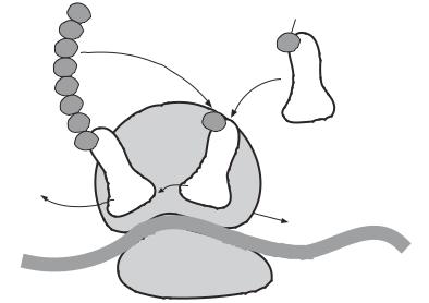
Elongation. Elongation is a process by which amino acids are added to the initiation fMet-tRNA one at a time. Such process is regulated by several protein elongation factors (EFs), including EF-Tu, EF-Ts, and EF-G. The elongation factor EF-Tu regulates the attachment of aminoacyl-tRNAs to a specific mRNA codon localized to an rRNA site adjacent to the P site, known as the A site. The binding of GTP to the aminoacyl-tRNA is required in this process for supplying energy. When GTP is hydrolyzed, elongation factor EF-Ts attaches to the ribosome, regulating the release of the EF-Tu-GDP complex, leaving tRNA at the A site. Note that at this state the first amino acid or a synthesized partial peptide is attached to the P site. An enzyme called peptidyltransferase catalyzes a process that transfer the amino acid or partial peptide from the P to the A site. At the same time, the elongation factor EF-G initiates a process that moves the mRNA molecule by three base pairs in the 5′ → 3′ direction, which is associated with the release of the tRNA at the P site and the transfer of the peptide together with the associated tRNA from the A to the P site (Fig. 1.9).
Termination. Termination of protein translation is initiated when one of the three stop codons of the mRNA, specifically UAG, UAA, and UGA, appears at the A site. At least three regulatory proteins, known as release factors (RFs) 1, 2, and 3, regulate translation termination. These release factors can recognize and bind to a stop codon at the A site of the ribosome, inducing the release of synthesized peptide from the P site. The ribosome is then dissociated into two free subunits. Synthesized peptides undergo protein splicing and folding processes, eventually forming proteins with a three-dimensional structure.
5'
3
AUG
Amino acid
2 |
|
|
|
|
|
|
|
|
|
|
Peptidyltransferase |
|
|
|
|
|
|
|
tRNA |
|
|
|
|
|
|
|
|
|
|
|
|
|
|
|
|
|
|
1 |
|
|
|
|
|
P |
|
A |
|
|
|
|
|
U C G |
|
|
|
|
|
|
|
|
|
|
|
||
|
4 |
|
|
|
|
|
|
|
|
|
G |
C U |
A |
|
|
4 |
|
|
|
|
|
U |
|
|
|
|
|
|
|
|
|
|
A |
G |
A |
|
|
|
|
|
|
|
|
C |
|
|
|
|
|
|
|
|
||
A |
|
U |
AG |
|
|
|
|
UU |
3' |
|
U |
|
|
|
|
|
|
|
|||
|
|
|
|
C |
|
|
|
G |
||
C |
|
|
|
|
UC |
|
|
C |
|
|
|
|
|
|
|
G |
A |
|
|||
G |
|
|
|
|
|
|
||||
|
|
|
|
|
|
A |
|
|
||
C |
|
|
|
|
|
|
|
|
|
|
Figure 1.9. Schematic representation of protein translation. Several steps are involved in protein elongation: (1) recruitment of an aminoacyl-tRNA molecule to site A; (2) transfer of a partial peptide from site P to site A; (3) removal of the tRNA at site P; (4) movement of the peptide–tRNA complex from site A to site P; (5) movement of the rRNA complex toward the 3′ direction by three base pairs (based on bibliography 1.7).
PROTEINS 23
Protein Folding and Architecture [1.8]
All proteins are folded into a three-dimensional structure after being translated. A protein is composed of one or more peptides, which are sequences of linearly jointed amino acids via peptide bonds. Three atoms from each amino acid, including the nitrogen from the amino group, the α-carbon, and the carbon from the carboxyl group, join together to form a central structure for each peptide called the polypeptide backbone. A peptide chain is synthesized in a ribosome based on the codons of an mRNA. The peptide end with a free amino group is referred to as the N-terminus, where peptide synthesis begins, and the end with a carboxyl group is the C-terminus. The counting of the number of amino acids starts at the N-terminus. Most peptide bonds can rotate freely, giving the flexibility of forming a variety of different conformations for proteins.
A peptide is usually folded into a three-dimensional (3D) structure, resulting in a final conformation that exhibits maximal stability and functionality. A denatured protein molecule under harsh conditions, such as extreme pH and a high concentration of urea, lose not only its conformation but also function. However, proteins are able to refold back to their original conformation and regain their functions under restored physiological conditions. Various chemical and physical features of the amino acids in a peptide chain determine the process of protein folding and the 3D protein conformation. The hydrophobic and hydrophilic features of amino acids play a critical role in controlling protein folding and conformation. The uncharged hydrophobic nonpolar sidechains of the amino acids intend to pack themselves in the core of a protein to minimize exposure to water molecules. The core of a protein contains the most conservative amino acids. In contrast, most charged and polar sidechains of amino acids are localized to the surface of a protein and capable of interacting with water molecules.
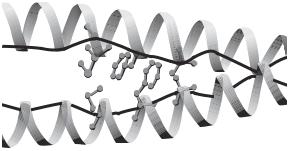
By X-ray diffraction and nuclear magnetic resonance spectroscopy, the structure of proteins can be determined to atomic accuracy. Protein structural analysis has demonstrated that proteins usually contain two types of secondary structure: α-helix and β-sheet. These structures are common in most proteins, although the overall conformation varies from protein to protein. An α-helix is a right-handed coiled structure with 3.6 residues per turn (Fig. 1.10). The helical structure is formed on the basis of hydrogen bonding between adjacent polar groups of the peptide backbone. A β-sheet is a structure containing several antiparallel segments of a single peptide, which is formed as a result
Figure 1.10. Crystallographic structure of a parallel α-helical coiled-coil dimer of the intermediate filament protein vimentin. (Reprinted from Burkhard P et al: Trends Cell Biol 11:82–8, 2001 by permission of Elsevier.)
24 STRUCTURE AND FUNCTION OF MACROMOLECULES
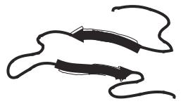
Figure 1.11. Schematic representation of the structure of a β-sheet-containing protein (based on bibliography 1.8).
of the back-and-forth turning of the peptide (Fig. 1.11). The antiparallel segments are linked together by lateral hydrogen bonds between the polar groups of the peptide backbone.
There exist peptide fragments that are not organized into regular structures like α- helices and β-sheets. These peptides exhibit disordered structures and can move more freely than a α-helix and β-sheet. Such structures are often found at the N- and C-termini. Collectively, α-helices, β-sheets, and irregular fragments are referred to as secondary structures (note that the amino acid sequences are known as the primary structure). These structures can be further organized into higher levels of 3D conformation. Examples of 3D protein structures include the coiled-coil structure, which is a complex of paired α- helices interacting laterally via hydrophobic bonds (Fig. 1.10), and the β-barrel structure, which is a cylindrical structure formed by a number of β-sheets. The formation of these superstructures enhances the stability of proteins. Furthermore, most proteins are composed of multiple peptides, which are integrated into a protein molecule. Proteins also form complexes such as dimmers and trimers. All these forms are essential for the functionality of proteins.
Changes in Protein Conformation [1.8]
Proteins undergo dynamic changes in their conformation. The atoms of a protein may move in a very fast speed in the order of 100 m/s within a nanometer range. Protein– protein interactions may induce conformational changes on the molecular scale. Such conformational changes play a critical role in the regulation of molecular activities. For instance, the binding of a growth factor to its receptor may cause a conformational change in the receptor, initiating autophosphorylation of the cytoplasmic receptor tyrosine kinase, which is associated with most growth factors. Conformational changes and autophosphorylation are critical processes for the activation of mitogenic signaling pathways. In the contractile apparatus of muscular cells, a conformational change in the myosin head, on the activation of a myosin molecule, is an essential process that initiates the sliding of actin filaments and the generation of forces. Conformational changes in protein kinases and phosphatases, such as the Src protein tyrosine kinase and the Src homology 2 domaincontaining protein tyrosine phosphatase, control the state of molecular activation. It is commonly received that a protein conformational change is an essential process for the regulation of protein functions.
LIPIDS 25
LIPIDS
There are various types of lipid molecules in mammalian cells, including phospholipids, glycolipids, steroids, and triglycerides. These lipid molecules play important roles in the construction of cellular structures and in the regulation of cellular functions. Lipids are the basic building blocks for cell and subcellular membranes, serve as hormones and intracellular signaling molecules, and contribute to the production of energy. The structure and function of common lipids are briefly described in the following sections.
Phospholipids [1.9]
Phospholipids are the primary constituents of cell membrane. A phospholipid contains several basic elements, including an alcohol, two fatty acid chains, and a phosphate group, which can be bonded with another alcohol group. Based on the alcohol structure, phospholipids can be further divided into two subgroups: phosphoglycerides and sphingolipids. A phosphoglyceride contains a glycerol as an alcohol group, whereas a sphingolipid contains a sphingosine.
Phosphoglycerides. In a phosphoglyceride molecule, two of the three —OH groups of the alcohol (glycerol) molecule are bonded with fatty acids via ester bonds, and the remaining —OH group is esterified by a phosphate group. The phosphate group can be bonded with another chemical groups, which can be either inositol, serine, ethanolamine, or choline (Fig. 1.12). Each combination gives rise to a distinct phosphoglyceride, including phosphatidylinositol, phosphatidylserine, phosphatidylethanolamine, or phosphatidylcholine, respectively. It is important to note that some of these phosphoglycerides not only contribute to the construction of cell membrane, but also serve as signaling molecules. For instance, phosphatidylinositol (Fig. 1.13), when phosphorylated into phosphatidylinositol 3,4 biphosphates, play a critical role in the regulation of G-protein receptor-initiated signal transduction (see Chapter 5).
A phosphoglyceride molecule contains a polar alcohol head and nonpolar fatty acid tails, which render the molecule amphipathic in nature, that is, hydrophilic at the head and hydrophobic at the tail. Such a modular arrangement determines the form of phosphoglyceride aggregation while they are mixed in an aqueous solution. The hydrophilic head of phosphoglyceride interacts with the water molecules, while the hydrophobic tail intends to interact with the hydrophobic tails from different phosphoglyceride molecules, resulting in the spontaneous formation of a lipid bilayer with two water-contacting surfaces composed of hydrophilic heads and an internal layer composed of hydrophobic tails (Fig. 1.14). Indeed, all cell membranes are established on the basis of such a principle.
Sphingolipids. Sphingolipids are composed of a sphingosine, two fatty acid chains, and a phosphate group (Fig. 1.15). These molecules possess properties similar to those of phosphoglycerides and can be found in the membrane of many cell types. Sphingolipids can aggregate into microdomains in cell membranes, which may play a role in targeting specific proteins to the plasma membrane and in organizing membrane-associated signaling pathways. For instance, K+ and other ion channels are localized to lipid microdomains on the cell surface. Sphingolipids can interact with ion channels and mediate the localization of the ion channels. Sphingolipids are also involved in the regulation of cellular