

Bioregenerative Engineering Principles and Applications - Shu Q. Liu
..pdf6
FUNDAMENTAL CELLULAR FUNCTIONS
Dynamic rearrangement of chromosomes and microtubules during mitosis. Hela cells are cultured and arrested by treatment with nocodazole, which results in the synchronization of the cells in prometaphase of mitosis. Following the removal of nocodazole, cells start to enter mitosis. Microtubules (green) and chromosomes (blue) undergo dynamic rearrangement during mitosis as shown at 60 and 90 min after the removal of nocodazole. [Reprinted by permission from Macmillan Publishers Ltd: Yasuda S et al: Nature 428:767–71, copyright 2004.] See color insert.
Bioregenerative Engineering: Principles and Applications, by Shu Q. Liu
Copyright © 2007 John Wiley & Sons, Inc.
256
CELL DIVISION |
257 |
The cell is capable of conducting a number of basic activities, including cell division (cell proliferation and differentiation), migration, adhesion, and apoptosis. These activities play critical roles in the development, morphogenesis, and remodeling of tissues and organs. Cell differentiation, proliferation, and migration are essential processes that contribute to the formation of specialized tissues and organs during development and to the regeneration of malfunctioned and lost cells in injury. Cell-to-cell and cell-to-matrix adhesion are critical processes for the formation of coherent tissue and organ systems. Cell apoptosis is a process that eliminates unnecessary cells, ensuring appropriate morphogenesis and development of tissues and organs. These basic activities are precisely controlled to achieve specialized functions for a living tissue, organ, and system. In a multicellular organism, various cellular activities may occur at different locations and levels. These activities must be precisely coordinated between different systems at the cellular, tissue, and organ levels, which is an essential mechanism for the survival and function of the entire organism. In this chapter, these fundamental cellular activities are briefly reviewed.
CELL DIVISION
Cell division is a process of cell reproduction, which generates progeny with identical genotypes. In mammalian cells, there are two types of division: mitosis and meiosis. Mitosis is a cell division process for nongerm cells. This process transmits identical copies of all genes from parent cells to daughter cells. Meiosis is a cell division process for germ cells, which produces gametes with half or 23 of the chromosomes.
Mitosis [6.1]
Cell division via mitosis may result in two consequences: cell proliferation and differentiation. Cell proliferation is a process of cell division, which results in the reproduction of progeny with identical phenotypes, that is, physical, chemical, and physiological characteristics. Cell differentiation is a process of cell division, which results in the production of specialized cells with phenotypes different from the mother cells. These are two fundamental processes, which determine the morphogenesis of tissues and organs during embryonic development and the structure and function of tissues and organs during physiological and pathological remodeling.
Cell proliferation and differentiation may coexist at a given time. The relative activity of cell proliferation and differentiation is dependent on the stage of development. During the embryonic and fetal stages, the two processes occur simultaneously. The differentiation of embryonic stem cells gives rise to various types of specialized cells for various tissue and organ systems, whereas the proliferation of a specialized cell type contributes to cell multiplication and the construction of a specialized tissue and organ. After birth, the activity of cell differentiation is relatively reduced, while cell proliferation remains prominent for tissue and organ growth. After reaching maturation, both cell differentiation and proliferation are reduced significantly. However, a basal level of cell proliferation and differentiation remains for the replacement of malfunctioned, lost, or aging cells. Although cell proliferation and differentiation result in different cell fates, both undergo a common cell division cycle.
258 FUNDAMENTAL CELLULAR FUNCTIONS
Cycle of Mitotic Cell Division
A cell division cycle is defined as the period between two mitotic cell divisions. A cell cycle is composed of two major periods: the interphase and mitotic phase (M). The interphase is defined as the period from the end of the prior M phase to the beginning of the next M phase and is traditionally divided into the G1 (gap 1), S (synthetic), and G2 (gap 2) phases. The M phase is the period during which cells divide, whereas the interphase is the period during which cells prepare for cell division. These different phases are corresponding to highly ordered and discrete molecular processes that regulate cell division.
The length of a cell cycle is dependent on the stage of development. During the early embryonic stage, the G1 and G2 phases are short or even missing. Embryonic cells primarily undergo alternations of the S and M phases. The duration of a cell cycle is <1 h. The short cell cycle is in coordination with the rapid growth of the early embryo. The length of a cell cycle increases with maturation. In a fully developed mammal, a typical cell cycle may last for 12–24 h with four discrete phases. Each phase of a standard cell cycle is briefly discussed here.
G1 Phase. The G1 phase is the interval during which the cell is prepared for DNA synthesis. It is 6–12 h in length, starting from the end of the M phase to the beginning of the S phase. During this phase, the cell size increases, due to the synthesis of proteins and lipids.
S Phase. The S phase is the period of DNA replication. It lasts for 7–8 hrs, starting from the end of G1 phase to the beginning of the G2 phase. The total DNA content and the number of chromatids are doubled by the end of the S phase. The nucleus increases in size apparently. In addition to DNA synthesis, other cellular components, including RNA and proteins, are also synthesized. However, DNA synthesis occurs only during the S phase, other components are synthesized continuously through the cell cycle. By the beginning of mitosis, the mass of the mother cell is doubled with cellular components equally apportioned for the two daughter cells.
G2 Phase. The G2 phase is the interval of cell preparation for cell mitosis. It is defined as the period (3–4 h) from the end of the S phase to the beginning of the M phase. During this phase, the cell possesses two complete diploid sets of chromosomes and is prepared for entering the mitotic phase. One of the major cellular activities during the G2 phase is the proofreading of the synthesized DNA. The detection of abnormal, damaged, or unreplicated DNA fragments can activate a protein kinase cascade of the G2 DNA damage checkpoint. The consequence of such activity is the inhibition of cyclin-dependent kinases, which are required for the initiation of mitosis, leading to a delay or blockade of entering the M phase.
M Phase. The M phase is the period of chromosome separation ( 1 h), staring from the end of the G2 phase to the beginning of the G1 phase. During this phase, the chromosomes are separated into two equal parts for the two daughter cells. The cytoplasm is separated via cytokinesis. Chromosome separation is accomplished through several stages, including the prophase, prometaphase, metaphase, anaphase, and telophase (Fig. 6.1).
During the prophase, chromatins, which are composed of DNA and associated proteins and are dispersed in the nucleus during the non-M phases, are condensed into discrete
CELL DIVISION |
259 |
Prophase |
Prometaphase |
Centrosome 

 Microtubule


 Kinetochore


 Chromosome
  Nuclear envelope
Metaphase
Telophase
Anaphase
Cytokinesis
Newly formed
nuclear envelope Microtubule Cleavage furrow
Figure 6.1. Schematic representation of cell mitosis. Mitosis is a process of cell division and consists of several defined phases, including prophase, prometaphase, metaphase, anaphase, telophase, and cytokinesis. Before a cell enters the prophase, the content of DNA is doubled. During the prophase, the chromosomes are organized from dispersed chromatins, the nuclear envelope is disassembled, the two centrosomes are deployed to the two mitotic poles, and microtubules are organized into a spindle-like network. During prometaphase, the chromosomes attach to the spindle microtubules and move toward the cell equator. During metaphase, the chromosomes are aligned along the cell equator and are ready for separation. During anaphase, the two chromatids of each chromosome complex are separated and move toward the opposite mitotic poles. During telophase, the chromosomes approach the mitotic poles, the nuclear envelope of the two daughter nuclei starts to form, and the cell is ready for cytokinesis. Cytokinesis is a process by which the cytoplasm is divided into two equal parts, each with a daughter nucleus. The formation of the daughter cells indicates the end of the cell division cycle. Based on bibliography 6.1.
chromosomes, each containing a pair of chromatids that are connected at the centromere. At the same time, the cytoskeletal microtubules start to reassemble into a mitotic spindle between two centrosomes outside the nucleus.
During the prometaphase, the nucleus envelope is disrupted, and the spindle microtubules are redistributed in the nucleus. The kinetochore protein, which forms a complex with the centromere of each chromatid, attaches to a selected spindle microtubule, establishing a kinetochore microtubule. Since each chromosome is composed of a pair of chromatids that are joined by two centromeres, each chromosome contains two kinetochore complexes. The kinetochore-free spindle microtubules are known as polar microtubules.
During the metaphase, with the help of the kinetochore microtubules, the chromosomes are aligned midway between the two spindle poles on a plane perpendicular to the spindle
260 FUNDAMENTAL CELLULAR FUNCTIONS
microtubules, which are connected to the spindle poles. This chromosome plane is defined as the metaphase plate.
During the anaphase, the kinetochore–centromere complex is separated and the two chromatids of each chromosome are pulled toward opposite spindle poles at a speed of1 μm/min. These activities are induced by the dynamic shortening of the kinetochore microtubules, which connect the chromatids. At the same time, the two spindle poles move away from each other and the polar microtubules, which are not connected to chromosomes, elongate in the spindle direction.
During the telophase, separated chromatids, now referred to as chromosomes, approach the two spindle poles symmetrically, the kinetochore microtubules gradually disappear, and the nucleus envelope appears around each group of chromosomes surrounding each spindle pole. The chromatin is gradually dispersed in the nucleus and the typical interphase nucleus starts to form, indicating the end of the M phase and beginning of the G1 phase.
Cytokinesis [6.2]
Cytokinesis is the process of cytoplasmic cleavage or segregation, which takes place immediately following chromosomal separation. During the late telophase, the cytoskeletal actin filaments and myosin are deployed along the cell equator, forming a contractile ring or cleavage furrow. The contractile ring gradually constricts the cell until the two daughter nuclei are completely separated with equal cytoplasm contents. This indicates the completion of the cell division cycle.
The formation of the cleavage furrow has always fascinated scientists. This is a complex process, which involves several structures, including the microtubule spindle, actin filaments and myosin, and cell membrane. It is now understood that the assembly of the contractile actin–myosin ring is regulated by several types of molecule. These include microtubule spindle-associated molecules, the RhoA guanosine triphosphatase (GTPase), myosin II, actin and actin-associated factors, and molecules mediating the fusion of membrane structures. The coordinated actions of all these structures and regulatory molecules contribute to the formation and performance of the contractile ring during cytokinesis.
Cell membrane fusion is an important part of cytokinesis. When the cytoplasm is separated by the constriction of the contractile ring, the cytoplasm of each daughter cell must be completely covered with the cell membrane. Two mechanisms are involved in the formation of the membrane of the daughter cells: (1) the daughter cells are able to generate additional cell membrane, which is deployed to the cleavage furrow to cover the open cytoplasm; and (2) membrane vesicles can be produced in the daughter cells. These vesicles can be transported to the cleavage furrow to bridge the open surface of the cytoplasm. Membrane fusion is required for the formation of a complete cell membrane.
Control of Cell Division [6.3]
The mitotic events described above are precisely controlled in a highly coordinated manner. The order and completeness of these events, which are critical to successful cell division, are controlled by two mechanisms: constitutive and extracellular. The constitutive mechanism is intrinsic in nature and ensures the initiation, progression, and completion of cell division. The extracellular mechanism is dependent on the stimulation of extracellular factors and ensures that cell division takes place in coordination with the global function and physiological status of a specialized tissue or organ. In other words,
CELL DIVISION |
261 |
the extracellular mechanism controls whether, when, and to what degree cell division occurs. Once a cell cycle is triggered by an extracellular cue, the constitutive mechanism controls the progression of cell division.
Both constitutive and extracellular mechanisms are implemented via the mediation of cell cycle regulatory molecules, including cyclins, cyclin-dependent kinases (CDKs), and inhibitors of cyclin-dependent kinases (CKIs). A common feature of cell cycle regulation is the occurrence of oscillatory changes in the activity of regulatory factors, which determine the cyclic events of cell division. In particular, periodic phosphorylation/dephosphorylation of CDKs and cyclic increase and decrease in the level of cyclins play key roles in the initiation, progression, and cessation of cell division.
Constitutive Control of Cell Division. The constitutive control mechanism is dependent on several “checkpoints.” These checkpoints inspect and control the progression of cell division and eliminate errors, if any. Checkpoints have been identified in all phases of the cell cycle. Several proteins, including p53, and p21, have been found to play a role in these checkpoints.
In the G1 phase, a major checkpoint is known as the restriction point, which controls the initiation of cell division. A cell can pass the G1 phase only if the prior mitosis is complete, DNA is undamaged, and the cell reaches a critical size. DNA damage, if any, must be repaired before a new cell cycle is initiated. Unrepaired DNA may result in the halt of the cell cycle. Also, a critical size of cell mass is required for cell division. The G1 phase is the longest period with the largest variation in length among all cell cycle phases. Thus, the G1 phase is timely flexible to adjust the rate of cell division in response to various stimulations.
The S-phase checkpoints assess the integrity of DNA. The checkpoints prevent the cell from synthesizing DNA, if the DNA is damaged or disrupted. All DNA molecules in the genome must be completely replicated, which is a prerequisite for the continuation of cell division to the G2 phase. The cell is arrested in the S phase if damaged DNA is found. Once a DNA replication process is initiated, it must be completed, or the cell cycle is stopped.
During the G2 phase, the newly replicated DNA is examined by the G2-phase checkpoints to ensure that the DNA molecules have been correctly duplicated. DNA repair factors can be activated to correct replication errors. These checkpoints determine whether the cell can move to the M phase. If the DNA molecules are not completely replicated, the cell is arrested before the M phase.
The M-phase checkpoints are responsible for detecting problems that potentially influence cell division. The checkpoints assess the completeness of proceeding preparatory events as well as the mitotic events. For example, unattached kinetochore can be detected by the checkpoints, leading to the halt of cell cycle progression. The completeness of the entire mitosis is also assessed by the M-phase checkpoints. Any incomplete events may result in cell arrest.
Some cells, especially terminally differentiated cells, are not committed to cell division and enter a phase known as the G0 phase, which resembles the G1 phase in certain aspects. These calls cannot pass the restriction points and cannot proceed to the S phase. However, under appropriate extracellular stimulations, some of these cells can be stimulated to pass the restriction points, initiating cell division.
The discovery of the cell cycle checkpoints has led to a significant advance in the understanding of the control mechanisms of cell division. All key processes of cell
262 FUNDAMENTAL CELLULAR FUNCTIONS
division are assessed by the checkpoints. The presence of continuous checkpoints through the cell cycle ensures the initiation, progression, and completion of cell division and the accuracy of cell reproduction.
Extracellular Control of Cell Division. There are a variety of extracellular factors that regulate the initiation and progression of cell division. These include growth factors, nutrient supplies, cell–cell interactions, and mechanical forces. Growth factors, such as platelet-derived growth factor, epidermal growth factor, fibroblast growth factor, activate cyclins and CDKs and promote the cell to enter the S phase and reduce the length of the G1 phase. In contrast, a growth inhibitor, such as transforming growth factor β, activates cell division inhibitors, such as p21, p27, and p57, and induces cell arrest. A reduction or depletion of nutrient supplies may promote the cell to enter the G0 phase. An increase in cell density or cell–cell contact may result in cell arrest. Mechanical forces have also been found to mediate cell mitosis. A decrease in bloodflow or fluid shear stress may activate the division cycle of vascular smooth muscle cells. In contrast, an increase in mechanical stretch or tensile stress in the wall of blood vessels induces mitosis of vascular smooth muscle cells. These mechanical factors may directly regulate cell mitosis or influence cell mitosis via the mediation of mitogenic factors.
Signaling Events of Cell Cycle Control. The progression of the cell cycle is regulated by a cascade of signaling molecules (Fig. 6.2). In the G1 phase, cells either enter the G0 phase or are committed to enter the S phase, initiating the cell division cycle. An increase in growth factors stimulates the cell to enter the S phase by inducing the expression of cyclin D. The level of cyclin D remains high through the G1, S, and G2 phases and rapidly reduces during the M phase via ubiquitination-mediated degradation (see Chapter 5). Increased cyclin D in the G1 phase promotes the formation of the cyclin D-CDK4/6 complex. Activated cyclin D-CDK4/6 complex stimulates cell growth, which is required for the passing of the G1 restriction point.
The cyclin D-CDK4/6 complex can phosphorylate retinoblastoma tumor suppressor (Rb), which contains the transcriptional factor E2F (elongation factor). The phosphorylation of Rb induces the release of E2F, which stimulates the expression of the cyclin E gene, resulting in a transient increase in cyclin E during the transition period from the G1 to S phase. Cyclin E forms a complex with CDK2 and the cyclin E/CDK2 complex is in turn activated by CDC25A-mediated dephosphorylation of CDK2 on the Thr14 and Tyr15 residues. Activated cyclin E-CDK2 complex promotes the cell to pass the G1 restriction point and enter the S phase. (See Table 6.1.)
During the early S phase, the transcriptional factor E2F stimulates the expression of cyclin A, which forms a complex with CDK2. The cyclin A-CDK2 and cyclin E-CDK2 complexes are capable of phosphorylating critical components that initiate and regulate DNA replication. The cyclin E-CDK2 complex is dissociated due to the degradation of cyclin E by the ubiquitin–proteasome system (see Chapter 5) during the early S phase, whereas the cyclin A-CDK2 complex remains active through the S and G2 phases.
During the S phase, another cyclin molecule, cyclin B, is gradually accumulated and forms a complex with cell division cycle protein (CDC)2. The cyclin B/CDC2 complex is known as the M-phase-promoting factor (MPF). MPF can be activated by CDC25B/C- mediated dephosphorylation at the G2/M transition. Activated MPF can phosphorylate a number of substrate proteins, including lamin, vimentin, and caldelsom, leading to cell mitosis. The phosphorylation of lamin is thought to induce the disruption of the cell

CELL DIVISION |
263 |
Growth factors
|
Cyclin D |
|
+ |
CDK4/6 |
|
|
|
|
|
|
|
|||||||
|
|
|
|
|
|
|
|
|||||||||||
|
|
|
|
|
|
|
|
|||||||||||
|
|
|
|
|
|
|
|
|
|
|
|
|
|
|
|
|||
Cyclin D-CDK4/6 |
|
|
|
|
|
|
|
|||||||||||
|
|
|
|
|
|
|
|
|
|
|
+ Rb |
|
||||||
Rb/E2F |
|
|
|
|
|
|||||||||||||
|
|
|
|
|
|
|
E2F |
|
||||||||||
|
|
|
|
|
|
|
|
|
|
|
|
|
|
|
|
|
|
G1 |
|
|
|
|
|
|
|
|
|
|
|
|
+ |
|
CDK2 |
|
|||
|
|
|
|
|
|
|
Cyclin E |
|
|
|
|
|||||||
|
|
|
|
|
|
|
|
|
|
|
|
|
|
|||||
|
|
|
|
|
|
|
|
Cyclin E/CDK2 |
|
P |
|
|||||||
|
|
|
|
|
|
CDC25A |
|
|
|
|
|
|
|
|||||
|
|
|
|
|
|
|
|
|
|
|
|
|
|
P |
|
|||
|
|
|
|
|
|
|
|
|
|
|
|
|
||||||
|
|
|
|
|
|
|
Cyclin E/CDK2 |
|
|
|
|
|
||||||
|
|
|
|
|
|
|
|
|
|
|
||||||||
|
|
|
|
|
|
|
|
|
|
|
|
|
||||||
|
|
|
|
|
|
|
DNA synthesis |
|
|
|
|
|
||||||
Expression |
Expression |
of cyclin D |
of cyclin E |
5' |
3' |
3' |
5' |
Figure 6.2. Schematic representation of the regulatory mechanisms of cell mitosis. Based on bibliography 6.3.
nucleus. The phosphorylation of vimentin is responsible for dynamic changes in microtubules and the formation of the spindle. The phosphorylation of caldelsom induces the interaction of actin filaments with myosin molecules, leading to the formation of the cleavage furrow and cell cytokinesis. MPF is deactivated by ubi-quitination of cyclin B during the M phase, which indicates the end of cell mitosis. (See Table 6.2.)
Inhibition of Cell Division Cycle. A family of proteins, including p15(INK4B), p16(INK4), p18(INK4C), and p19(INK4D), exerts an inhibitory effect on the activity of the cyclin D/CDK4/CDK 6 complex, and induces cell arrest during the G1 phase. The suppression of these inhibiting molecules leads to uncontrolled cell proliferation.

264
TABLE 6.1. Characteristics of Selected Cell Cycle Regulatory Molecules*
|
|
Amino |
Molecular |
|
|
Proteins |
Alternative Names |
Acids |
Weight (kDa) |
Expression |
Functions |
|
|
|
|
|
|
Cyclin D1 B-cell leukemia 1, BCL1 |
295 |
34 |
Liver, kidney, intestine, |
Binding to CDK4 and CDK6, |
|
|
|
|
|
uterus, pancreas, placenta |
mediating the activity of these |
|
|
|
|
|
CDKs, regulating the transition |
|
|
|
|
|
of the cell division cycle from the |
|
|
|
|
|
G1 to S phase, and promoting cell |
|
|
|
|
|
division and tumorigenesis |
Cyclin E1 Cyclin E, G1/S-specific cyclin E1 |
410 |
47 |
Lung |
Binding to CDK2, mediating the |
|
|
|
|
|
|
activity of this CDK, regulating |
|
|
|
|
|
the transition of cell division cycle |
|
|
|
|
|
from the G1 to S phases, and |
|
|
|
|
|
promoting cell division and |
|
|
|
|
|
tumorigenesis |
CDK2 |
Cyclin-dependent kinase 2, cell division |
298 |
34 |
Kidney, prostate, |
A serine/threonine protein kinase that |
|
kinase 2, p33 protein kinase, cell |
|
|
lymphocytes, skin |
regulates the transition from G1 to |
|
division protein kinase 2 |
|
|
|
S phases |
CDK4 |
Cyclin-dependent kinase 4, cell division |
303 |
34 |
Ubiquitous |
A serine/threonine protein kinase that |
|
kinase 4 |
|
|
|
phosphorylates the retinoblastoma |
|
|
|
|
|
protein and regulates the transition |
|
|
|
|
|
from G1 to S phase |
CDK6 |
Cyclin-dependent kinase 6, cell division |
326 |
37 |
Ubiquitous |
A serine/threonine protein kinase that |
|
protein kinase 6 |
|
|
|
forms a complex with CDK4 and |
|
|
|
|
|
regulates the transition from the |
|
|
|
|
|
G1 to S phases |
E2F1 |
E2F transcription factor 1, transcription |
437 |
47 |
Pancreas, connective tissue, |
Serving as a transcriptional factor, |
|
factor E2F, retinoblastoma associated |
|
|
skin |
stimulating gene transcription, and |
|
protein 1, and retinoblastoma-binding |
|
|
|
regulating cell division, |
|
protein 3 |
|
|
|
proliferation, differentiation, and |
|
|
|
|
|
apoptosis |
CDC25 |
Cell division cycle 25A, M-phase inducer |
523 |
59 |
Brain |
A phosphatase that activates CDC2 by |
|
phosphatase 1, dual-specificity |
|
|
|
dephosphorylation and regulates the |
|
phosphatase Cdc25A |
|
|
|
progression of cell division cycle |
|
|
|
|
|
from the G1 to S phases |
|
|
|
|
|
|
*Based on bibliography 6.3.
CELL DIVISION |
265 |
The p15 protein may be activated in response to TGF-β, which suppresses the proliferation of several cell types, including epithelial cell and smooth muscle cell. Another group of proteins, including p21, p27, and p57, may inhibit the activity of the cyclin D/CDK4/CDK6 and cyclin A/CDK2 complexes, and induce cell arrest in the G1 phase. The tumor suppressor protein p53 induces activation of p27, leading to cell arrest. (See Table 6.3.)
Meiosis [6.4]
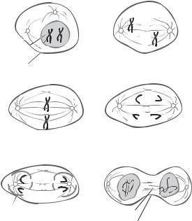
Meiosis is the process of gametogenesis or germ cell division. During such a process, a diploid germ progenitor cell undergoes DNA synthesis and two division events to produce four daughter cells with a haploid set of chromosomes. Each germ progenitor cell contains homologous pairs of chromosomes. Each homologous pair of chromosomes is composed of a maternal chromosome and a paternal chromosome, which can be identical or allelic (not completely identical). In response to the stimulation of signals for gametogenesis, the germ progenitor cell enters the S phase and initiates DNA synthesis, yielding two identical chromatids for each chromosome. The cell then enters two consecutive division processes, designated as meiosis I and meiosis II, to produce daughter germ cells. The meiosis I process is usually divided into several stages, including prophase I, metaphase I, anaphase I, and telophase I. The meiosis II process is divided into metaphase II, anaphase II, and telophase II. By the end of telophase II, four haploid daughter cells are produced from a single diploid germ progenitor cell (Fig. 6.3).
During meiosis prophase I, the nucleus envelope is reorganized, degraded, and disappeared. Centrosomes and microtubule spindles start to form. Scattered chromosomes are organized into apparently double-chromatid structures. The chromosomes can be clearly recognized under an optical microscope. Crossing over of chromatid fragments may occur between the maternal and paternal chromosomes, resulting in homologous recombination (Fig. 6.4). During meiosis metaphase I, a complete spindle and centrosome system is established. The chromosomes are aligned in the equator region. The centromeres of the chromosomes are connected to the spindles. During meiosis anaphase I, the chromosomes remain paired and are pulled toward the poles of the cell. Note that the chromosomal segregation process of meiosis is different from that of mitosis. In mitosis, the pair of chromatids for each chromosome is separated during the anaphase. In meiosis, the paired chromatids of each chromosome are not separated during anaphase I. During meiosis telophase I, the chromosomes are moved to the poles and rearranged. The germ progenitor cell is divided into two cells. However, no nucleus envelope is developed.
Meiosis II is the process by which the two daughter cells are further divided to produce haploid germ cells. The chromosomal separation in meiosis II is similar to that in mitosis. During meiosis metaphase II, the chromosomes in each daughter cell are aligned in the equator region. The centromeres are connected to the microtubule spindles. During meiosis anaphase II, the two identical chromatids of each chromosome are separated and pulled to the cell poles in opposite directions. During meiosis telophase II, each daughter cell is further divided into two granddaughter cells with a haploid set of chromosomes (a single copy of each chromosome from either the mother or the father). The chromosomes are rearranged and enveloped within the nucleus.
Experimental Assessment of Cell Division [6.5]
Cell division can be assessed by detecting DNA synthesis, which occurs only during cell division. There are two basic approaches for the detection of DNA synthesis: measuring